Overview
This chapter (Anatomy of Flowering Plants) introduces internal structure of angiosperms and explains how tissues and tissue systems are organized to perform functions such as support, transport, storage and protection. Starting from meristematic and permanent tissues, students study major tissue systems — epidermal, ground and vascular — and their constituent cells (parenchyma, collenchyma, sclerenchyma, tracheids, vessels, sieve elements and companion cells). The chapter compares anatomical features of monocot and dicot roots, stems and leaves through transverse sections, and explains secondary growth in dicotyledonous stems and roots (vascular cambium, cork cambium, secondary xylem and phloem, formation of annual rings). Understanding stomata, root cap, endodermis, pericycle and modifications of tissues for storage and protection is emphasized. Importance: anatomy links structure to function, aids identification of plant parts, has practical implications for forestry, agriculture, medicine and wood technology. After studying this chapter, students will be able to recognize and describe plant tissues microscopically and diagrammatically, compare monocot and dicot anatomy,…
Learning Objectives
- Define meristematic and permanent tissues and give examples of each in flowering plants
- Differentiate between simple and complex permanent tissues with two characteristic features and examples
- Describe the structure and functions of parenchyma, collenchyma and sclerenchyma cells
- Explain the structure, types and functions of xylem and phloem elements in vascular bundles
- Compare the transverse anatomy of monocot and dicot roots, stems and leaves and list key distinguishing features
- Draw and accurately label transverse sections of a dicot root, dicot stem and dicot leaf from memory
- Identify tissues and cell types in given prepared slides or labeled diagrams of root, stem and leaf
- Explain the process of secondary growth in dicot roots and stems by describing the role of vascular cambium and cork cambium
Topics in this chapter
30 topics · tap a topic title to jump straight to it.
Introduction
Definition and scope
Anatomy of flowering plants (angiosperms) is the study of the internal structure and organization of plant organs (root, stem, leaf, flowers) at tissue and cellular levels. The introductory ideas cover levels of organization, tissue systems, types of meristems, primary and secondary growth, common histological techniques and the practical importance of anatomy.
Levels of organisation
- Cell > Tissue (simple: parenchyma, collenchyma, sclerenchyma; complex: xylem, phloem) > Organ (root, stem, leaf) > Tissue system (dermal/epidermal, ground/fundamental, vascular).
Meristems and growth
- Meristems are regions of dividing cells: apical meristems (primary growth — increase in length), lateral meristems (vascular cambium and cork cambium — secondary growth, increase in girth).
- Primary tissues are formed by apical meristems; secondary tissues (secondary xylem and phloem, periderm) are products of lateral meristems.
Key anatomical features of organs (primary)
- Root: epidermis (root hairs), cortex with endodermis (Casparian strip), vascular cylinder with xylem and phloem arranged centrally.
- Stem: epidermis, cortex (may contain collenchyma), vascular bundles (arranged in ring in many dicots, scattered in monocots), pith.
- Leaf: dorsiventral or isobilateral structure; upper and lower epidermis, mesophyll (palisade and spongy), vascular bundles (veins).
Differences commonly emphasised (monocot vs dicot)
- Monocot stem: vascular bundles scattered, no typical cambium between xylem and phloem (limited secondary growth).
- Dicot stem: vascular bundles in a ring, cambium present between xylem & phloem enabling true secondary growth (wood formation).
Methods and techniques
- Sectioning (microtome), maceration, clearing, staining (e.g., safranin–fast green) and microscopic observation; modern methods include SEM and confocal microscopy.
Significance
Anatomical knowledge helps in plant identification, understanding transport and mechanical support, improving crop varieties, timber quality assessment, and diagnosing diseases. It links structure with function — e.g., presence of thick-walled xylem for conduction and support; cork and lenticels for protection and gaseous exchange.
- Maize (Zea mays) — typical monocot stem: scattered vascular bundles, no vascular cambium; demonstrates why most monocots lack secondary thickening.
- Sunflower (Helianthus annuus) or Pea (Pisum sativum) — dicot stem: vascular bundles in a ring with cambium allowing secondary growth.
- Mango (Mangifera indica), Teak (Tectona grandis) — trees showing pronounced secondary growth; growth rings visible in wood.
- Root hairs in bean or mustard — increase root surface area for water and mineral absorption; illustrates functional anatomy of roots.
- Bamboo — a monocot with massive culms but no true secondary growth (uses primary thickening and fibrous tissues for strength).
- Surface area to volume ratio (important for cells and absorption): SA:V = Surface area / Volume. (Example: for a sphere SA = 4πr^2, V = 4/3πr^3 ⇒ SA:V = 3/r.)
- Hagen–Poiseuille relation (applies conceptually to flow in xylem vessels): Q = (π r^4 ΔP) / (8 η l). This shows flow rate Q is proportional to the fourth power of vessel radius r — small increases in vessel diameter greatly increase hydraulic conductance.
Meristems
Meristems
Definition: Meristems are regions of actively dividing, undifferentiated plant cells responsible for growth. Cells in meristems remain embryonic and give rise to all primary and secondary tissues.
Characteristics of meristematic cells
- Small, isodiametric cells with dense cytoplasm and large prominent nucleus.
- Thin primary cell walls (no thick secondary walls) and very little or no vacuole.
- High mitotic activity (frequent cell divisions).
- Cells are totipotent/undifferentiated and can divide periclinally and anticlinally.
Classification (by position and origin)
- Apical meristems (at shoot and root tips) — responsible for primary (length) growth. Shoot apical meristem (SAM) and root apical meristem (RAM). SAM zones: central (stem cells), peripheral (gives rise to lateral organs), rib zone (gives stem tissues).
- Primary meristems (derived from apical meristem):
- Protoderm → epidermis
- Procambium → primary xylem and phloem
- Ground meristem → cortex and pith
- Lateral meristems (cylindrical, cause secondary growth/girth):
- Vascular cambium (produces secondary xylem inward and secondary phloem outward)
- Cork cambium / phellogen (produces periderm: phellem/cork outward and phelloderm inward)
- Intercalary meristems (at base of internodes or leaf bases in many monocots like grasses) — allow rapid elongation/regrowth after grazing or mowing.
Functions
- Produce new cells for primary and secondary growth (length and girth).
- Form all permanent tissues by differentiation.
- Participate in wound healing, regeneration, and formation of adventitious organs (roots/shoots).
- Serve as the basis for vegetative propagation and tissue culture techniques.
Regulation of meristem activity
Plant hormones and signals regulate meristem behaviour. Key regulators include:
- Auxins — maintain apical dominance and influence vascular differentiation;
- Cytokinins — promote cell division and shoot formation;
- Balance of auxin : cytokinin influences whether cells divide or differentiate;
- Genetic regulatory networks (e.g., WUSCHEL and CLAVATA in SAM) maintain stem cell populations.
Applications and significance
- Tissue culture and micropropagation: callus formation from meristematic cells and regeneration of whole plants.
- Horticulture: pruning, grafting and layering exploit meristem activity and wound-induced meristems.
- Crop resilience: intercalary meristems in grasses allow regrowth after grazing/mowing.
- Wood production: vascular cambium activity produces secondary xylem (wood).
Microscopy & experimental uses
Meristematic regions (e.g., onion root tip, Allium cepa) are widely used to study mitosis and calculate mitotic index.
Summary
Meristems are the plant’s growth centers — undifferentiated, actively dividing cells that generate primary and secondary tissues. Their position (apical, lateral, intercalary), regulation by hormones, and practical uses (tissue culture, propagation, wood formation) make them central to plant development and agriculture.
- Shoot apical meristem (SAM) at the tip of a shoot — produces leaves and flowers.
- Root apical meristem (RAM) at root tip — produces root cap and root tissues.
- Vascular cambium in woody stems — produces secondary xylem (wood) and secondary phloem (bark inner tissues).
- Cork cambium (phellogen) producing cork (phellem) in tree bark.
- Intercalary meristem in grasses (e.g., wheat, rice, sugarcane) — allows regrowth after mowing or grazing.
- Onion (Allium cepa) root tip used in laboratories to observe mitosis in meristematic cells.
- Mitotic index (%) = (Number of dividing cells / Total number of cells observed) × 100
- Relative Growth Rate (RGR) = (ln W2 − ln W1) / (t2 − t1), where W = plant mass or size at times t1 and t2
- Percentage increase in girth (over a period) = ((G2 − G1) / G1) × 100, where G1 and G2 are stem diameters at two dates
Tissue Systems
What is a tissue system? In flowering plants (angiosperms) tissues with related structure and function are grouped into three major tissue systems: the epidermal (dermal) system, the ground (fundamental) system and the vascular system. Each system extends throughout the plant and comprises several tissue types that arise from meristematic tissues and become permanent (differentiated).
Meristematic vs permanent tissues (brief):
- Meristematic tissues: composed of actively dividing, thin-walled cells (apical meristems, intercalary meristems, lateral meristems—vascular cambium and cork cambium).
- Permanent tissues: derived from meristems; cells have specific forms and functions and include simple and complex tissues.
Classification of permanent tissues
- Simple tissues: formed of similar cells. Examples: parenchyma (storage, photosynthesis, repair), collenchyma (flexible support), sclerenchyma (mechanical support; thick lignified walls; fibres and sclereids).
- Complex tissues: formed of different kinds of cells working together. Examples: xylem (water conduction and support) and phloem (organic solute conduction).
Epidermal (Dermal) Tissue System
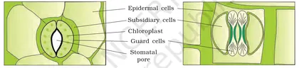
- Components: epidermis (single-cell layer in primary plant body), cuticle (waxy layer), stomata (guard cells + pore), trichomes (hair-like outgrowths), root hairs; in secondary growth the periderm (cork, phelloderm) replaces epidermis.
- Functions: protection against water loss, pathogens and mechanical injury; regulation of gas exchange (stomata); absorption in roots (root hairs); secretion in some trichomes.
Ground (Fundamental) Tissue System
- Parenchyma: living, thin-walled, often large intercellular spaces. Roles: storage (e.g., potato tuber), photosynthesis (leaf mesophyll), wound repair and regeneration.
- Collenchyma: living, unevenly thickened primary walls, provides flexible support in young stems and petioles (e.g., strands under the epidermis of celery petiole provide flexibility).
- Sclerenchyma: dead at maturity, thick lignified secondary walls; two forms—fibres (long, slender; support and tensile strength) and sclereids (variable shapes; hard seed coats, nutshells).
Vascular Tissue System
- Xylem (complex, conducts water & minerals; provides support): elements include tracheids, vessels (continuous tubes in angiosperms), xylem parenchyma, xylem fibres. Water movement is largely unidirectional (root → shoot) and assisted by cohesion–tension and root pressure.
- Phloem (complex, conducts organic solutes—mainly sucrose): sieve tube elements (living but enucleate in maturity in angiosperms), companion cells, phloem parenchyma, phloem fibres. Transport is bidirectional (sources → sinks) by pressure-flow mechanism.
- Arrangement differences: in dicot stems vascular bundles are arranged in a ring (allowing secondary growth via vascular cambium), while in monocot stems vascular bundles are scattered.
Integration and functions of tissue systems
- Tissue systems work together: epidermis reduces water loss while stomata and xylem/phloem manage gas exchange and transport; ground tissues store and support; vascular tissues create mechanical strength and transport pathways that integrate the whole plant.
- Growth: apical meristems give primary tissues (primary growth = increase in length); lateral meristems (vascular cambium, cork cambium) give secondary tissues (secondary growth = increase in girth).
Important concepts students should remember
- Distinguish simple vs complex tissues and meristematic vs permanent tissues.
- Know components and functions of each tissue system and typical examples (e.g., parenchyma in cortex and pith, collenchyma in petiole margins, sclerenchyma in seed coats, xylem vessels in woody stems, phloem sieve tubes in stems and leaves).
- Recognize tissue-system patterns in transverse sections of root, stem and leaf (e.g., epidermis outside, ground tissue in middle, vascular bundles centrally or in a ring).
Practical/observation tips: In practicals and diagrams, identify epidermis (outermost), cortical parenchyma, vascular bundles (xylem toward center, phloem toward outside in stems), and pith. Note stomata on lower leaf surface and root hairs on young roots.
- Epidermis and cuticle on leaves: reduces water loss — seen in xerophytic plants like cactus (thickened cuticle, sunken stomata).
- Root hairs (epidermal outgrowths) increase surface area for water and mineral absorption in most terrestrial plants.
- Parenchyma in potato tuber stores starch (amyloplast-rich cells).
- Collenchyma in celery stalks (petiole) provides flexible support — you can see the tough strands when you tear a celery stalk.
- Sclerenchyma fibres in jute provide tensile strength and are used commercially as fibre crops.
- Xylem vessels in woody stems conduct water; their arrangement in annual rings reflects secondary growth.
- Water potential: Ψ = Ψs + Ψp + Ψg (Ψ: total water potential; Ψs: solute/osmotic potential; Ψp: pressure potential; Ψg: gravitational potential)
- Surface area to volume ratio (approx. for a sphere): SA/V ≈ 6 / d (d = diameter). Relevant because cells/tissues exchange with environment based on SA:V.
- Fick's law (diffusion): J = -D (dC/dx) (J: flux, D: diffusion coefficient, dC/dx: concentration gradient). Used for movement of gases and solutes across tissues.
- Hagen–Poiseuille relation (flow in a tube): Q ∝ r^4 (Q: volumetric flow rate; r: vessel radius). Explains why wider xylem vessels conduct water much more efficiently.
- Pressure-flow (qualitative): mass flow rate ∝ (ΔP) / resistance. In phloem, pressure difference between source and sink drives bulk flow of sap.
Meristematic Tissues
Definition: Meristematic tissues are groups of actively dividing, undifferentiated plant cells that are responsible for growth (formation of new cells) and the production of primary and secondary tissues. They retain the ability to divide and give rise to various permanent tissues.
General characteristics of meristematic cells:
- Small, isodiametric cells with thin primary cell walls composed mainly of cellulose.
- Dense cytoplasm and prominent nucleus; few or no vacuoles.
- Closely packed cells with little intercellular space.
- High metabolic and mitotic activity (frequent cell division).
- Cells are embryonic/undifferentiated and capable of giving rise to various permanent tissues.
Types of meristems (based on position):
- Apical meristems: Located at the tips of roots and shoots; responsible for primary growth (increase in length). Example: shoot apical meristem (SAM), root apical meristem (RAM). RAM contains a quiescent centre surrounded by actively dividing initials.
- Lateral meristems: Cylindrical meristems responsible for secondary growth (increase in girth). Main examples: vascular cambium and cork cambium (phellogen).
- Intercalary meristems: Found at the base of internodes or leaf bases (common in monocots like grasses); allow rapid elongation and regrowth after grazing or mowing.
Types based on origin and function:
- Primary meristems: Derive from apical meristems and give rise to primary tissues (protoderm, procambium, ground meristem).
- Secondary meristems: Form later (e.g., vascular cambium) and produce secondary tissues (secondary xylem and phloem).
Organization concepts:
- Histogen theory: (Korshinsky) Proposes three histogens in shoot apex—tunica (surface layers), corpus (inner mass), and dermatogen (in some older formulations). Modern view refines these as tunica–corpus organization in many shoots: tunica divides anticlinally, corpus divides in various planes.
- Quiescent centre: A region in the root apical meristem with very slow cell division that helps maintain stem cell activity and organization of surrounding initials.
Functions:
- Generate new cells for plant growth—apical meristems for length, lateral meristems for thickness.
- Form all permanent tissues (dermal, ground, vascular) through cell division and differentiation.
- Permit regeneration and vegetative propagation (wound healing, callus formation in tissue culture).
Cell cycle and division: Meristematic cells divide by mitosis. The balance between cell division and subsequent cell enlargement/differentiation establishes organ formation (e.g., leaves, roots).
Practical and ecological significance:
- Commercial propagation: Meristem culture and micropropagation use meristematic explants for virus-free planting material.
- Tree growth rings: Result from seasonal activity of vascular cambium (a lateral meristem).
- Rapid regrowth of grasses after mowing or grazing is due to intercalary meristems.
Microscopic identification: Under the microscope meristematic tissue appears as a compact mass of small cells with dense cytoplasm, conspicuous nuclei, and thin walls. Staining often highlights large nuclei and abundant cytoplasm.
Summary: Meristematic tissues are the growth centres of plants—small, undifferentiated cells that divide actively to form primary and secondary tissues. Their position (apical, lateral, intercalary) determines the kind of growth (length, girth, or localized elongation) they produce.
- Shoot apical meristem (SAM) produces leaves and shoot tissues—example: shoot tip of a sunflower.
- Root apical meristem (RAM) at the tip of a root—with a quiescent centre—example: root tip of a bean.
- Vascular cambium (lateral meristem) produces secondary xylem and phloem—example: growth rings in a tree trunk (oak, pine).
- Cork cambium (phellogen) forms protective bark—example: cork oak (Quercus suber).
- Intercalary meristem at the base of grass internodes allows regrowth after cutting—example: lawn grass, sugarcane.
- Meristem tissue in tissue culture (callus) used for micropropagation and producing disease-free plants—example: banana or potato micropropagation.
- Cell doubling: N = N0 × 2^n (N = final number of cells, N0 = initial number of cells, n = number of mitotic divisions)
- Mitotic index (MI) = (Number of dividing cells / Total number of observed cells) × 100 (gives fraction/percent of cells in mitosis in a sample of meristematic tissue)
- Relative growth rate (mass or length basis): RGR = (ln W2 − ln W1) / (t2 − t1) (W = plant mass or organ length at times t1 and t2; useful to quantify growth driven by meristem activity)
Tissues — classification
Overview
In plants, a tissue is a group of cells similar in origin, structure and function. Plant tissues are broadly classified into two major categories: meristematic tissues (actively dividing) and permanent tissues (derived from meristems and matured).
1. Meristematic tissues
Characteristics: small, thin primary cell wall, dense cytoplasm, prominent nucleus, no vacuole, ability to divide. Function: growth by cell division. Based on position:
- Apical meristems — tips of root and shoot; produce primary tissues (primary growth).
- Intercalary meristems — at internodes or leaf bases (common in grasses); allow elongation after injury or grazing.
- Lateral meristems — along length; responsible for secondary growth: vascular cambium (produces secondary xylem and phloem) and cork cambium (phellogen, produces cork).
2. Permanent tissues
Permanent tissues are non-dividing and are of two types: simple and complex.
Simple permanent tissues (composed of similar cells):
- Parenchyma — living cells with thin primary walls, intercellular spaces. Functions: storage (starch in potato), photosynthesis (chlorenchyma in leaf mesophyll), aeration (aerenchyma). Examples: pith, cortex, mesophyll.
- Collenchyma — living cells with unevenly thickened primary walls (cellulose/pectin); provide flexible support to young stems and petioles. Example: strands in celery petiole.
- Sclerenchyma — thick, lignified secondary walls; usually dead at maturity; two forms: fibres (long, slender; e.g., jute, flax) and sclereids (short, variable shape; e.g., gritty texture of pear, nutshell).
Complex permanent tissues (composed of different types of cells working together):
- Xylem — conducts water and minerals; elements include tracheids, vessels (vessel members), xylem fibres and xylem parenchyma. Xylem cells are generally dead at maturity and have lignified walls.
- Phloem — translocates organic solutes (mainly sugars); elements include sieve tube elements (with sieve plates), companion cells, phloem fibres and phloem parenchyma. Sieve elements are alive but have reduced organelles.
Functional notes
- Meristems generate new cells that differentiate into various tissues according to positional cues and hormonal signals (auxin, cytokinin).
- Mechanical support in plants is provided by collenchyma (flexible) and sclerenchyma (rigid).
- Vascular tissues (xylem and phloem) form vascular bundles; arrangement differs in monocots vs dicots (scattered in monocot stem; ring in dicot stem).
Key identification markers (microscopy)
- Meristem: small dense cells, no vacuoles.
- Parenchyma: isodiametric cells with large vacuole and intercellular spaces.
- Collenchyma: corners thickened; no lignin.
- Sclerenchyma: heavily lignified walls; narrow lumen.
- Xylem vessels: thickened, with perforation plates; tracheids with pits; phloem sieve tubes with sieve plates and companion cells adjacent.
Summary classification (short)
Meristematic tissues: apical, intercalary, lateral. Permanent tissues: simple (parenchyma, collenchyma, sclerenchyma) and complex (xylem, phloem).
- Parenchyma: potato tuber (storage parenchyma); leaf mesophyll (chlorenchyma) for photosynthesis.
- Collenchyma: 'strings' in celery stalks and the flexible ridges of young stems and petioles.
- Sclerenchyma fibres: jute, flax (linen) and hemp fibres used for ropes and textiles.
- Sclereids: gritty texture in pear flesh; hard seed coats and nutshells.
- Xylem: woody parts of trees — vessels and tracheids conducting water; visible as wood rings formed by secondary xylem.
- Phloem: companion cells and sieve tube elements in vascular bundles translocating sugars (e.g., from leaves to developing fruits).
- Surface area and volume of a spherical cell: SA = 4πr², V = (4/3)πr³ → SA/V = 3/r. (Important: smaller cells have higher SA:V favoring diffusion.)
- Fick's first law of diffusion (qualitative relevance to transport in tissues): J = -D (ΔC/Δx), where J = flux, D = diffusion coefficient, ΔC = concentration difference, Δx = distance. (Explains why thin walls / small distances speed diffusion.)
- Hagen–Poiseuille relation (applies qualitatively to flow in vessels): Q ∝ r⁴, where Q = volumetric flow rate and r = radius of the vessel. (Small increases in vessel radius greatly increase flow capacity.)
Permanent Tissues — Simple
Definition: Simple permanent tissues are groups of structurally similar cells that have lost the capacity to divide and perform a common function. They arise from meristematic tissues by differentiation.
General characteristics:
- Made of one type of cell (morphologically similar).
- Cells may be living or dead at maturity.
- Usually have thin or thickened cell walls depending on function.
- No intercellular space in some types (dense packing) or large spaces in others (aerenchyma).
Major types:
1. Parenchyma
- Cell structure: Isodiametric/oval cells with thin, primarily cellulose walls and large central vacuole; living at maturity.
- Intercellular spaces: Often present.
- Functions: Photosynthesis (chlorenchyma), storage (starch in tubers), secretion, wound healing, and sometimes mechanical support by turgidity.
- Modifications: chlorenchyma (contains chloroplasts, e.g., leaf mesophyll), aerenchyma (large air spaces in aquatic plants like water hyacinth, rice), storage parenchyma (potato tuber cortex), transfer cells (with wall ingrowths for solute transport).
2. Collenchyma
- Cell structure: Living cells with unevenly thickened primary walls rich in pectin and cellulose; usually elongated.
- Location: Beneath the epidermis in stems and petioles (e.g., celery stalk—those crunchy strands are collenchyma).
- Functions: Flexible mechanical support for growing organs, allows stretching during growth while resisting bending.
3. Sclerenchyma
- Cell structure: Cells with thick, lignified secondary walls; usually dead at maturity (lack protoplast).
- Two main forms: fibres (elongated, e.g., jute, flax, hemp) and sclereids (short, variable shape; give hardness — stone cells in pear, grit in guava, shells of coconut).
- Functions: Rigid mechanical support, protection, and strengthening of mature tissues; fibers provide tensile strength to stems and are used as commercial fibres.
Comparative summary (short):
- Parenchyma: thin walls, living, metabolic/storage/photosynthesis.
- Collenchyma: uneven thick walls, living, flexible support in growing parts.
- Sclerenchyma: thick lignified walls, usually dead, rigid support and protection.
Microscopy & identification tips: Parenchyma: large vacuolated cells with nucleus near periphery; Collenchyma: angular or lamellar thickening visible with cellulose stains; Sclerenchyma: very thick walls that stain strongly for lignin (phloroglucinol + HCl gives red coloration).
Biological significance: Simple permanent tissues form the bulk of ground tissue in plants and provide storage, metabolic sites, and mechanical support adapted to growth stage and environment (flexible support in young tissues vs. rigid support in mature tissues).
- Parenchyma — Storage parenchyma in potato tubers; chlorenchyma in leaf mesophyll (spinach, banyan leaf palisade/spongy parenchyma); aerenchyma in water hyacinth and submerged parts of rice.
- Collenchyma — The ‘strings’ along the ribs of celery and petiole of bougainvillea; hypodermal collenchyma in sunflower stem.
- Sclerenchyma — Fibres in jute, flax, hemp and paper-making plants; sclereids in pear (stone cells), guava (grit), and coconut shell (hard endocarp).
- Surface area of sphere: SA = 4πr². Volume of sphere: V = (4/3)πr³. Surface-area-to-volume ratio (SA/V) = 3/r. (Useful concept: smaller cells have higher SA/V facilitating exchange — relevant to parenchyma cell function.)
- Porosity (useful for aerenchyma) = (Volume of air / Total volume) × 100%.
- Mechanical stress relation (applies to supportive fibres): Stress (σ) = Force (F) / Cross-sectional area (A). (Explains why fibre thickness/area affects load-bearing capacity.)
Simple tissues
Definition: Simple tissues are groups of similar cells that perform common functions in plants. Each simple tissue is composed of only one type of cell (morphologically similar) and together they form the fundamental ground tissues of plants.
Main types:
- Parenchyma
- Structure: Cells are isodiametric to elongated, thin primary cell walls (cellulose + hemicellulose + pectin), large central vacuole, living at maturity, intercellular spaces often present.
- Functions: Photosynthesis (chlorenchyma), storage of food and water (storage parenchyma — e.g., potato tuber), gas exchange and buoyancy (aerenchyma in aquatic plants), wound healing and regeneration.
- Special types: Chlorenchyma (contains chloroplasts), Aerenchyma (large air spaces), Transfer cells (with wall ingrowths for short-distance transport).
- Locations: Cortex, pith, mesophyll of leaves, pulp of fruits, storage tissues.
- Collenchyma
- Structure: Living cells with unevenly thickened primary walls rich in pectin and cellulose; no lignin; usually elongated with narrow lumens.
- Functions: Flexible mechanical support for growing parts — provides tensile strength without restricting growth; contributes to turgor-based stiffness.
- Types (based on wall thickening): Angular collenchyma (thickening at cell corners), Lamellar (tangential) collenchyma (under epidermis in cylinders), Lacunar collenchyma (around intercellular spaces).
- Locations: Beneath the epidermis in stems, petioles, leaf veins (e.g., strings in celery stalks).
- Sclerenchyma
- Structure: Cells have thick, lignified secondary walls; usually dead at maturity (no protoplast). Two forms: fibres (long, slender) and sclereids (variable shapes, short).
- Functions: Rigid mechanical support, protection, and strengthening; resistance to compression and tension; forms hard seed coats and nutshells.
- Examples: Fibres used in fibers (jute, flax, hemp); sclereids produce gritty texture in pear, hardness of coconut shell, stone of peach.
- Locations: Phloem bundles, vascular bundles, seed coats, nutshells, cortex in older stems.
Key distinguishing features:
- Living at maturity: Parenchyma and collenchyma are living; sclerenchyma cells are usually dead.
- Cell wall: Parenchyma — thin primary walls; Collenchyma — unevenly thickened primary walls (no lignin); Sclerenchyma — thick lignified secondary walls.
- Function: Parenchyma (metabolic, storage, photosynthesis), Collenchyma (flexible support), Sclerenchyma (rigid support, protection).
Physiological relevance: Parenchyma cells with intact protoplasts maintain turgor pressure (Ψp) which provides mechanical stiffness to soft tissues. Collenchyma combines turgor and wall-thickening to support elongating organs. Sclerenchyma provides permanent mechanical strength after cells die because of lignified walls.
Practical importance / Uses: Plant fibers (sclerenchyma fibres) are raw materials for ropes, textiles (flax, hemp, jute). Parenchyma storage tissues are sources of food (potato tuber, sugar beet). Sclereids determine fruit texture (pear grit) and provide protective hard coverings (nuts).
Summary (one-line): Simple tissues—parenchyma (living, thin-walled, metabolic/storage), collenchyma (living, uneven-thickened walls, flexible support), sclerenchyma (dead, lignified walls, rigid support).
- Parenchyma: Mesophyll cells in leaves (chlorenchyma) that perform photosynthesis; storage parenchyma in potato tuber (starch grains).
- Aerenchyma (a type of parenchyma) in water lilies and rice — provides buoyancy and internal gas exchange.
- Collenchyma: The strings in a celery stalk and the ridged green petiole of Hibiscus — flexible support in young stems and petioles.
- Sclerenchyma fibers: Jute, flax, and hemp fibers used for ropes and textiles (phloem fibers).
- Sclereids: Gritty stone cells in pear flesh; hardness of coconut shell and seed coats (e.g., peach/almond stone).
- Water potential: Ψ = Ψs + Ψp (Ψs = solute potential, Ψp = pressure/turgor potential) — relevant to turgor-driven support in parenchyma and collenchyma.
- Surface area to volume ratio (SA:V) = Surface area / Volume — smaller cells have higher SA:V, affecting exchange and metabolic rate (important for parenchyma cell function).
- Stress (mechanical) = Force / Area — useful to compare how wall thickness and lignification (sclerenchyma) resist mechanical stress.
- Relative mechanical strength ∝ cell wall thickness × degree of lignification (qualitative relation used in biomechanics of plant tissues).
Complex tissues
Definition: Complex tissues are plant tissues composed of more than one type of cells that work together to perform a common function. In flowering plants the two main complex tissues are xylem and phloem.
Overview: Xylem and phloem form the vascular system. Xylem primarily conducts water and dissolved minerals from roots to shoots and provides mechanical support. Phloem translocates organic solutes (mainly sugars) from sources (e.g., leaves) to sinks (e.g., roots, fruits, growing shoots).
Xylem — structure and components:
- Tracheary elements: tracheids (elongated, tapered cells) and vessels (shorter, wide tubes formed by end-to-end vessel elements with perforation plates). These cells are dead at maturity and have lignified secondary walls.
- Xylem parenchyma: living cells that store food and help lateral transport and radial conduction.
- Xylem fibres (sclerenchyma): provide mechanical strength.
- Functions: upward transport of water and minerals, mechanical support, storage.
Phloem — structure and components:
- Sieve tube elements (in angiosperms): elongated living cells with sieve plates connecting end to end; cytoplasm reduced and nucleus absent or degraded at maturity.
- Companion cells: closely associated with sieve tubes (derived from the same mother cell); retain nucleus and metabolic activity to support sieve-tube function (loading/unloading of solutes).
- Phloem parenchyma: storage and lateral transport.
- Phloem fibres (bast fibres): provide support and are often used commercially (e.g., jute, hemp).
- Functions: translocation of organic solutes, signalling molecules, some storage and mechanical support.
Origin and growth: Primary vascular tissues arise from procambium; secondary vascular tissues (secondary xylem and phloem) arise from vascular cambium during secondary growth. Cambium produces secondary xylem (wood) toward the inside and secondary phloem toward the outside.
Mechanisms of transport:
- Xylem: water ascent is best explained by the cohesion-tension theory — transpiration from leaves generates negative pressure (tension) that pulls a continuous water column up through xylem conduits.
- Phloem: translocation follows the pressure-flow (Münch) hypothesis — osmotically generated pressure differences between source and sink drive bulk flow of phloem sap in sieve tubes, assisted by companion cells.
Key structural/functional differences (summary):
- Xylem cells are mostly dead at maturity; phloem sieve elements are alive (with companion cells).
- Xylem conducts water and minerals (mostly upward, unidirectional); phloem transports organic solutes (bi-directional, from sources to sinks).
- Xylem walls are heavily lignified (mechanical support); phloem walls are thinner.
Modifications and economic importance:
- Wood (secondary xylem) is used for timber, paper, fuel. Sapwood (functional xylem) vs heartwood (non-functional, durable).
- Phloem fibres (bast) yield materials like jute, flax, hemp (rope, textiles).
- Grafting success depends on compatible vascular connections (xylem and phloem reconnection).
Microscopic identification tips: In stained transverse sections xylem vessels/tracheids often appear with thick, patterned walls and may stain for lignin; sieve tubes are recognized by sieve plates and are associated with companion cells.
Teaching points to emphasize: cell types and their maturity (living vs dead), mechanism differences (cohesion-tension vs pressure-flow), arrangement of vascular bundles in monocots vs dicots, and the role of cambium in forming secondary tissues.
- Wood used in construction and furniture is mainly secondary xylem (timber).
- Sugarcane and other phloem-rich stems: transported sugars in phloem make these stems sweet and are extracted as juice.
- Bast fibres (phloem fibres) from jute and hemp are used to make ropes, sacks and textiles.
- Maple syrup tapping utilizes seasonal pressure changes in the vascular system (sap flow through xylem in spring).
- Ring-barking (removal of a ring of phloem) kills trees by interrupting phloem transport of photosynthates to roots.
- Paper is produced from plant xylem fibres (pulp from wood).
- Poiseuille's law (flow in a cylindrical conduit; relevant to flow in vessels/tracheids as an approximation): Q = (π r^4 ΔP) / (8 η l), where Q = volumetric flow rate, r = radius of conduit, ΔP = pressure difference, η = viscosity, l = length. (Shows strong dependence of flow on vessel radius.)
- Simplified pressure-flow relation for phloem (conceptual): Flow ∝ ΔP / R, where ΔP = pressure difference between source and sink and R = resistance of the pathway. In the Münch model ΔP is generated osmotically by loading/unloading of solutes.
- Cohesion–Tension concept (qualitative relation): Transpiration rate ↑ ⇒ tension (negative pressure) in xylem ↑ ⇒ upward water potential gradient ↑ ⇒ xylem sap ascent ↑. (No single simple scalar formula routinely used in CBSE-level text.)
Permanent Tissues — Complex (Vascular Tissues)
Overview
Complex permanent tissues are groups of more than one type of cell that work together to perform a common function. In plants the main complex permanent tissues are the vascular tissues — xylem and phloem — responsible for long‑distance transport, mechanical support and storage.
Xylem
Xylem conducts water and dissolved mineral salts from roots to shoots and provides mechanical support. It is composed of four principal cell types:
- Tracheary elements — tracheids (elongated, tapered cells with pits) and vessel elements (shorter cells joined end‑to‑end to form continuous vessels). Both are dead at maturity and have lignified walls.
- Xylem parenchyma — living cells that store food and help lateral transport.
- Xylem fibres — long, thick‑walled cells that give mechanical strength.
Key structural features: lignified secondary wall patterns (annular, spiral, reticulate, scalariform, pitted), perforation plates in vessels, bordered pits in tracheids. Gymnosperms mainly have tracheids; most angiosperms have vessels plus tracheids.
Phloem
Phloem translocates organic solutes (mainly sucrose) from sources (leaf mesophyll) to sinks (growing tissues, roots, storage organs). Its major components are:
- Sieve tube elements — elongated, living cells that lack nucleus at maturity and bear sieve plates; conduct sap.
- Companion cells — nucleated cells closely associated with sieve tubes; they manage loading/unloading and metabolic support.
- Phloem parenchyma — storage and lateral transport.
- Phloem fibres (bast) — provide mechanical strength.
Phloem loading may be symplastic or apoplastic and translocation is commonly explained by the pressure‑flow (Münch) hypothesis: active loading of solutes at source raises osmotic pressure, drawing water in and generating a pressure gradient that drives bulk flow toward sinks where unloading occurs.
Development and arrangement
Xylem and phloem originate from procambium (primary growth) and vascular cambium (secondary growth). Vascular bundles may be collateral (xylem and phloem on same radius), bicollateral, concentric, or radial. In dicot stems, vascular bundles are arranged in a ring and often develop secondary xylem (wood) and phloem; monocots have scattered vascular bundles and no true secondary growth.
Functional adaptations & physiological notes
- Ascent of sap in xylem is explained by cohesion‑tension theory (transpiration pull + cohesion of water molecules and adhesion to vessel walls). Root pressure and capillarity also contribute under some conditions.
- Cavitation and embolism: air bubbles can block vessels; plants have repair mechanisms (root pressure, refilling) or anatomical strategies (redundant pathways).
- Girdling (removing a ring of bark/phloem) blocks downward translocation of photosynthates and demonstrates phloem’s role in carbon allocation.
Summary of differences (brief)
- Xylem: mostly dead cells, upward transport of water/minerals, lignified walls, provides support.
- Phloem: living conducting cells, bidirectional transport of organic solutes, associated companion cells, less lignified.
Key learning points: identify cell types in prepared slides (e.g., TS of stem/root), understand structure–function relationships (vessel diameter vs conductivity), and be able to explain the cohesion‑tension and pressure‑flow hypotheses at a conceptual level.
- Xylem in trees (oak, teak): forms wood (secondary xylem) that conducts water and provides mechanical support.
- Gymnosperms (pine): mainly tracheids for conduction and support—no vessels.
- Sugarcane and beet: phloem translocates and stores sucrose (economic importance — harvesting phloem sap in sugar processing).
- Maple syrup tapping: sap flow in xylem/phloem influenced by freeze–thaw cycles and pressure differences.
- Girdling experiments in horticulture: removal of phloem (bark) halts carbohydrate transport and causes root starvation.
- Water potential: Ψ = Ψs + Ψp (where Ψ is total water potential, Ψs is solute/osmotic potential, Ψp is pressure potential).
- Hagen–Poiseuille law (flow through a cylindrical tube — approximation for a single vessel): Q = (π r^4 ΔP) / (8 η l). Explanation: flow rate Q depends strongly on vessel radius r (proportional to r^4), pressure difference ΔP, fluid viscosity η and vessel length l.
- Darcy’s law (porous flow, qualitative use in plant hydraulics): Q = -K A (ΔP / L), where K is hydraulic conductivity, A cross‑sectional area, ΔP pressure difference and L length.
- Simplified pressure‑flow relation (qualitative): Bulk flow rate ∝ (P_source - P_sink) / Resistance, where P_source is pressure at loading site and P_sink at unloading site.
Tissue systems
Definition: Tissue systems are groups of tissues in plants that perform common functions and occur throughout the plant body. In flowering plants (angiosperms) three major tissue systems are recognised: epidermal (dermal), ground (fundamental) and vascular (conducting).
Overview
- Epidermal (Dermal) tissue system: the outer protective layer of primary plant organs (roots, stems, leaves).
- Ground (Fundamental) tissue system: fills the bulk of the plant body and is involved in photosynthesis, storage and support.
- Vascular tissue system: conducts water, minerals and organic solutes and provides mechanical support (xylem and phloem).
1. Epidermal tissue system
- Main components: epidermal cells, guard cells (stomata), trichomes (hair), root hairs; in secondary growth replaced by periderm (cork).
- Functions: protection against physical injury and pathogens, regulation of gas exchange and transpiration (stomata), secretion (cuticle) and absorption (root hairs).
- Special features: cuticle (waxy layer) reduces water loss; stomata open/close to regulate gas exchange.
2. Ground (fundamental) tissue system
- Main tissues: parenchyma, collenchyma and sclerenchyma.
- Parenchyma: living cells with thin walls, large vacuoles; functions—storage (starch, oils), photosynthesis (mesophyll), secretion and wound repair. Examples: potato tuber (starch storage), pith and cortex.
- Collenchyma: living cells with unevenly thickened primary walls; provide flexible mechanical support in growing regions (e.g., ‘strings’ in celery petiole beneath the epidermis).
- Sclerenchyma: dead at maturity with thick lignified secondary walls; provide rigid support and protection (fibres and sclereids). Examples: fibres in jute and hemp; stone cells in pear and nutshells.
- Specialized ground tissues and regions: endodermis (inner cortex of root with Casparian strip) regulates radial flow of water; pericycle (just inside endodermis) is meristematic and gives rise to lateral roots and contributes to secondary growth.
3. Vascular tissue system
- Consists of xylem and phloem arranged in vascular bundles; collectively they form the stele (vascular cylinder) in roots and stems.
- Xylem: conducts water and dissolved minerals from root to shoot; major elements—tracheids, vessels (angiosperms), xylem parenchyma and xylem fibres. Xylem also provides mechanical strength.
- Phloem: translocates organic solutes (mainly sucrose) from sources (leaves) to sinks (roots, fruits, growing tips); composed of sieve tube elements, companion cells, phloem parenchyma and phloem fibres.
- Arrangement differences: in dicot stems vascular bundles are arranged in a ring (allowing formation of vascular cambium and secondary growth); in monocots bundles are scattered (no typical secondary thickening).
- Vascular cambium: a lateral meristem that produces secondary xylem (wood) inward and secondary phloem outward during secondary growth; cork cambium (phellogen) forms periderm (protective layer) replacing epidermis in older stems/roots.
Integration and functions of the three tissue systems
- Together they form continuous layers/systems throughout the plant: epidermis is outermost, ground tissue occupies the bulk, and vascular tissues form continuous strands/bundles to transport materials.
- Main plant-level functions: protection (epidermal), metabolism/storage/support (ground), transport/support (vascular).
Important structural examples by organ
- Root cross-section: epidermis with root hairs (epidermal), cortex and endodermis (ground), pericycle and vascular cylinder with xylem and phloem (vascular).
- Leaf cross-section: upper and lower epidermis (epidermal), palisade and spongy mesophyll (ground) for photosynthesis, vascular bundles (veins) with xylem on upper side and phloem on lower side.
- Young stem: epidermis often with cuticle and stomata on leaves, cortex (collenchyma under epidermis in dicots), vascular bundles/ring and pith.
Practical/biological significance: Understanding tissue systems explains how plants absorb water (root hairs), transport it (xylem), manufacture organic food (mesophyll), distribute it (phloem), and protect themselves (epidermis and periderm). This underpins agriculture (selecting storage organs, improving transport), forestry (wood formation), and horticulture (grafting, pruning).
Summary: The three tissue systems—epidermal, ground and vascular—are functional units made up of different tissue types (simple and complex). They are arranged in a coordinated way to support growth, metabolism, transport and protection of the plant.
- Parenchyma: Potato tuber—cells store starch in parenchymatous tissue (tuber is mostly parenchyma).
- Collenchyma: Celery stalk strings—collenchyma cells under the epidermis give flexible support.
- Sclerenchyma: Coconut shell and nutshells—sclereids and fibres provide hard, protective tissue.
- Xylem: Wood in trees—secondary xylem (annual rings) formed by vascular cambium transports water and provides structural support.
- Phloem: Sugarcane stem—phloem tissue translocates sucrose from leaves to storage tissues.
- Epidermis/trichomes: Mint leaf hairs—reduce herbivory, reflect light and reduce water loss; root hairs increase surface area for water absorption.
- Water potential: Ψ = Ψs + Ψp (Ψ = total water potential; Ψs = solute/osmotic potential; Ψp = pressure potential)
- Hagen–Poiseuille relation (idealised flow in xylem vessels): Q = (π r^4 ΔP) / (8 η l) (Q = volumetric flow; r = vessel radius; ΔP = pressure difference; η = viscosity; l = length). Used to explain how small changes in vessel radius greatly affect conductivity.
- Fick's law for diffusion (relevant to gas/water movement in tissues): J = -D (ΔC/Δx) (J = flux; D = diffusion coefficient; ΔC/Δx = concentration gradient)
- Surface area to volume concept (qualitative): higher surface area/volume ratio increases exchange efficiency (e.g., root hairs, thin leaves).
Vascular Bundle Types and Arrangement
Overview
Vascular bundles are the transport units in vascular plants, containing xylem (water/mineral conduction) and phloem (organic food conduction). Their type and arrangement vary with organ (root, stem, leaf), plant group (monocot/dicot) and developmental capacity (secondary growth).
Basic components
A typical vascular bundle contains xylem (vessels, tracheids, xylem parenchyma), phloem (sieve tubes, companion cells, phloem parenchyma) and sometimes a cambium or bundle sheath. Presence/absence of cambium determines if the bundle is open (has cambium → can form secondary xylem/phloem) or closed (no cambium → no secondary growth from that bundle).
Types of vascular bundles (based on relative position of xylem & phloem)
- Radial — xylem and phloem occur in separate radial patches on different radii; typical of roots (e.g., dicot root with xylem arms and phloem between them).
- Conjoint — xylem and phloem together on same radius. Conjoint bundles are common in stems and leaves and have three principal subtypes:
- Collateral — xylem on the inner side, phloem on the outer side (can be open if cambium present, e.g., dicot stem; or closed, e.g., many monocot stems).
- Bicollateral — phloem on both outer and inner sides of the xylem (seen in some families like Cucurbitaceae and Solanaceae; example: pumpkin/cucurbita).
- Concentric — one tissue surrounds the other; two subtypes:
- Amphicribral (hadrocentric) — xylem surrounded by phloem (phloem external). Often seen in certain pteridophytes and some specialised stems.
- Amphivasal (leptocentric) — phloem surrounded by xylem (phloem central). Found in some parasitic and monocot taxa.
Arrangement in different organs
- Stem (Dicot) — vascular bundles are discrete and arranged in a ring (collateral, often open with cambium). The ring separates cortex and pith and allows secondary growth (formation of wood and bark).
- Stem (Monocot) — vascular bundles are numerous and scattered throughout the ground tissue (generally collateral and closed; monocots usually lack vascular cambium → no secondary thickening).
- Root — vascular bundles are radial; xylem typically forms a central star or cross (dicyclic/ tetrarch/ polyarch depending on number of xylem poles) with phloem between arms. Roots usually show endarch/centripetal maturation patterns (protoxylem location relative to metaxylem).
- Leaf — bundles are conjoint, collateral and usually closed; each bundle is surrounded by a bundle sheath (sclerenchymatous or parenchymatous). Venation pattern differs: reticulate (dicots) vs parallel (monocots).
Functional significance
- Arrangement affects mechanical support: ring arrangement (dicot stem) gives better flexural strength; scattered bundles (monocots) relate to different mechanical strategies.
- Open bundles with cambium enable secondary growth (wood formation) and increased conduction capacity over time.
- Vascular bundle patterns correlate with leaf venation and overall plant habit (e.g., vines, trees, grasses).
Terminology related to maturation and polarity
Endarch: protoxylem towards center and metaxylem towards periphery (typical in stem). Exarch/centrifugal/centripetal/mesarch describe positions of protoxylem/metaxylem in different organs and taxa and are used to interpret developmental patterns.
Summary table (short)
Radial — roots; Conjoint/collateral open — dicot stems (ring) with secondary growth; Conjoint/collateral closed — monocot stems (scattered) without cambium; Bicollateral — some families (Cucurbitaceae); Concentric (amphicribral/amphivasal) — special cases (ferns, some parasitic/monocot taxa).
- Dicot stem (e.g., sunflower/Helianthus): collateral, conjoint, usually open bundles arranged in a ring; allows secondary growth.
- Monocot stem (e.g., maize/Zea mays or bamboo): collateral, conjoint, usually closed bundles scattered throughout ground tissue (no secondary growth).
- Dicot root (e.g., radish/Raphanus or bean): radial vascular bundles with xylem forming a central star and phloem between xylem arms (polyarch in many monocot roots).
- Bicollateral bundle example: Pumpkin (Cucurbita) — phloem present both outside and inside the xylem.
- Concentric (amphicribral/amphivasal): seen in some ferns and specialised stems or parasitic plants (e.g., certain species of Cuscuta and others).
- Area of a cylindrical vessel (cross-section): A = πr² — useful to estimate conductive cross-sectional area of a vessel.
- Hagen–Poiseuille law (simplified relevance to xylem flow): Q = (π ΔP r⁴) / (8 η l) - Q = volumetric flow rate, ΔP = pressure difference, r = vessel radius, η = fluid viscosity, l = length of vessel. (Shows flow scales with radius⁴.)
- Vascular bundle density (simple measure): D = N / A - N = number of bundles in cross-section, A = area of cross-section (e.g., mm²).
- Xylem:Phloem area ratio: R = A_xylem / A_phloem — used to compare conductive investment between tissues.
Anatomy of root
Overview: The root is the underground organ of a plant primarily responsible for anchorage, absorption of water and minerals, conduction to the shoot, storage of food and secondary growth in many dicots. Roots show distinct zones and internal tissue organization adapted for these functions.
External zones (longitudinal):
- Root cap: Protects the tip and helps perception of gravity (statocytes with statoliths).
- Meristematic (apical) zone: Contains actively dividing cells that add new cells to the root.
- Zone of elongation: Cells elongate, pushing the root tip forward.
- Zone of differentiation (maturation): Cells differentiate; root hairs develop here for absorption.
Primary internal anatomy (transverse section of young root):
- Epidermis: Single outer layer; in the root hair zone it produces root hairs (extensions of epidermal cells) that increase absorptive surface area.
- Cortex: Several layers of parenchymatous cells with intercellular spaces; stores food; may contain amyloplasts.
- Endodermis: Innermost cortical layer characterized by Casparian strips (suberin bands) in radial and transverse walls; controls apoplastic flow into the stele and forces selective entry via symplast.
- Pericycle: A thin layer of meristematic cells internal to the endodermis; origin of lateral roots (endogenous) and contributes to vascular cambium during secondary growth.
- Vascular cylinder (stele): Arrangement differs in monocots and dicots:
- Dicot (primary root): Xylem typically star-shaped with phloem patches between arms; central region usually lacks pith.
- Monocot (primary root): Vascular bundles arranged in a ring around a central pith; xylem and phloem alternate.
Secondary growth in roots (mainly in dicots): Secondary (lateral) meristems arise — vascular cambium (from procambium and pericycle) produces secondary xylem inward and secondary phloem outward, increasing girth. Cork cambium (phellogen) forms periderm that replaces epidermis.
Special tissues & features: Root hairs (increase absorption), Casparian strip (selective barrier), root nodules (in legumes — symbiotic N2 fixation by Rhizobium), mycorrhizae association (fungal symbiosis improving absorption).
Functions: Anchorage, absorption of water and minerals, conduction to shoot, storage of food and water, vegetative propagation, synthesis of growth regulators (e.g., cytokinins, some gibberellins), formation of symbiotic structures for nitrogen fixation.
Root modifications (brief): Many roots are modified to perform special functions — e.g., storage (carrot, radish), support/prop roots (Ficus, maize brace roots), respiratory pneumatophores (mangroves), haustorial roots of parasites (mistletoe), adventitious storage roots (sweet potato).
Practical notes for investigations: In a transverse section of a young root under microscope look for epidermis, cortex, endodermis with Casparian strip, pericycle, and central vascular bundle. Lateral roots originate from the pericycle and grow outward by breaking cortex and epidermis.
- Carrot (Daucus carota) and radish (Raphanus sativus): storage taproots that store carbohydrates.
- Gram/Pea (legumes): roots form nodules with Rhizobium for biological nitrogen fixation.
- Maize (Zea mays): fibrous root system with numerous adventitious roots; brace roots provide extra support.
- Banyan (Ficus benghalensis): prop/aerial roots that grow down to support large branches.
- Mangrove (e.g., Avicennia): pneumatophores—specialized aerial roots for gas exchange in waterlogged soils.
- Sweet potato (Ipomoea batatas): tuberous adventitious roots storing starch.
- Lateral surface area of a cylindrical root ≈ 2πrh (r = radius, h = length) — useful to estimate absorptive surface
- Volume of a cylindrical root segment ≈ πr²h (r = radius, h = length)
- Root growth rate = (L2 − L1) / (t2 − t1) (change in length over time)
- Relative growth rate (% per unit time) = [(ΔL / L1) / Δt] × 100
- Root:Shoot ratio = (dry mass of root) / (dry mass of shoot) — indicates allocation of biomass
Stele Types
Definition: The stele is the central vascular cylinder of a stem or root comprising xylem, phloem and sometimes pith. Stele types describe the different structural arrangements of vascular tissues in the axis of vascular plants.
Main categories and distinguishing features:
- Protostele – Solid core of xylem surrounded by phloem (no pith). It is the most primitive stele. Subtypes:
- Haplostele: simple cylindrical xylem core (seen in some early lycophytes).
- Actinostele: star-shaped xylem core (e.g., some ferns/lycophytes).
- Plectostele: xylem as strands intermingled with phloem.
- Siphonostele – Xylem forms a cylinder or ring with a central parenchymatous pith. Siphonosteles show leaf gaps where leaf traces leave the cylinder. Two main variants:
- Soleno- or solenostele: continuous vascular cylinder with one or more leaf gaps (common in many ferns).
- Dictyostele: cylinder broken into a network of separate vascular strands (common in many leptosporangiate ferns).
Phloem location relative to xylem: ectophloic (phloem outside xylem) or amphiphloic (phloem on both sides of xylem).
- Eustele – Discrete vascular bundles arranged in a ring around a distinct pith (typical of most seed-bearing gymnosperms and dicotyledonous angiosperms). Leaf traces originate from specific bundles and form leaf gaps in the ring.
- Atactostele – Vascular bundles scattered irregularly through ground tissue; there is no continuous cylinder or distinct pith (characteristic of most monocot stems such as grasses, maize, coconut).
Evolutionary sequence (generalized): Protostele → Siphonostele (solenostele/dictyostele) → Eustele → Atactostele. This represents an increase in complexity and specialization, often correlated with larger body size and more complex leaves.
Functional significance:
- Presence of pith (in siphonostele/eustele) permits storage and lighter, larger stems.
- Ringed bundle arrangement in eustele confers better secondary growth potential (wood formation) in many dicots.
- Scattered bundles in atactostele aid flexibility and wound healing in monocots, which lack typical secondary thickening.
- Leaf gaps in siphonostele/eustele are developmental consequences of leaf trace departure and are important for leaf-stem vascular connectivity.
How to recognize in cross-section:
- Protostele: continuous solid xylem core; no central pith.
- Siphonostele: central pith surrounded by ring/ cylinder of vascular tissue; look for leaf gaps in longitudinal view.
- Eustele: discrete vascular bundles in a single ring separated from central pith.
- Atactostele: many separate vascular bundles scattered randomly in the ground tissue.
Practical tips for students: When observing transverse sections under the microscope, first locate the pith. If there is none and xylem forms a solid core → protostele. If there is a pith and vascular tissue forms a ring → look if bundles are continuous (siphonostele) or discrete (eustele). If bundles are scattered → atactostele.
Summary table (textual): Protostele = primitive, solid xylem; Siphonostele = pith + continuous vascular cylinder (leaf gaps present); Eustele = ring of discrete bundles (seed plants/dicots); Atactostele = scattered bundles (monocots).
- Protostele: Lycopodium (club mosses), some primitive vascular plants.
- Siphonostele (solenostele/dictyostele): Many ferns such as Pteris (solenostele), Adiantum (dictyostele).
- Eustele: Dicot stems like Helianthus (sunflower), Pisum (pea), and many gymnosperms.
- Atactostele: Monocot stems like Zea mays (maize/corn), Triticum (wheat; grasses), Cocos nucifera (coconut).
- Structural (non-mathematical) 'formulas' to summarize arrangements:
- Protostele: Xylem (solid core) + Phloem (surrounding) → No pith.
- Siphonostele: Pith + Vascular cylinder (Xylem ± Phloem) → Leaf gaps present (ectophloic or amphiphloic).
- Eustele: Pith + Ring of discrete vascular bundles (Xylem + Phloem in each bundle).
- Atactostele: Ground tissue + Scattered vascular bundles (no continuous ring or central pith).
- Note: There are no standard mathematical formulas for stele types; these are descriptive structural relations.
Anatomy of stem
Definition: The anatomy of stem deals with the internal structure of the stem (transverse and longitudinal sections) and how tissues are arranged to perform support, conduction and storage.
External features (visible): node, internode, leaf scar, bud (axillary and terminal), lenticels (in woody stems), nodes and internodes. Buds give rise to branches and leaves.
Primary structure — dicot (typical herbaceous dicot stem):
- Epidermis: single layer of cells; may have cuticle and stomata; protection.
- Cortex: multilayered region under epidermis. Hypodermis (outer layer) often collenchymatous for support; rest mainly parenchyma for storage and chlorenchyma in green stems.
- Endodermis: generally absent in stems (present in roots).
- Pericycle: sometimes present as a ring of sclerenchyma or parenchyma inside the cortex.
- Vascular bundles: arranged in a ring; typically conjoint, collateral and open (having cambium between xylem and phloem). Xylem faces inward (vessels, tracheids, fibres) and phloem faces outward (sieve elements, companion cells, phloem parenchyma).
- Pith: central parenchymatous tissue for storage; large in herbaceous stems.
Primary structure — monocot stem:
- Epidermis over ground tissue; ground tissue not differentiated into cortex and pith (large parenchymatous mass).
- Vascular bundles scattered throughout ground tissue, usually closed (no cambium) and conjoint (xylem and phloem together). Each bundle may have sclerenchymatous bundle sheath.
- No regular ring of bundles, so monocots generally lack secondary growth (no normal cambium).
Secondary growth (dicots & gymnosperms): Vascular cambium (a lateral meristem) forms between primary xylem and phloem; it produces secondary xylem (wood) inward and secondary phloem outward. This increases girth. In temperate woody trees, seasonal variation in xylem produces annual rings (earlywood + latewood) that can be used to estimate age.
Anomalous secondary growth: Some plants show atypical cambial activity or successive cambia (e.g., Boerhaavia, Dracaena, and Bougainvillea-like patterns) and certain monocots show cambium-like thickening. These are exceptions to the simple monocot/dicot rule.
Tissues and functions (summary):
- Epidermis: protection
- Collenchyma: mechanical support in young regions
- Parenchyma: storage, photosynthesis (chlorenchyma)
- Xylem: conduction of water & minerals, mechanical strength
- Phloem: conduction of organic food
- Cambium: secondary growth
- Pith: storage
Practical/biological significance: Wood (secondary xylem) is used for timber, paper and fuel. Stem anatomy explains transport of water (xylem vessels) and food (phloem), mechanical support, and adaptations such as succulence in cactus stems, or adventitious roots from stems (e.g., banyan).
Microscopy tips (for Class 11 practicals): Prepare TS (transverse section) of young dicot stem and monocot stem. Identify epidermis, cortex, vascular bundles (ring vs scattered), cambium (if present) and pith. For woody stems find growth rings in secondary xylem.
Key terms to remember: conjoint collateral bundle, open bundle (with cambium), closed bundle (without cambium), pith, cortex, hypodermis, collenchyma, sclerenchyma, cambium, annual ring.
- Dicot herb: Sunflower (Helianthus) — ring of vascular bundles; open bundles with cambium in young stems.
- Woody dicot/tree: Mango, Teak — extensive secondary growth; visible annual rings in cross-section.
- Monocot: Maize, Coconut — scattered vascular bundles; closed bundles (no cambium); no normal secondary thickening.
- Stem modification: Potato (stem tuber), Ginger (rhizome), Strawberry (stolon/runner), Cactus (photosynthetic succulent stem).
- Anomalous secondary growth examples: Dracaena (monocot with secondary thickening), Boerhaavia (dicot with successive cambia).
- Lateral surface area of a cylindrical stem (approx. bark area) = 2 × π × r × h (r = radius, h = length).
- Volume of a stem segment (approx.) = π × r^2 × h.
- Percent area of a tissue in cross-section = (Area of tissue / Total cross-sectional area) × 100.
- Average spacing of vascular bundles around circumference ≈ (2 × π × r) / N (where N = number of bundles; useful for approximating distribution).
- Growth rate of stem diameter = (D2 − D1) / (t2 − t1) (D in mm/cm, t in years or months).
Root Anatomy — Primary Structure
Overview
Primary structure of a root is the organisation of tissues produced during primary growth from the root apical meristem. A typical young root (tap root or fibrous root) shows a longitudinal organization into the root cap, meristematic region, zone of elongation and zone of maturation (root-hair zone), and a transverse (cross) sectional organization of distinct tissue layers: epidermis, cortex, endodermis, pericycle and vascular cylinder (stele).
Longitudinal zones
- Root cap — protects the apical meristem; secretes mucilage to ease movement through soil.
- Meristematic (apical meristem) — small, actively dividing cells that give rise to primary tissues.
- Zone of elongation — cells elongate and increase root length.
- Zone of maturation (differentiation) — cells differentiate; root hairs form here to increase absorptive surface.
Transverse sectional organization (from outside in)
- Epidermis — usually single layer of thin-walled cells; root hairs (unicellular) arise from specialised epidermal cells and greatly increase absorptive surface.
- Cortex — many layers of parenchyma (storage and passage of water); may contain intercellular spaces to help diffusion.
- Endodermis — single-layered innermost cortical cells with Casparian strips (suberin and lignin) in their radial and transverse walls; regulates apoplastic movement and forces selective uptake into symplast.
- Pericycle — just inside endodermis; made of one or more layers of meristematic cells. Important for lateral root initiation and (in dicots) for forming the vascular cambium during secondary growth.
- Stele (vascular cylinder) — contains primary xylem and phloem arranged in a radial pattern in roots. Xylem is typically exarch (protoxylem toward periphery) and phloem lies between xylem arms. In monocot roots a central pith may be present and xylem/phloem are arranged in a ring.
Key anatomical differences: Dicot vs Monocot primary roots
- Dicot root — xylem usually forms a central cross or star-shaped structure (with a limited number of xylem "arms"); phloem occurs between xylem arms; typically no pith; pericycle often gives rise to lateral roots and cambium.
- Monocot root — vascular tissue forms a ring around a central pith; often many xylem and phloem patches (polyarch); pericycle is present but cambium formation for secondary growth is rare.
Functional significance
- Root hairs increase surface area for absorption of water and minerals.
- Casparian strip in endodermis enforces selective uptake, preventing uncontrolled apoplastic flow into the stele.
- Pericycle enables lateral root formation, essential for branching and anchorage.
- Cortex stores food and often aids gaseous diffusion; stele conducts water and solutes to the shoot.
Additional notes
Primary root anatomy is established by the activity of the apical meristem. Many adaptive modifications (storage roots, pneumatophores, adventitious roots) are built upon this basic organization. Histological staining (e.g., staining for lignin/suberin) and transverse sections under a microscope are standard methods to study primary root structure.
- Pea (Pisum sativum) root — typical dicot root: xylem forms a cross-shaped pattern with phloem between the arms; root hairs on epidermis.
- Maize (Zea mays) root — typical monocot root: ring of vascular bundles around a central pith; numerous xylem/phloem patches (polyarch).
- Carrot (Daucus carota) and beet — storage roots formed by thickening of primary root cortex/parenchyma (modified primary structure).
- Wheat/rice root — fibrous root systems with abundant root hairs, increasing soil contact and absorption efficiency.
- Perennial dicots (e.g., many woody plants) — pericycle contributes to formation of vascular cambium and secondary growth.
- Lateral surface area of a cylindrical root segment ≈ 2πrh (r = radius, h = length). Useful to estimate absorptive surface (root hairs add to this).
- Volume of a cylindrical root segment = πr^2h (useful for estimating storage capacity of root tissue).
- Surface area to volume ratio (approx.) for a long cylinder ≈ 2/r — smaller radius gives higher SA:V, improving absorption per unit tissue.
- Fick's first law (diffusion; qualitative application): J = -D (dC/dx), where J is flux, D diffusion coefficient, dC/dx concentration gradient — explains passive movement of ions/water over short distances in root tissues.
Anatomy of leaf
Overview: The leaf is the primary photosynthetic organ of most plants. Leaf anatomy deals with external features (lamina, veins, petiole, margin, apex, base) and internal structure (epidermis, mesophyll, vascular tissues and special cells) that together perform gas exchange, light capture, water regulation and transport.
External features
- Lamina (blade): flattened surface for light capture.
- Midrib and veins: vascular supply (xylem and phloem) and mechanical support; pattern = reticulate (dicots) or parallel (monocots).
- Petiole: stalk that connects lamina to stem; may be absent (sessile leaves).
- Leaf margin, apex, base: shape features used in identification.
Typical internal structure (transverse section)
- Epidermis: single-layered outer covering on both surfaces. Cells are compact, covered by a cuticle (waxy layer) to reduce water loss.
- Stomata: pores in epidermis surrounded by guard cells; regulate gas exchange and transpiration. Stomatal distribution varies (amphistomatic = both surfaces, hypostomatic = lower only, epistomatic = upper only).
- Mesophyll: tissue between epidermises with abundant chloroplasts.
- Palisade parenchyma (usually upper side in dorsiventral leaves): columnar tightly packed cells rich in chloroplasts—main site of photosynthesis.
- Spongy parenchyma (lower side): loosely arranged cells with intercellular air spaces for gaseous exchange.
- Vascular bundles (veins): composed of xylem (water) and phloem (food). Each bundle often has a surrounding bundle sheath (parenchymatous or sclerenchymatous cells) for support and in some plants (C4) for biochemical compartmentalization.
Dorsiventral (heterophyllous) vs Isobilateral leaves
- Dorsiventral (typical dicots): distinct upper (adaxial) palisade layer and lower (abaxial) spongy layer; stomata mainly on lower surface (hypostomatic). Example: Hibiscus.
- Isobilateral (typical many monocots and some xerophytes): mesophyll not differentiated into palisade/spongy; similar tissues on both sides; stomata often on both surfaces (amphistomatic). Example: Maize leaf cross-section.
Special features and adaptations
- Xerophytic adaptations (dry habitats): thick cuticle, sunken stomata, epidermal hairs, multiple epidermal layers, thickened palisade, reduced surface area, succulence (water storage). Examples: Nerium, Opuntia (modified leaves/spines).
- Hydrophytic adaptations (aquatic): thin/no cuticle, large thin-walled air-filled spaces (aerenchyma), stomata often on upper surface (epistomatic in floating leaves). Examples: Nymphaea (water lily), Vallisneria.
- C4 (Kranz) anatomy: concentric arrangement where bundle sheath cells are large, chloroplast-rich and closely surround vascular bundles; mesophyll cells surround bundle sheath—specialization for spatial separation of CO2 fixation (example: maize, sugarcane).
Functional significance
- Cuticle and epidermis limit water loss and protect against pathogens.
- Palisade parenchyma maximizes light capture and photosynthetic output.
- Spongy parenchyma provides CO2 diffusion pathways.
- Stomata regulate gas exchange and transpiration to balance photosynthesis and water conservation.
- Vascular bundles transport water, minerals and photosynthates and provide mechanical support.
Key terminology: stomatal index, stomatal density, aerenchyma, bundle sheath, palisade and spongy parenchyma, dorsiventral, isobilateral, Kranz anatomy.
- Mesophytic leaf (dorsiventral) — Hibiscus: distinct palisade and spongy layers, stomata mainly on lower surface.
- Monocot leaf (isobilateral) — Maize: mesophyll not differentiated; Kranz anatomy in C4 varieties with prominent bundle sheath cells.
- Xerophytic leaf — Nerium: thick cuticle, sunken stomata and multiple epidermal layers to reduce transpiration.
- Hydrophytic leaf — Nymphaea (water lily): large air spaces, thin cuticle, stomata on upper surface.
- Stomatal index (SI) = [S / (E + S)] × 100, where S = number of stomata per unit area, E = number of epidermal cells in same area.
- Stomatal density = Number of stomata per unit leaf area (e.g., stomata per mm²).
- Specific leaf area (SLA) = Leaf area (cm²) / Leaf dry mass (g) — used in ecology to compare leaf thickness and investment.
Stem Anatomy — Primary Structure
Overview
Primary structure of a stem is the arrangement of tissues produced by the apical meristem during primary growth. It includes the outer protective layer, ground tissue (cortex and pith), and primary vascular bundles (xylem and phloem). Primary tissues are responsible for conduction, support and storage before any secondary growth begins.
Major tissue zones (from outside to inside)
- Epidermis: Single layer of cells, may have cuticle, stomata and trichomes. Protects and reduces water loss.
- Cortex: Several layers of ground tissue between epidermis and vascular bundles. Often differentiated into hypodermis (may be collenchymatous for support), outer parenchyma, and sometimes an endodermis.
- Endodermis/Pericycle: In some stems an inner boundary (endodermis) and a pericycle of sclerenchyma or parenchyma cells occur; these regulate movement to vascular tissue and can give rise to lateral modifications.
- Vascular bundles: Contain primary xylem (water-conducting vessels/tracheids, protoxylem & metaxylem) and primary phloem (sieve elements, companion cells). Arrangement differs between dicots and monocots.
- Pith: Central parenchymatous tissue for storage; size varies with species.
Dicot (typical herbaceous) stem — key features
- Vascular bundles arranged in a ring around the pith.
- Each bundle is open (contains a cambial layer) so it can produce secondary xylem and phloem; cambium lies between xylem and phloem.
- Xylem is oriented towards the center, phloem toward the periphery; xylem is endarch (protoxylem towards the centre, metaxylem on the outer side).
- Cortex is distinct from pith; collenchyma under the epidermis gives flexible support.
Monocot stem — key features
- Vascular bundles are numerous and scattered throughout the ground tissue (no ring); they are usually closed (lack cambium), so typical monocots do not undergo secondary thickening by a vascular cambium.
- Ground tissue is not clearly differentiated into cortex and pith.
- Bundles often surrounded by sclerenchymatous sheath; large air spaces occur in some monocots.
Important internal details
- Protoxylem vs Metaxylem: Protoxylem elements mature early and are smaller, often annular/spiral; metaxylem has larger vessels with pitted walls.
- Protophloem vs Metaphloem: Protophloem functions early (smaller elements), metamphloem matures later and persists as the main conducting phloem.
- Medullary rays: Parenchymatous radial files connecting pith and cortex; they facilitate lateral transport.
Functional significance
- Vascular bundles provide longitudinal transport of water, minerals and organic solutes; arrangement affects mechanical strength and flexibility.
- Endodermis/pericycle and bundle sheath influence selective transport and mechanical support.
- Xylem vessel diameter and number determine hydraulic conductivity (important in plant water relations).
Typical developmental notes
Primary structure is established by the shoot apical meristem (SAM). In dicots, procambial strands differentiate into vascular bundles in a ring; later a continuous cambium may form between bundles enabling secondary growth. In monocots, procambial bundles remain scattered and cambium is absent.
Common anatomical variations / adaptations
- Hypodermal collenchyma in stems of climbing plants for extra support.
- Sclerenchymatous sheath around bundles in grasses for rigidity.
- Sunken stomata, thick cuticle, or multi-layered epidermis in xerophytic stems to reduce water loss.
Summary (concise)
Primary stem anatomy revolves around epidermis, cortex, vascular bundles and pith. Dicots: vascular bundles in ring, open bundles (cambium present). Monocots: scattered bundles, closed (no cambium), ground tissue undifferentiated. Xylem and phloem structure and arrangement determine conduction and mechanical properties.
- Dicot example: Sunflower (Helianthus) or Bean stem — shows vascular bundles in a ring and a distinct pith and cortex.
- Monocot example: Maize (Zea mays) stem — scattered vascular bundles in undifferentiated ground tissue; bundle sheaths of sclerenchyma.
- Climbing plant: Bryonia or pea — hypodermal collenchyma beneath epidermis gives flexibility and support.
- Xerophyte adaptation: Cactus stem (succulent) — thick epidermis, reduced leaves, large water-storage parenchyma (modified primary tissues).
- Area of circular vessel (A) = π × r² (use to estimate conductive cross-sectional area)
- Percentage area of a tissue = (area of that tissue / total cross-sectional area) × 100
- Hagen–Poiseuille law (flow through a cylindrical vessel): Q = (π × r^4 × ΔP) / (8 × η × l) — so Q ∝ r^4 (hydraulic conductance highly sensitive to vessel radius)
- Vulnerability index (empirical) = mean vessel diameter / vessel frequency per mm² (used to compare embolism risk among species)
Leaf Anatomy
Introduction: A leaf is the primary photosynthetic organ of the plant. Leaf anatomy (internal structure) explains how tissues are organised to perform photosynthesis, gas exchange, transport and protection.
External parts: lamina (blade), petiole (when present), midrib and veins (venation: reticulate in dicots, parallel in monocots).
Tissues (transverse section, typical dorsiventral leaf):
- Epidermis: single (or multiple) layers of cells on adaxial (upper) and abaxial (lower) surfaces. Covered by a cuticle to reduce water loss.
- Stomata: pores in epidermis for gas exchange; guarded by guard cells (contain chloroplasts). Types of stomatal arrangement commonly studied: anomocytic, anisocytic, paracytic, diacytic, tetracytic.
- Trichomes: unicellular or multicellular hairs; may reduce transpiration, protect from herbivory.
- Mesophyll: ground tissue between epidermises, differentiated in dorsiventral leaves into
- Palisade parenchyma: columnar, chloroplast-rich cells just below the upper epidermis (main site of photosynthesis).
- Spongy parenchyma: loosely arranged cells with intercellular air spaces to facilitate gas exchange; located toward lower epidermis.
- Vascular bundles (veins): xylem (toward adaxial side) and phloem (toward abaxial side) with a bundle sheath of parenchyma or sclerenchyma. Bundle type: collateral (common), bicollateral, concentric in some plants.
- Bundle sheath cells: may be parenchymatous (dicots) or specialised (C4 plants).
Dorsiventral (bifacial) vs Isobilateral (equifacial) leaves:
- Dorsiventral: distinct upper (palisade) and lower (spongy) mesophyll — typical of dicots (e.g., pea).
- Isobilateral: similar mesophyll on both surfaces (often no distinct palisade); stomata present on both surfaces — typical of many monocots (e.g., grasses).
Kranz anatomy (C4 plants): characteristic concentric arrangement around veins — bundle sheath cells are enlarged, chloroplast-rich and encircle vascular bundles; mesophyll cells surround bundle sheath in a wreath-like (kranz) arrangement. This specialization helps the C4 carbon-fixation pathway (e.g., maize, sugarcane).
Hydrophyte vs Xerophyte leaf adaptations:
- Hydrophytes (water plants, e.g., Nymphaea): thin/no cuticle, large air spaces (aerenchyma), stomata often only on upper surface, reduced vascular tissue.
- Xerophytes (dry habitats, e.g., Nerium, Opuntia): thick cuticle, sunken stomata, multiple epidermis, reduced surface area, extensive sclerenchyma, water-storage parenchyma.
Functional correlation: Palisade cells maximize light capture; spongy tissue and stomata permit CO2 diffusion; xylem supplies water, phloem transports photosynthates; cuticle and epidermis limit water loss; Kranz anatomy minimises photorespiration in C4 species.
Microscopy and study tips: Identify epidermis, stomata, palisade and spongy parenchyma, vascular bundle and bundle sheath in transverse sections. Compare monocot vs dicot leaf sections and hydrophytic vs xerophytic adaptations.
- Dorsiventral leaf (dicot): Pea (Pisum sativum) — distinct upper palisade layer and lower spongy layer with stomata on lower surface.
- Isobilateral/monocot leaf: Maize (Zea mays) — mesophyll not clearly differentiated; parallel venation; stomata on both surfaces.
- Hydrophyte: Water lily (Nymphaea) — large air spaces (aerenchyma), stomata on upper surface, thin cuticle.
- Xerophyte: Nerium (oleander) and Opuntia — thick cuticle, sunken stomata or reduced leaf (spines), water-storage tissues.
- C4 plant (Kranz anatomy): Maize and sugarcane — bundle sheath cells rich in chloroplasts forming concentric arrangement around veins.
- Stomatal Index (SI) = (Number of stomata / (Number of epidermal cells + Number of stomata)) × 100
- Stomatal Frequency (Density) = Number of stomata per mm² of leaf surface
- Transpiration rate (simple experimental form) = (Loss of mass of plant or leaf sample in mg) / (Leaf area × Time) — units often mg cm⁻² hr⁻¹
- Vein density = Total length of veins per unit leaf area (mm mm⁻² or mm mm⁻² often expressed as mm mm⁻² = mm of vein per mm² leaf)
Vascular bundles — types and arrangement
Definition: A vascular bundle is an integrated strand of conducting tissues (xylem and phloem) with associated tissues; it serves transport of water, minerals and photosynthates and contributes to mechanical support in vascular plants.
Components: Xylem (vessels/tracheids, xylem parenchyma, fibres), phloem (sieve elements, companion cells or albuminous cells, phloem parenchyma, fibres), and sometimes cambium (vascular cambium between xylem and phloem in open bundles).
Classification by relative position of xylem and phloem:
- Conjoint bundle (xylem and phloem together in the same strand)
- Collateral: Phloem on the outer side of xylem. If cambium is present between them it is an open collateral bundle (enables secondary growth); if cambium is absent it is a closed collateral bundle. Common in stems.
- Bicollateral: Phloem both external and internal to xylem (phloem–xylem–phloem). Found in some families (e.g., Cucurbitaceae).
- Concentric: One tissue surrounds the other
- Amphicribral (hadrocentric): Xylem in centre surrounded by phloem.
- Amphivasal (leptocentric): Phloem at centre surrounded by xylem.
- Radial bundle (xylem and phloem occur as separate strands arranged alternately around the centre). Typical of roots.
Classification by presence of cambium:
- Open vascular bundles: Contain cambium between xylem and phloem; capable of producing secondary xylem and phloem (secondary growth). Typical of many dicot stems (ring arrangement).
- Closed vascular bundles: Lack cambium; no secondary growth in those bundles (typical of most monocot stems).
Arrangement in plant organs (typical patterns):
- Root: Vascular bundles are radial — xylem and phloem form alternate radiating patches. Number of xylem arms may vary: diarch, triarch, tetrarch, polyarch; many monocot roots are polyarch.
- Stem (dicot): Vascular bundles are usually conjoint, collateral and open; bundles are arranged in a ring near the periphery. This ring allows formation of continuous vascular cambium and secondary growth (woody stems).
- Stem (monocot): Vascular bundles are usually conjoint, collateral and closed; bundles are scattered throughout the ground tissue (no ring), which typically prevents secondary thickening.
- Leaf: Vascular bundles form the veins (midrib, lateral veins); bundles may be collateral or bicollateral depending on species.
Functional significance:
- Efficient transport of water/minerals (xylem) and organic solutes (phloem).
- Mechanical support — arrangement and presence of fibres contribute to stem strength.
- Open bundles with cambium permit secondary growth (increase in girth) in dicots.
Key features to identify types on cross-section:
- Collateral: phloem towards periphery, xylem towards centre.
- Bicollateral: outer phloem + xylem + inner phloem.
- Radial: xylem and phloem in alternating radiating patches (root).
- Concentric: one tissue encircles the other (identify which is central).
- Open vs closed: presence of a ring of cambial initials between xylem and phloem = open.
Quick comparison (stem): Monocot stem = scattered closed conjoint bundles (no secondary growth). Dicot stem = ring of open conjoint collateral bundles (secondary growth possible).
- Monocot stem (maize, wheat, coconut): vascular bundles scattered, conjoint, collateral, closed.
- Dicot stem (sunflower, pea, hibiscus): vascular bundles arranged in a ring, conjoint, collateral, usually open (with cambium) — allows secondary growth.
- Root (many angiosperms): radial vascular bundles; number of xylem arms may be diarch/triarch/tetrarch/polyarch depending on species.
- Bicollateral example: Cucurbita (pumpkin, cucumber family) often shows bicollateral bundles.
- Concentric examples: Amphicribral bundles in some fern stems; amphivasal bundles reported in certain monocots like Dracaena (specialized cases).
- Vascular bundle density = (Number of vascular bundles) / (Cross-sectional area of stem) — useful for comparative anatomy studies.
- Fractional vascular area (%) = (Total area occupied by vascular bundles / Cross-sectional area of organ) × 100.
- Hagen–Poiseuille relation (relevant to xylem flow): Volumetric flow rate Q ∝ r^4, more precisely Q = (π ΔP r^4) / (8 η L). This shows small increases in vessel radius greatly increase flow capacity.
- Conductive capacity of xylem ∝ sum of vessel radii^4 (so vessel diameter distribution strongly affects water transport).
Endodermis and Casparian Strip
Definition
Endodermis: the innermost single cell-layer of the cortex in roots (and in some stems/leaves) that forms a selective barrier between the cortex and stele. Casparian strip: a belt-like band of hydrophobic material (mainly suberin, often with lignin) deposited in the radial and transverse walls of endodermal cells.
Structure and location
- Endodermal cells are compact, with thickened cell walls where the Casparian strip is present. Cells facing the protoxylem may remain unsuberized and are called passage cells.
- The Casparian strip appears as a continuous band encircling each endodermal cell in the transverse and radial walls; in older roots, the entire inner/outer walls may become suberized (forming an exodermis or fully suberized endodermis).
Composition
The Casparian strip is mainly composed of suberin (a fatty, hydrophobic polyester) and often lignin; this makes the strip impermeable to apoplastic (cell-wall) movement of water and solutes.
Functions
- Forces symplastic control: the Casparian strip blocks apoplastic flow into the stele so water and solutes must enter endodermal cells (symplast) via membranes/transporters — enabling selective uptake of ions.
- Prevents backflow: helps stop loss of valuable ions from the stele back into the soil and limits uncontrolled entry of toxic ions.
- Maintains root pressure: by restricting apoplastic leakage, it helps establish and maintain hydraulic and osmotic gradients needed for root-generated pressure (guttation under some conditions).
- Adaptive role: degree of suberization changes with age and environment (e.g., increased in xerophytes or saline conditions), modifying water and ion permeability.
Transport pathways in root (role of endodermis/Casparian strip)
- Apoplastic route: movement through cell walls and intercellular spaces — blocked at the Casparian strip.
- Symplastic route: movement through cytoplasm via plasmodesmata — required to cross the endodermis into the stele.
- Transmembrane route: repeated crossing of membranes and cell walls by specific transporters/channels.
Passage cells
Some endodermal cells remain unsuberized (passage cells); these facilitate lateral transport of water and solutes into the stele and are often opposite protoxylem poles.
Developmental and ecological notes
Young root tips (meristem and zone of elongation) lack fully developed Casparian strips — apoplastic movement is possible near the tip. Environmental stresses (drought, salinity) often induce earlier or stronger suberization to control water loss and ion entry.
Key takeaways
The endodermis with its Casparian strip is a crucial selective barrier in roots that enforces membrane-mediated control of water and solute entry into the vascular system, contributing to nutrient selectivity, water relations, and overall plant homeostasis.
- Guttation in some plants: Root pressure that drives guttation is maintained because the Casparian strip prevents loss of ions/water from the stele back into the soil.
- Salt exclusion in mangroves and some halophytes: Increased suberization of endodermis/exodermis helps restrict entry of Na+ and Cl– into the vascular cylinder.
- Xerophytic adaptation: In dry-soil conditions, plants increase suberization of endodermal cell walls to reduce uncontrolled water loss and regulate uptake.
- Water potential: Ψ = Ψs + Ψp (Ψ = total water potential; Ψs = solute/osmotic potential; Ψp = pressure potential) — explains driving forces for water movement through symplast and xylem.
- Darcy's law for flow through porous tissues (radial water flow approximation): J = -K (dΨ/dx) (J = flux; K = hydraulic conductivity; dΨ/dx = water potential gradient) — useful to think about how barriers like the Casparian strip change K.
- Hagen–Poiseuille equation (xylem flow approximation for capillary tubes): Q = (π r^4 ΔP) / (8 η l) (Q = volumetric flow; r = vessel radius; ΔP = pressure difference; η = viscosity; l = length) — helps relate how changes in hydraulic architecture affect bulk flow once water passes the endodermal barrier.
Secondary growth
Definition
Secondary growth is the increase in girth (thickness) of stems and roots due to the activity of lateral meristems — primarily the vascular cambium and the cork cambium (phellogen). It produces secondary vascular tissues (secondary xylem and secondary phloem) and periderm, replacing the epidermis in older regions.
Origin and types of cambium
- Vascular cambium: formed by two sources — intrafascicular cambia (from the procambium of vascular bundles) and interfascicular cambium (from dedifferentiated cells of the medullary rays). These join to form a continuous cambial ring.
- Cork cambium (phellogen): arises in the outer cortex or phloem and produces cork (phellem) outward and sometimes phelloderm inward, together forming the periderm.
Activity and products
- The vascular cambium is a meristematic ring of radially flattened cells. It divides periclinally to produce secondary xylem (wood) toward the inside and secondary phloem toward the outside.
- Secondary xylem accumulates year after year and forms the bulk of the woody stem. Secondary phloem is continuously renewed but is much thinner because older phloem is crushed and sloughed off.
- The cork cambium produces multiple layers of dead, suberized cork cells externally that protect against water loss and mechanical injury; lenticels are interruptions in cork to allow gas exchange.
Seasonality and annual rings
In temperate climates, cambial activity is seasonal. Rapid early-season growth makes wide thin-walled vessels, called early wood (spring wood). Later in the season, growth slows and produces narrow, thick-walled vessels (late wood). The abrupt change produces an annual ring; counting rings estimates age (dendrochronology).
Anatomical features related to secondary growth
- Rays: radial files of parenchyma that extend through secondary xylem and phloem for radial transport and storage.
- Sapwood vs Heartwood: sapwood (outer secondary xylem) is functional in water conduction; heartwood (inner) is often darker, non-conducting, impregnated with resins or tannins.
- Growth rings: indicate yearly increments and reflect environmental conditions (rainfall, temperature).
Anomalous secondary growth
Not all plants follow the typical pattern. Monocots generally lack a vascular cambium and so do not show normal secondary growth (e.g., palm stems). Some plants (Dracaena, Yucca, Bignoniaceae) show anomalous secondary thickening via different cambial activity or successive cambia.
Control and significance
- Hormones: Auxin promotes cambial activity; cytokinins and gibberellins also influence cell division and differentiation.
- Environmental effects: water availability, temperature and nutrients affect ring width and wood properties.
- Importance: wood & timber production, transport of water and nutrients, mechanical support and protection.
Summary (stepwise)
- Formation of cambial ring from procambium and interfascicular cells.
- Periclinal divisions produce secondary xylem (inwards) and phloem (outwards).
- Continuous addition of secondary xylem increases girth and forms wood layers/annual rings.
- Cork cambium forms periderm, replacing epidermis; lenticels provide gas exchange.
- Common trees with pronounced secondary growth: Mango (Mangifera indica), Neem (Azadirachta indica), Oak (Quercus spp.), Pine (Pinus spp.).
- Conifers (gymnosperms) like pines and firs show clear annual rings used in dendrochronology.
- Monocots that show anomalous thickening: Dracaena and Yucca (secondary thickening by a different cambial activity).
- Practical example: Tree-ring width correlates with yearly rainfall — narrow rings in drought years, wide rings in wet years.
- Circumference of stem: C = 2πr (useful to relate change in radius to change in girth).
- Cross-sectional area: A = πr^2; increase in wood area after growth from radius r to R: ΔA = π(R^2 - r^2).
- Approximate annual radial increment (if measured): Δr_year ≈ (ring width measured radially for that year). (No universal constant; measured from wood cross-section.)
Anomalous secondary growth
Definition: Anomalous secondary growth refers to any deviation from the typical secondary growth produced by a continuous vascular cambium and cork cambium in normal dicot stems and roots. It results in unusual patterns of thickening such as multiple cambia, internal (intraxylary) phloem, dilated bundles or special monocot secondary thickening.
Why it occurs: It is an adaptive modification related to storage (storage roots/tubers), climbing habit (lianas), or growth form (monocots that cannot form a conventional ring-forming cambium). The underlying cause is variation in meristem activity — e.g., formation of additional cambia, activity of a secondary thickening meristem (SVM) in monocots, or unusual differentiation of primary tissues.
Main types and mechanism (with typical tissue patterns):
- Successive (multiple) cambia: A series of cambia are formed one after another. Each cambium produces a ring of xylem and phloem; when a new cambium forms outside the old one, concentric rings or zones of storage parenchyma and vascular tissues appear. Common in many storage roots. (Mechanism: repeated initiation of cambial rings from parenchyma.)
- Intraxylary or internal phloem (interxylary phloem): Patches or continuous bands of phloem occur inside the xylem, often surrounding islands of xylem. Seen in many lianas and some stems. (Mechanism: anomalous differentiation of procambial strands or cambia producing phloem internally.)
- Dilated or anomalous vascular bundles: Vascular bundles become unusually large or break into patches; the cambial ring may be discontinuous, producing wedge-shaped xylem/ phloem patches. Observed in some shrubs and climbers.
- Monocot secondary thickening (SVM activity): Monocots (e.g., Dracaena, Yucca) lack a normal vascular cambium. A secondary thickening meristem (a lateral meristem different from the dicot cambium) produces parenchyma and new vascular bundles leading to stem thickening. This is a distinct, anomalous form of secondary growth.
Microscopic features to note: concentric rings of xylem and phloem (successive cambia), internal phloem patches surrounded by xylem, discontinuous cambial strips, multiple vascular cambium layers, and presence of abundant storage parenchyma.
Significance: Provides increased girth or storage capacity (root crops), flexibility and mechanical adaptation for climbers, and thickening in monocot stems where normal cambium is absent. It represents evolutionary modification to meet ecological/physiological needs.
How to recognize on slides: In transverse section look for concentric rings (storage roots), internal islands of phloem (intraxylary phloem), numerous isolated vascular bundles produced by an SVM in monocots, or discontinuous cambial strips rather than a continuous ring.
- Bougainvillea (stem shows anomalous cambial activity and disconnected vascular patches)
- Bignonia (climbers with internal phloem/intraxylary phloem)
- Nyctanthes (anomalous cambial patterns in stem)
- Beta vulgaris – beetroot (successive cambia in root forming concentric rings used for storage)
- Ipomoea batatas – sweet potato (storage root showing anomalous secondary growth)
- Dracaena and Yucca (monocots with secondary thickening meristem producing stem thickening)
- Circumference of stem/root: C = 2πr (useful to convert radial growth Δr into girth change)
- Cross‑sectional area: A = πr² (gives change in conductive/supporting tissue area as radius changes)
- Approximate incremental area for small radial change Δr: ΔA ≈ 2πr · Δr (useful to estimate additional tissue produced per unit radial growth)
- Radial growth rate: growth rate = Δr / Δt (change in radius per unit time) or ΔC / Δt for change in circumference
Pericycle
Definition: The pericycle is the outermost layer of cells of the stele (vascular cylinder) in roots, located just internal to the endodermis.
Location and structure: In a transverse section of a typical angiosperm root the pericycle forms a continuous cylinder immediately inside the endodermis. It is usually one cell-layer thick in most roots and is composed mainly of thin‑walled parenchyma cells that are capable of cell division; in some species one or more layers may be present. The pericycle is of procambial origin (i.e., derived from the primary meristem).
Cellular features: Pericycle cells are living, contain plastids and reserve substances, and retain the ability to divide (become meristematic). Divisions may be anticlinal (increasing circumference) or periclinal (contributing to inner or outer tissues).
Major functions:
- Initiation of lateral (secondary) roots: Specific pericycle cells, generally opposite the protoxylem, become meristematic and form lateral root primordia that grow outward, breaking through cortex and epidermis.
- Contribution to secondary growth: In dicot roots, pericycle cells participate in the formation of the vascular cambium (by periclinal divisions) and sometimes cork cambium, contributing to radial thickening.
- Support and storage: Pericycle may provide mechanical support and store food reserves (starch) in some species.
- Regeneration role: Because pericycle cells are meristematic, they can regenerate tissues after injury and are important in root organogenesis in plant tissue culture and cutting propagation.
Physiological control: Lateral root initiation from the pericycle is controlled by plant hormones (auxin plays a central role), local cell signalling and positional cues (commonly near protoxylem poles).
Comparison note: The pericycle is distinct from the endodermis (which has Casparian strips); it lies immediately internal to the endodermis and external to vascular tissues (xylem and phloem).
Summary (concise): The pericycle is a meristematic layer inside roots that initiates lateral roots and contributes to secondary thickening, acting as a site of organogenesis and tissue regeneration.
- Formation of lateral roots in garden bean (Phaseolus) and pea (Pisum): lateral root primordia arise from pericycle cells opposite protoxylem.
- Secondary growth in dicot tree roots (e.g., neem, Ficus/peepal): pericycle contributes cells to the vascular cambium during radial thickening.
- Root regeneration in cuttings and in vitro cultures: pericycle cells can dedifferentiate and produce new root primordia under auxin treatment.
- There are no specific biochemical 'pericycle formulas'. Useful related measurements for anatomy/lab work:
- Root cross-sectional area (approx.) = π × r² (r = radius of root)
- Percentage contribution of pericycle area = (area_of_pericycle / area_of_stele) × 100
- Lateral root density (per unit length) = number_of_lateral_roots ÷ root_length
Cambium — Types and Origin
Definition: Cambium is a lateral meristem — a thin, usually single- to multi-cell-layered ring of meristematic cells — responsible for secondary growth (increase in girth) in stems and roots of many vascular plants. Cambial activity produces secondary xylem (wood) toward the inside and secondary phloem toward the outside.
Main types of cambium:
- Vascular cambium (responsible for secondary vascular tissues). It has two components:
- Fascicular (intrafascicular) cambium: arises from procambium inside vascular bundles (present as patches in the primary stem).
- Interfascicular cambium: originates by dedifferentiation of parenchyma cells of medullary rays between vascular bundles; it connects the fascicular cambium patches to form a continuous cambial ring.
- Cork cambium (phellogen) — produces protective tissues of the periderm: phellem (cork) outward and phelloderm inward. Cork cambium is also a type of lateral meristem but distinct from vascular cambium.
Cambial initials (cell types within cambium): Two kinds of meristematic initials are present:
- Fusiform initials: elongated cells producing vertical files of conducting cells (secondary xylem and phloem elements).
- Ray initials (or ray mother cells): isodiametric cells producing radial files (medullary rays) that conduct substances laterally and store food.
Origin of vascular cambium — stems vs roots:
- In stems (typical dicot stem): vascular cambium arises from the procambium within the vascular bundles (fascicular cambium). Parenchyma cells between bundles (medullary ray cells) dedifferentiate to form interfascicular cambium. Fusion of both types creates a continuous cambial ring beneath the cortex.
- In roots (dicot root): vascular cambium originates primarily from the procambium located between the primary xylem and phloem and also from the pericycle (cells opposite the protoxylem), which become meristematic and join with procambial cambium to form a ring.
Function and outward signs: Active cambium produces secondary xylem inward (major contributor to wood and tree rings) and secondary phloem outward (inner bark). Seasonal variation in cambial activity (high in spring, lower in summer, dormant in winter) causes differences in cell size and wall thickness that produce annual rings in temperate climates.
Other notes and exceptions:
- Most dicots and gymnosperms have a vascular cambium and show secondary growth; most monocots do not form a continuous vascular cambium and therefore generally lack typical secondary growth (exceptions: some monocots like Dracaena have an anomalous secondary thickening meristem).
- Cork cambium (phellogen) arises from cortex or hypodermis cells in stems, and from pericycle in roots.
- Cambial activity is regulated by plant hormones (notably auxin) and environmental factors (temperature, water availability).
Practical importance / applications: Wood formation (timber, paper), cork production (cork oak, Quercus suber), and understanding tree rings for age and climatic studies (dendrochronology).
- Formation of woody stem in mango (Mangifera indica) — vascular cambium produces wood (secondary xylem) and inner bark (secondary phloem).
- Cork production from cork oak (Quercus suber) — phellogen (cork cambium) generates thick cork layers harvested for wine stoppers.
- Annual rings in temperate trees (oak, pine) — result of seasonal cambial activity, useful in estimating tree age (dendrochronology).
- Anomalous secondary growth in some monocots (Dracaena) — a secondary thickening meristem, not a typical vascular cambium, produces girth increase.
- Age estimation from rings: Age (years) = number of distinct annual rings visible in transverse wood section.
- Average radial growth per year = (final radius − initial radius) / number of years
- Average circumferential (girth) increase per year = (final circumference − initial circumference) / number of years (circumference = 2π × radius)
Stomata and epidermal structures
Overview
The epidermis is the outermost single layer of cells in leaves, stems and other primary plant organs. It forms a protective interface between the plant and its environment and is often covered by a cuticle (waxy layer) that reduces water loss. Epidermal structures include stomata, trichomes (hair-like outgrowths), glands and hydathodes.
Epidermis — structure & functions
- Usually a single layer of compactly arranged cells with a thick cuticle on aerial parts.
- Functions: mechanical protection, prevention of excessive water loss, gas exchange regulation (via stomata), secretion (glandular trichomes) and absorption (in roots, the epidermis forms root hairs).
Stomata: definition and components
A stoma (plural stomata) is a pore in the epidermis that facilitates gas exchange (CO2 in for photosynthesis, O2 out) and transpiration (water vapour loss). Each stomatal complex consists of:
- Stomatal pore — the opening in the epidermis.
- Guard cells — a pair of specialised epidermal cells that surround and control the pore. In most dicots they are kidney (reniform) shaped; in many monocots (grasses) they are dumbbell-shaped with specialised subsidiary cells.
- Subsidiary (accessory) cells — neighbouring epidermal cells that assist guard cells in function (not always distinct).
Classification of stomata (common types by subsidiary cell arrangement)
- Anomocytic (irregular-celled): no distinct subsidiary cells — common example: Ranunculus (often cited in texts).
- Anisocytic (3 unequal subsidiary cells): characteristic of Brassicaceae (e.g. Brassica).
- Paracytic (one or two subsidiary cells parallel to pore): common in many families (often cited example: Psidium/Gossypium in some texts).
- Diacytic (subsidiary cells with common wall at right angles to pore): seen in Caryophyllaceae.
- Actinocytic (radial arrangement of subsidiary cells): reported in some Asteraceae members.
Distribution of stomata on leaf surfaces
- Hypostomatic: stomata only on lower surface (common in many dicots).
- Epistomatic: stomata only on upper surface (rare — e.g. some floating aquatic leaves).
- Amphistomatic: stomata on both surfaces (common in many monocots, grasses).
Mechanism of stomatal opening and closing
- Opening is an active, turgor-driven process of guard cells. Steps (simplified):
- Blue light activates H+-ATPase pumps in guard cell membranes.
- H+ efflux hyperpolarizes the membrane, opening inward K+ channels.
- K+ (and accompanying anions: Cl−, malate2−) accumulate in guard cells; solute concentration rises.
- Water enters by osmosis, guard cell turgor increases, cell shape changes (guided by radial cellulose microfibrils) and the pore opens.
- Closing occurs when K+ and anions are pumped out (darkness, ABA during drought), reducing turgor; ABA (abscisic acid) triggers channel changes leading to rapid closure to conserve water.
- Monocot dumbbell-shaped guard cells open and close faster because of specialized subsidiary cells and mechanical features.
Adaptive modifications related to environment
- Xerophytes (dry habitats): sunken stomata, fewer stomata per unit area, stomatal crypts, thick cuticle, reduced leaves (e.g. Nerium has sunken stomata; succulents store water and reduce transpiration).
- Hydrophytes (aquatic plants): stomata often only on upper surface of floating leaves (e.g. Nymphaea); submerged leaves may lack stomata entirely.
- Mesophytes: typical stomatal distribution and density for moderate environments.
Physiological significance
- Regulate CO2 uptake for photosynthesis and release O2.
- Control transpiration, influencing water relations and leaf temperature.
- Balance between CO2 gain and water loss is central to plant productivity and drought responses.
Quantitative measures used in study
- Stomatal density: number of stomata per unit leaf area (e.g. stomata mm−2).
- Stomatal index (SI): SI = [S / (E + S)] × 100, where S = number of stomata per unit area and E = number of other epidermal cells in the same area. (A standard CBSE/NCERT formula.)
Practical notes for observation
Stomata are commonly observed with epidermal peels (abaxial surface for hypostomatic leaves) or clear nail-polish impressions. Under microscope, note guard cell shape, number and arrangement of subsidiary cells, and stomatal aperture.
- Hydrophyte: Nymphaea (water lily) — stomata present only on upper surface of floating leaves.
- Xerophyte: Nerium (oleander) — sunken stomata and thick cuticle to reduce transpiration.
- Dicot stomata type: Anisocytic stomata common in Brassica (family Brassicaceae).
- Dicot stomata type: Anomocytic stomata often cited in Ranunculus.
- Monocot stomata: Grasses have dumbbell-shaped guard cells, allowing rapid opening and closing.
- Stomatal index: SI = [S / (E + S)] × 100, where S = number of stomata per unit area and E = number of other epidermal cells in the same area.
- Stomatal density: D = number of stomata counted / area (e.g., stomata mm⁻²).
- Water potential (basic relation): Ψ = Ψp + Ψs (Ψ = total water potential, Ψp = pressure potential, Ψs = solute/osmotic potential).
- Transpiration (simplified relation): E ≈ g_s × VPD, where E = transpiration rate, g_s = stomatal conductance and VPD = vapour pressure deficit (es − ea).
- Fick's law (diffusion basis for gas exchange): J = −D × (ΔC / Δx), where J = flux, D = diffusion coefficient, ΔC = concentration difference and Δx = diffusion path length.
Secondary Growth in Dicot Stem
Overview
Secondary growth is the increase in girth (thickness) of stems and roots due to activity of lateral meristems — primarily the vascular cambium and cork cambium (phellogen). It is typical of dicotyledonous stems and gymnosperms and results in formation of wood (secondary xylem) and bark (secondary phloem + periderm).
Steps and structure
- Formation of vascular cambium: At the end of primary growth, the cambium is formed by fusion of fascicular cambium (from vascular bundles) and interfascicular cambium (from dedifferentiated cells of the medullary rays). Together they form a continuous cambial ring.
- Cambial initials and derivatives: The vascular cambium consists of two types of initials — fusiform initials (long, give rise to vertical elements: vessels, tracheids, sieve tubes, fibers) and ray initials (isodiametric, give rise to radial parenchyma = xylem and phloem rays).
- Production of secondary tissues:
- On the inner side of cambium: secondary xylem (wood) — produced in greater quantity.
- On the outer side: secondary phloem (part of inner bark).
- Annual rings: Seasonal variation in cambial activity (active in favorable season, slow in unfavorable) produces concentric annual rings in secondary xylem: earlywood (springwood) — large vessels, thin walls; latewood (summerwood) — smaller vessels, thick walls. Counting rings gives tree age (in temperate climates).
- Formation of periderm (bark): As secondary tissues accumulate, the epidermis and cortex rupture. A cork cambium (phellogen) arises in the cortex or pericyclic region and produces:
- Outwards: phellem (cork) — cells suberized and dead, protect and prevent water loss.
- Inwards: phelloderm — living parenchyma.
- Rays: Xylem and phloem rays (radial files of parenchyma) connect inner and outer tissues and store/reserve transport materials radially.
- Heartwood and sapwood: With age, inner secondary xylem becomes heartwood (dark, filled with deposits, non-functional in conduction but adds strength), outer xylem remains sapwood (functional in conduction).
Key characteristics to remember
- Vascular cambium = lateral meristem producing wood and inner bark.
- Secondary xylem (wood) > secondary phloem in amount; wood contributes most to increase in thickness.
- Periderm replaces epidermis; cork is suberized and protective.
- Annual rings reflect seasonal cambial activity; width influenced by climate.
- Monocots generally lack a vascular cambium and typical secondary growth (exceptions: anomalous secondary thickening).
Significance
Secondary growth provides mechanical strength and support for taller growth, increases conductive tissue for transport of water and food, and forms bark for protection. It allows perennial plants (trees/shrubs) to live for many years.
Simple experimental/observation notes
Transverse section (T.S.) of an older dicot stem shows concentric rings of secondary xylem, rays cutting across, and outer bark with periderm. Longitudinal radial and tangential sections reveal vessel elements, fibers, and rays.
- Mango tree (Mangifera indica) — clear secondary growth forming wood and rings
- Neem (Azadirachta indica) — dicot tree with pronounced secondary xylem and bark
- Guava (Psidium guajava) — shows annual rings and periderm formation
- Teak (Tectona grandis), Banyan (Ficus benghalensis) — common trees illustrating secondary growth
- Age estimation (temperate trees): Age ≈ number of visible annual rings
- Circumference C = 2πr (relates girth to radius); radial increment Δr = ΔC / (2π)
- Basal area (cross-sectional area at breast height) BA = πr^2
- Basal area increment (BAI) over period: BAI = π(r2^2 − r1^2) (useful for growth studies)
- Average annual radial growth = (r_final − r_initial) / years
Secretory structures and inclusions
Overview
Secretory structures are plant tissues and cells that synthesize, accumulate and/or release a variety of organic or inorganic substances (nectar, latex, resins, essential oils, mucilage, gums, etc.). Inclusions are stored materials inside cells (starch, proteins, lipids, crystals, tannins) usually serving reserve, metabolic or defensive roles.
Classification of secretory structures
- External secretory structures: glandular trichomes (hair-like glands), floral nectaries, hydathodes, salt glands. These release substances to the plant surface.
- Internal secretory structures: laticifers (latex-bearing cells), resin ducts/ canals, secretory cavities (oil or mucilage cavities), glandular pockets and specialized cells (secretory idioblasts). These store or secrete substances inside tissues.
Mode of formation of internal cavities/ducts
- Schizogenous: intercellular spaces formed by separation of cells along middle lamella; lined by secretory epithelium (e.g., many resin ducts).
- Lysigenous: cavities formed by breakdown (lysis) of cells; products occupy the space (common in oil cavities of Citrus).
- Schizolysigenous: combination of both processes.
Types of laticifers
- Non-articulated (simple): originate from a single cell that elongates and becomes multinucleate (e.g., some Euphorbiaceae).
- Articulated: formed from a series of cells with perforated walls; may be branched or unbranched (e.g., latex system in Hevea, Papaver).
Secretory cells and epithelia
Secretory ducts/cavities are usually lined with a layer of epithelial cells that synthesize and secrete products into the lumen. Glandular trichomes have a head (secretory cells) and a stalk; floral nectaries have specialized secretory tissue supplying nectar to pollinators.
Inclusions — common types and locations
- Starch grains (amyloplasts): energy reserves in storage organs, seeds, endosperm; give blue-black with iodine.
- Proteins (protein bodies, aleurone grains): seed storage proteins.
- Lipids/oils (oleosomes): in seeds and oil glands (e.g., oil glands of Citrus peel).
- Crystals (calcium oxalate — raphides, druses, styloids; or calcium carbonate): commonly in idioblasts for defense and regulation of calcium.
- Tannins and phenolic compounds: in vacuoles for defense and astringency.
Functions
- Ecological: attract pollinators (nectar), deter herbivores (latex, tannins, crystals), antimicrobial (resins, essential oils).
- Physiological and storage: reserve food (starch, proteins, oils), store secondary metabolites.
- Economic: sources of rubber, resins, essential oils, pharmaceuticals.
Identification and simple tests
- Iodine test: starch → blue-black (starch–iodine complex).
- HCl test: calcium carbonate CaCO3 effervesces with dilute HCl (CO2 release).
- Polarized light: many crystals (calcium oxalate) are birefringent and visible under polarizing microscope.
- Microscopy: secretory ducts/cavities appear as lumina lined with epithelial cells; laticifers as elongated tubes or branched networks filled with latex.
Examples of economically important secretions: rubber from Hevea (latex), opium alkaloids from Papaver laticifers, turpentine/resin from Pinus (resin ducts), essential oils from Citrus peel oil glands, menthol from Mentha glandular trichomes.
- Hevea brasiliensis — laticifers producing rubber (polyisoprene).
- Euphorbia spp. — latex from non-articulated laticifers (milky sap).
- Papaver somniferum — articulated laticifers containing opium alkaloids.
- Pinus (conifers) — resin ducts (schizogenous) producing resin/turpentine.
- Citrus spp. — oil glands (lysigenous cavities) in peel storing essential oils (limonene).
- Salvia, Ocimum (Lamiaceae) — glandular trichomes producing essential oils and aromatic compounds.
- Starch + I2 → starch–iodine complex (blue-black coloration) — diagnostic test for starch.
- CaCO3 + 2HCl → CaCl2 + CO2↑ + H2O — effervescence indicates presence of calcium carbonate crystals.
- Polyisoprene (main constituent of natural rubber) — (C5H8)n (cis-1,4-polyisoprene).
- Amylase-catalyzed hydrolysis: (C6H10O5)n + n H2O --(amylase)--> n maltose/glucose — starch breakdown.
Secondary Growth in Dicot Root
What is secondary growth? Secondary growth is the increase in girth (thickness) of plant organs due to the activity of lateral meristems: vascular cambium and cork cambium (phellogen). In dicot roots of woody plants this results in formation of secondary vascular tissues (secondary xylem and secondary phloem) and periderm, replacing the epidermis and cortex.
Origin of cambium in dicot root
- Primary vascular cylinder in dicot roots is radial (xylem and phloem in alternate arms).
- Vascular cambium originates partly from the pericycle (cells just inside the endodermis) opposite the protoxylem, and partly from remnants of procambium between primary xylem arms. These cambial strips join to form a continuous cambial ring.
Activity of vascular cambium
- Once continuous, the vascular cambium is a ring of meristematic cells that divides periclinally to produce secondary xylem (centripetally, toward the center) and secondary phloem (centrifugally, toward the periphery).
- Secondary xylem accumulates year after year (wood). Secondary phloem is produced but much of it is crushed and incorporated into the bark; only a thin layer remains active.
Formation of periderm (cork cambium)
- The pericycle (or sometimes cortical cells) gives rise to a cork cambium (phellogen). Phellogen produces phellem (cork) outward and phelloderm inward, together forming the periderm that replaces epidermis and cortex.
- Cork cells become suberized and provide protection and reduce water loss.
Anatomical changes during secondary growth
- Cortex and endodermis are gradually crushed and sloughed off as the stele increases in diameter.
- Secondary xylem shows wood elements (vessels, tracheids, fibers, xylem parenchyma); secondary phloem shows sieve tubes, companion cells, phloem parenchyma and fibers.
- Radial systems (xylem rays) may develop that conduct laterally.
Annual rings
- In temperate climates cambial activity is seasonal. Early in the growth season cambium produces large thin-walled vessels (earlywood); later it produces smaller thick-walled elements (latewood). The contrast forms annual rings — used in dendrochronology to estimate age and past climate.
Differences from secondary growth in stem
- Origin of cambium: in stems cambium arises from interfascicular and fascicular regions; in roots it mainly arises from pericycle and procambial remnants.
- Arrangement of primary tissues is radial in roots and collateral in stems, so the pattern of secondary tissues differs.
Functional significance
- Provides mechanical support to withstand greater loads and allows transport of larger volumes of water and solutes.
- Produces protective periderm that prevents desiccation and infection.
Important points to remember (summary)
- Vascular cambium in dicot roots originates largely from the pericycle and procambial cells.
- Secondary xylem forms toward the center and accumulates as wood; secondary phloem forms toward outside and is often crushed.
- Cork cambium (from pericycle) forms periderm; cortex is sloughed off.
- Annual rings form due to seasonal variation in cambial activity.
- Woody dicot roots of trees such as mango (Mangifera indica), banyan (Ficus benghalensis) and neem (Azadirachta indica) show pronounced secondary growth producing woody roots.
- Annual ring formation in tree roots used with stem rings in dendrochronology to estimate tree age and past climatic conditions (e.g., in oak and pine species).
- Contrast example: storage roots like carrot and sweet potato increase in girth mainly by primary thickening and storage parenchyma, not by typical vascular cambium activity — so they are not classical examples of cambial secondary growth.
- Percentage increase in girth = ((final girth - initial girth) / initial girth) × 100
- Circumference (girth) relation: C = 2πr (useful when converting radius change to increase in circumference)
- Incremental area of wood (approx.) = π(R2^2 - R1^2), where R2 and R1 are outer radii after and before a growth period (gives cross-sectional area added by secondary xylem)
- Ring width annual growth (mm/year) = outer radius(year n) - outer radius(year n-1) (used in dendrochronology)
Techniques and practicals in plant anatomy
Overview: Plant anatomy studies internal structure of plants. Proper techniques (fixation, sectioning, staining, clearing, mounting, observation) are essential to prepare specimens for light microscopy so tissues and cells are clearly visible.
1. Fixation: Preserves tissues and prevents decay. Common fixatives:
- FAA (Formalin–Acetic acid–Alcohol): e.g. 50 ml 50% ethanol + 10 ml formalin (37–40%) + 5 ml glacial acetic acid + 35 ml water (to 100 ml). Good for general plant tissues.
- Carnoy's fluid: ethanol : chloroform : glacial acetic acid (6:3:1). Rapid fixation for chromosomes and delicate tissues.
2. Dehydration and embedding:
- Dehydrate through an ethanol series (30%, 50%, 70%, 90%, absolute) before embedding.
- Paraffin embedding (paraffin melting point ≈58–60°C) for thin uniform sections using a rotary microtome (typical light-microscope sections 8–15 µm).
- Alternatives: freezing microtome (cryostat) for fresh tissue or hand/razor free-hand sections for quick observation (thicker, 10–50 µm).
3. Sectioning:
- Microtome sections (paraffin): very thin, even sections for detailed study.
- Free-hand/razor blade: used in school practicals for stems, roots, leaves; requires steady hands and thin slices.
- Thickness guideline: 8–15 µm (microtome), 10–50 µm (free-hand).
4. Clearing and maceration:
- Clearing makes tissues transparent: chloral hydrate (saturated), NaOH or bleaching with 5–10% NaOCl (household bleach) for temporary mounts.
- Maceration separates cells (e.g., Jeffrey's solution or concentrated nitric acid + chromic acid mixtures historically; careful use only in lab settings). Enzymatic maceration (cellulase/pectinase) is also used.
5. Staining:
- Safranin – stains lignified walls, nuclei red.
- Fast green – counterstain for cellulose/cytoplasm (green).
- Toluidine blue O – metachromatic; differentiates lignin and pectin (useful for routine anatomy).
- Phloroglucinol-HCl – detects lignin (red/pink colour).
- Sudan III/IV or Sudan Black – lipid/cuticle staining (e.g., suberin in cork).
6. Mounting and observation:
- Temporary mounts: water, glycerin, or clearing medium (chloral hydrate solution). Permanent mounts: mount in DPX or Canada balsam after dehydration and clearing (xylene).
- Microscope observation: start with low-power objective to locate fields, then switch to higher magnification. Record drawings and measurements.
7. Practical exercises commonly done in Class 11:
- Free-hand transverse sections (T.S.) of monocot stem (e.g., maize) and dicot stem (e.g., sunflower) to compare arrangement of vascular bundles.
- T.S. of dicot root and monocot root to study epidermis, cortex, endodermis, pericycle, xylem and phloem.
- T.S. of leaf or epidermal peel (e.g., peel of Tradescantia, onion) to observe stomata and epidermal cells; measure stomatal density and stomatal index.
- Onion root tip squash to observe mitotic stages (if included in practicals).
- Staining for lignin (phloroglucinol) to detect woody tissues; Sudan staining for cuticle/cork.
- Maceration of wood to isolate vessel elements and fibers for study of cell shape and diameter.
8. Safety and good practice:
- Use gloves and eye protection; handle formalin, xylene, chloroform, and chloral hydrate in a fume hood.
- Dispose chemical waste following school/lab rules.
- Label specimens clearly and note magnifications used.
9. Typical observations and interpretation:
- Compare monocot vs dicot stems: scattered vascular bundles (monocot) vs ringed vascular bundles with cambium and possibility of secondary growth (dicot).
- Identify tissues: epidermis, hypodermis, cortex, endodermis with Casparian strips, pericycle, vascular bundles (xylem vessels, tracheids, phloem sieve tubes), pith.
- Relate structure to function: thick-walled sclerenchyma for support, large vessel elements for efficient water conduction, stomatal distribution for gas exchange and transpiration control.
10. Recording and quantification: Measurements (cell size, stomatal counts) are made using an ocular micrometer calibrated with a stage micrometer. Record the magnification used and convert measurements to µm using the calibration.
- Stomatal density example: Compare stomatal density on upper (adaxial) and lower (abaxial) surfaces of a sunflower leaf — hydrophytic leaves often have fewer stomata and xerophytic leaves show reduced stomatal density or sunken stomata.
- Wood anatomy example: Counting annual growth rings in a cross section of a tree trunk to estimate age; phloroglucinol-HCl highlights lignified latewood vessels (red), helping distinguish ring boundaries.
- Root tip mitosis: Onion (Allium) root-tip squash practical — fix root tips in FAA, hydrolyse, squash, stain (acetocarmine or toluidine blue) and observe mitotic stages under microscope.
- Total magnification = ocular magnification × objective magnification
- Actual size of specimen = size of drawing (or measured image) / magnification of image
- Stomatal density = number of stomata counted / area observed (usually stomata per mm²)
- Stomatal index (SI) = [S / (S + E)] × 100, where S = number of stomata and E = number of epidermal cells in the same area
- Percentage area of a tissue = (area occupied by that tissue / total cross-sectional area) × 100
- Calibration: actual length per ocular division (µm) = (stage micrometer length represented by n divisions in µm) / (number of ocular divisions corresponding to n)
Periderm, Bark and Lenticels
Overview
Periderm, bark and lenticels are structures associated with secondary growth in woody stems and roots of dicots and gymnosperms. They protect internal tissues, reduce water loss, and permit gas exchange.
Periderm
Definition & origin: The periderm is a secondary protective tissue that replaces the epidermis when secondary growth begins. It is produced by a lateral meristem called the cork cambium or phellogen, which arises from the subepidermal tissues (often cortex) or pericycle.
Layers of periderm:
- Phellem (cork): outer layers of dead, suberized cells containing suberin (a waterproof, waxy polymer). These cells are compact and protect against water loss, pathogens and mechanical injury.
- Phellogen (cork cambium): a meristematic layer that divides periclinally to produce phellem outward and phelloderm inward.
- Phelloderm: living parenchyma-like cells produced inward by phellogen; thinner than phellem and involved in storage and limited metabolic activity.
Function: protection, insulation, reduced transpiration and defense. Suberin and sometimes waxes make cork impermeable to gases and water.
Successive periderms and rhytidome: As the stem expands, the first periderm may rupture; new periderms form deeper, producing a multilayered outer tissue called the rhytidome (dead outer bark).
Bark
Definition: All tissues outside the vascular cambium constitute the bark. Bark comprises:
- Inner bark: living secondary phloem (conducts food and stores).
- Outer bark: non-conducting tissues including periderm(s) and dead phloem.
Functions: transport and storage (inner bark), protection and mechanical support (outer bark). Economically important barks: cork (Quercus suber), cinnamon (inner bark of Cinnamomum), birch bark (papers, waterproofing).
Lenticels
Definition & origin: Lenticels are spongy areas in the bark (periderm) consisting of loosely packed cells with large intercellular spaces. They develop where the phellogen produces non-suberized cells or where suberized cells rupture; formation is schizogenous (separation of cells) and sometimes lysigenous (cell breakdown).
Structure & function: Lenticels form raised, often lens-shaped or elongated spots on bark and allow gas exchange (oxygen in, CO2 out) between internal living tissues (e.g., secondary phloem, parenchyma, and developing xylem) and atmosphere—important because cork is impermeable. They vary in size, shape, and density across species (e.g., prominent on apples and pear fruits, stems of woody plants).
Key physiological notes
- Suberin in cork cells reduces permeability—periderm is a barrier to water and gases; lenticels provide localized permeability.
- Repeated periderm formation with bark stretching leads to fissures, scales or plates of outer bark characteristic of tree species.
Microscopic & macroscopic identification (what to look for)
- Cross-section of stem: vascular cambium yields secondary xylem (wood) inward and secondary phloem outward; periderm develops outside the phloem replacing epidermis.
- Surface view: lenticels—raised pores often lighter in color; bark texture and fissuring pattern are diagnostic for species.
Practical importance
- Cork harvesting (Quercus suber) removes outer periderm without killing tree because phellogen regenerates new cork.
- Fruit lenticels: damaged lenticels increase susceptibility to pathogens (e.g., apple scab) and water loss.
- Bark products: cinnamon (aromatic inner bark), herbal medicines, tanning, mulches.
- Cork oak (Quercus suber): thick phellem harvested as commercial cork; phellogen regenerates new cork layers.
- Potato tuber skin: periderm formed by phellogen provides protection and is commonly called the skin.
- Cinnamon (Cinnamomum verum): inner bark (phloem layers) is collected as spice.
- Apple and pear fruits: conspicuous lenticels on the surface allow gas exchange; lenticel damage leads to rot and blemishes.
- Birch trees: thin papery outer bark due to characteristic periderm and rhytidome structure.
- Circumference of stem: C = π·d (useful for estimating bark area from diameter)
- Cross-sectional area of a stem: A_total = π·R^2, where R is outer radius.
- Bark (ring) cross-sectional area: A_bark = π·(R^2 - r^2), where R = outer radius and r = radius to the vascular cambium (wood boundary).
- Percentage of cross-section that is bark: %bark = [A_bark / A_total]·100.
- Simplified gas diffusion (Fick’s law) for lenticel-mediated exchange: Rate ∝ D·A·(ΔC/Δx), where D = diffusion coefficient, A = effective lenticel area, ΔC = concentration difference, Δx = diffusion path length. Lenticels increase A and reduce effective Δx.
Key Concepts
- Meristem
- Region of actively dividing undifferentiated cells responsible for plant growth.
- Apical meristem
- Meristem located at tips of roots and shoots that causes primary (length) growth.
- Lateral meristem
- Cylindrical meristem (e.g., vascular cambium, cork cambium) that produces secondary growth (thickness).
- Intercalary meristem
- Meristematic tissue occurring at base of internodes or leaf bases enabling regrowth in grasses.
- Epidermis
- Outer single-cell layer covering primary plant organs that protects and regulates gas exchange.
- Cuticle
- Waxy hydrophobic layer secreted by epidermal cells that reduces water loss.
- Stoma (plural: stomata)
- Pore on the epidermis flanked by guard cells that regulates gas exchange and transpiration.
- Trichome
- Hair-like outgrowth of epidermal cells that can protect against herbivores and reduce water loss.
- Parenchyma
- Fundamental, thin-walled living cells involved in storage, photosynthesis and tissue repair.
- Collenchyma
- Living cells with unevenly thickened primary walls providing flexible support to growing organs.
- Sclerenchyma
- Tissues of dead cells with thick lignified secondary walls that provide rigid support and protection.
- Xylem
- Vascular tissue that conducts water and dissolved minerals from roots to aerial parts; composed of tracheids, vessels, xylem parenchyma and fibers.
- Phloem
- Vascular tissue that transports organic solutes (mainly sucrose) from sources to sinks; includes sieve elements and companion cells.
- Tracheid
- Elongated, lignified xylem cell with tapered ends and pits for water conduction, common in gymnosperms.
- Vessel element
- Shorter, wider xylem cell joined end-to-end to form continuous vessels for efficient water transport; typical of angiosperms.
- Sieve tube element
- Elongated, living phloem cells arranged end-to-end with sieve plates for mass flow of phloem sap; lack nucleus at maturity.
- Companion cell
- Specialized parenchyma cell closely associated with sieve tube elements that supports their function and metabolism.
- Vascular bundle
- Discrete strand of xylem and phloem (with associated tissues) arranged in patterns like collateral or bicollateral.
- Secondary growth
- Increase in girth of stems and roots due to activity of lateral meristems (vascular cambium and cork cambium), producing secondary xylem and phloem.
- Periderm
- Protective tissue that replaces epidermis in mature stems and roots during secondary growth; includes cork (phellem), cork cambium (phellogen) and phelloderm.
End-of-Chapter Trial Paper & Test Questions
Topic-wise questions to test your understanding of every concept in this chapter.
-
Differentiate between simple and complex permanent tissues with one example of each. / सरल तथा जटिल स्थायी ऊतकों में एक-एक उदाहरण सहित अंतर बताएं।
Show answer
Simple permanent tissues are made of one type of cell, e.g. parenchyma, while complex permanent tissues are made of more than one type of cell working together, e.g. xylem (with tracheids, vessels, fibres and parenchyma). / सरल स्थायी ऊतक एक ही प्रकार की कोशिकाओं से बने होते हैं, जैसे पैरेन्काइमा, जबकि जटिल स्थायी ऊतक एक से अधिक प्रकार की कोशिकाओं से मिलकर बनते हैं जो साथ कार्य करती हैं, जैसे जाइलम (वाहिनिकाओं, वाहिकाओं, तंतुओं तथा पैरेन्काइमा सहित)।
-
Why is collenchyma well suited to provide support in young, growing stems? / कोलेन्काइमा युवा, बढ़ते तनों में सहारा प्रदान करने के लिए उपयुक्त क्यों है?
Show answer
Collenchyma consists of living cells with unevenly thickened primary walls rich in cellulose and pectin but no lignin, so it provides mechanical strength while remaining flexible and able to stretch as the organ grows. / कोलेन्काइमा जीवित कोशिकाओं से बना होता है जिनकी प्राथमिक भित्तियाँ असमान रूप से मोटी, सेलुलोज व पेक्टिन से समृद्ध परंतु लिग्निन रहित होती हैं, अतः यह यांत्रिक शक्ति देते हुए भी लचीला बना रहता है और अंग की वृद्धि के साथ खिंच सकता है।
-
List three features that distinguish a monocot stem from a dicot stem in transverse section. / अनुप्रस्थ काट में एकबीजपत्री तना को द्विबीजपत्री तना से अलग करने वाले तीन लक्षण लिखें।
Show answer
In a monocot stem the vascular bundles are scattered, closed (no cambium) and lack true secondary growth, whereas in a dicot stem the vascular bundles are arranged in a ring, open (with cambium) and undergo secondary growth. / एकबीजपत्री तने में संवहन बंडल बिखरे हुए, बंद (कैम्बियम रहित) होते हैं तथा सच्ची द्वितीयक वृद्धि नहीं होती, जबकि द्विबीजपत्री तने में संवहन बंडल वलय में व्यवस्थित, खुले (कैम्बियम सहित) होते हैं तथा द्वितीयक वृद्धि होती है।
-
Explain the role of the vascular cambium and cork cambium in secondary growth of a dicot stem. / द्विबीजपत्री तने की द्वितीयक वृद्धि में संवहन कैम्बियम तथा कॉर्क कैम्बियम की भूमिका समझाएं।
Show answer
The vascular cambium produces secondary xylem (wood) toward the inside and secondary phloem toward the outside, increasing girth, while the cork cambium (phellogen) forms the periderm—cork (phellem) outward and phelloderm inward—protecting the thickening stem. / संवहन कैम्बियम भीतर की ओर द्वितीयक जाइलम (काष्ठ) तथा बाहर की ओर द्वितीयक फ्लोएम बनाता है, जिससे परिधि बढ़ती है, जबकि कॉर्क कैम्बियम (फेलोजन) परिचर्म बनाता है—बाहर कॉर्क (फेलम) तथा भीतर फेलोडर्म—जो मोटे होते तने की रक्षा करता है।
-
Using the Hagen–Poiseuille relation, explain why a small increase in xylem vessel radius greatly increases water flow. / हेगन-पॉइज़्यूली संबंध का उपयोग करते हुए समझाएं कि जाइलम वाहिका त्रिज्या में थोड़ी वृद्धि जल प्रवाह को इतना अधिक क्यों बढ़ा देती है।
Show answer
By the Hagen–Poiseuille relation Q is proportional to r⁴, so the volumetric flow rate depends on the fourth power of the vessel radius; doubling the radius increases flow about sixteen-fold, making wider vessels far more efficient conductors. / हेगन-पॉइज़्यूली संबंध के अनुसार Q, r⁴ के समानुपाती होता है, अतः आयतनी प्रवाह दर वाहिका त्रिज्या की चौथी घात पर निर्भर करती है; त्रिज्या दुगुनी करने पर प्रवाह लगभग सोलह गुना बढ़ जाता है, जिससे चौड़ी वाहिकाएँ कहीं अधिक कुशल चालक बन जाती हैं।
-
How does the structure of a sieve tube element differ from a tracheary element, relating it to function? / चालनी नलिका तत्व की संरचना वाहिनी तत्व से किस प्रकार भिन्न होती है, इसे कार्य से जोड़ते हुए बताएं।
Show answer
Sieve tube elements are living cells (enucleate at maturity) with sieve plates and associated companion cells, suited for bidirectional translocation of organic solutes, whereas tracheary elements (tracheids and vessels) are dead at maturity with thick lignified walls, suited for unidirectional conduction of water and mechanical support. / चालनी नलिका तत्व जीवित कोशिकाएँ (परिपक्वता पर केंद्रकविहीन) होती हैं जिनमें चालनी पट्टिकाएँ तथा सहचर कोशिकाएँ जुड़ी होती हैं, जो कार्बनिक विलेयों के द्विदिशीय स्थानांतरण के लिए उपयुक्त हैं, जबकि वाहिनी तत्व (वाहिनिकाएँ व वाहिकाएँ) परिपक्वता पर मृत होती हैं जिनकी भित्तियाँ मोटी व लिग्निनयुक्त होती हैं, जो जल के एकदिशीय चालन तथा यांत्रिक सहारे के लिए उपयुक्त हैं।
-
Name the three types of meristem based on position and state the kind of growth each produces. / स्थिति के आधार पर तीन प्रकार के विभज्योतक के नाम बताएं तथा प्रत्येक द्वारा उत्पन्न वृद्धि का प्रकार बताएं।
Show answer
Apical meristems at root and shoot tips cause primary growth (increase in length), lateral meristems (vascular and cork cambium) cause secondary growth (increase in girth), and intercalary meristems at internode or leaf bases allow rapid elongation and regrowth, as in grasses. / जड़ व प्ररोह शीर्ष पर शीर्षस्थ विभज्योतक प्राथमिक वृद्धि (लंबाई में वृद्धि) करते हैं, पार्श्व विभज्योतक (संवहन व कॉर्क कैम्बियम) द्वितीयक वृद्धि (परिधि में वृद्धि) करते हैं, तथा पर्व या पर्ण आधार पर अंतर्वेशी विभज्योतक तीव्र दीर्घीकरण व पुनर्वृद्धि कराते हैं, जैसे घासों में।
-
Why is the Casparian strip in the root endodermis important for regulating water and mineral uptake? / जड़ की अंतस्त्वचा में कैस्पेरियन पट्टी जल तथा खनिज अवशोषण के नियमन के लिए क्यों महत्वपूर्ण है?
Show answer
The Casparian strip is a band of suberin in the endodermal cell walls that blocks the apoplastic (cell-wall) pathway, forcing water and minerals to pass through the selectively permeable cell membranes (symplast), allowing the plant to control and regulate what enters the vascular cylinder. / कैस्पेरियन पट्टी अंतस्त्वचा कोशिका भित्तियों में सुबेरिन की एक पट्टी है जो एपोप्लास्ट (कोशिका भित्ति) मार्ग को रोकती है, जिससे जल तथा खनिजों को वरणात्मक पारगम्य कोशिका झिल्लियों (सिम्प्लास्ट) से होकर गुजरना पड़ता है, और इस प्रकार पादप संवहन सिलेंडर में प्रवेश करने वाली वस्तुओं को नियंत्रित कर पाता है।
Related Laws & Principles
Explore allFoundational laws & principles behind this chapter. Each one opens a full page — what it says, why it matters, five practice questions and the mistakes to avoid.