Overview
This chapter explains chemical coordination and integration — how hormones act as chemical messengers to regulate physiological processes in animals and plants. It introduces the endocrine system of vertebrates (especially humans): endocrine glands, principal hormones they secrete, and how hormones reach and influence target cells. The chapter develops key concepts of hormone classification (peptide, steroid, amino-acid derivatives), modes of action (membrane receptors with second messengers, intracellular receptors altering gene expression), and regulation by feedback loops. It also covers plant growth regulators (auxins, gibberellins, cytokinins, ethylene, abscisic acid), tropic and nastic responses, and applications in agriculture and medicine. Importance is stressed through homeostasis, growth, development, reproduction and practical links to diseases (diabetes, thyroid disorders, adrenal disorders) and crop management. Students will learn to identify major endocrine glands and hormones, explain mechanisms of hormonal action, analyse feedback control, describe plant hormones and their effects, and apply this knowledge to solve numerical/diagrammatic and experimental problems.
Learning Objectives
- Define hormone and classify the major types of chemical messengers in animals and plants.
- Describe the structure and functions of major endocrine glands (hypothalamus, pituitary, thyroid, parathyroid, adrenal, pancreas) and their hormones.
- Explain the mechanisms of hormone action, including receptor binding, signal transduction and second messenger systems (cAMP, Ca2+).
- Differentiate between endocrine, paracrine, autocrine and neuroendocrine modes of chemical communication.
- Illustrate with examples how negative and positive feedback regulate hormonal secretion and maintain homeostasis.
- Outline the role of hypothalamus–pituitary axis in hormonal control of growth, reproduction and metabolism.
- Compare the physiological effects and applications of major plant hormones (auxins, gibberellins, cytokinins, abscisic acid, ethylene).
- State the causes, symptoms and basic diagnostic features of common endocrine disorders (diabetes mellitus, hypothyroidism, hyperthyroidism, goitre, Cushing’s syndrome).
Topics in this chapter
29 topics · tap a topic title to jump straight to it.
Introduction to Chemical Coordination
Definition: Chemical coordination is the regulation and integration of physiological activities in multicellular organisms by chemical messengers called hormones. Hormones are secreted by endocrine glands into the blood and act on specific target cells to maintain homeostasis, growth, reproduction and adaptive responses.
Key features of hormones
- Produced in specific endocrine glands (e.g., pancreas, thyroid, adrenal, pituitary).
- Released into blood (no ducts) and transported to distant targets; some are paracrine or autocrine.
- Act at very low concentrations but produce large effects (high potency and amplification).
- Action is specific because target cells have specific receptors (membrane or intracellular).
- Effects may be rapid (seconds to minutes) or slow (hours to days) depending on chemical nature and mechanism.
Types of chemical messengers
- Peptide/protein hormones (insulin, ADH, oxytocin): water-soluble, bind to cell-surface receptors, use second messengers (e.g., cAMP).
- Steroid hormones (cortisol, estrogen): lipid-soluble, diffuse across membranes, bind intracellular receptors to regulate gene transcription.
- Amino-acid derived hormones (thyroxine, adrenaline): thyroxine acts intracellularly; adrenaline acts on surface receptors.
Mechanisms of hormone action (overview)
- Peptide hormone pathway: hormone binds receptor → G-protein activation (in many cases) → adenylate cyclase activation → ATP → cAMP (second messenger) → protein kinase activation → altered enzyme activity and cell response. cAMP is degraded by phosphodiesterase to AMP.
- Steroid hormone pathway: steroid diffuses into cell → binds intracellular receptor → hormone–receptor complex acts as transcription factor → alters gene transcription → changes in protein synthesis and long-term effects.
Regulation: feedback control
- Negative feedback is most common: the hormone effect or the end-product inhibits further secretion (e.g., high blood thyroxine inhibits TSH release from pituitary).
- Positive feedback occurs in special cases (e.g., oxytocin during labour — uterine contraction increases oxytocin release until delivery).
Neuroendocrine integration
Hypothalamus links nervous and endocrine systems. It secretes releasing/inhibiting hormones to control anterior pituitary, and produces neurohormones (ADH, oxytocin) released by posterior pituitary.
Comparison with nervous coordination (brief)
- Nervous: fast, short-lived, electrical + chemical synapses, localized action.
- Endocrine: slower, longer-lasting, chemical by blood, widespread effects.
Importance — Chemical coordination maintains internal constancy: blood glucose (insulin/glucagon), water balance (ADH), metabolic rate (thyroxine), fight-or-flight (adrenaline), growth (growth hormone), reproduction (sex hormones), circadian rhythms (melatonin).
Tip for study: Learn representative hormones (source, target, major effect) and draw simple feedback loops — they appear frequently in exams.
- Insulin (pancreas): released after a meal; increases glucose uptake and glycogen synthesis, thereby lowering blood glucose — classic negative feedback with blood glucose as the controlled variable.
- Adrenaline (adrenal medulla): released during stress; increases heart rate, respiratory rate, and mobilises glucose and fatty acids — rapid fight-or-flight response via membrane receptors and second-messenger pathways.
- Thyroxine (thyroid): regulates basal metabolic rate; acts intracellularly by modulating gene transcription — long-term effects on development and metabolism.
- Antidiuretic hormone (ADH; posterior pituitary): increases water reabsorption in kidneys to concentrate urine and maintain body water balance.
- Oxytocin (posterior pituitary): causes uterine contractions during labour; example of a positive feedback mechanism (contractions stimulate more oxytocin release until delivery).
- ATP → cAMP + PPi (adenylate cyclase catalyses formation of the second messenger cAMP from ATP)
- cAMP → AMP (degradation by phosphodiesterase, terminating the signal)
- Receptor–ligand binding (Langmuir form): [HR] = (R0 × [H]) / (Kd + [H]) — fraction of receptors occupied depends on hormone concentration [H] and dissociation constant Kd.
- Dose–response (Hill equation, generalised): Response = (Emax × [H]^n) / (EC50^n + [H]^n) — describes how response depends on hormone concentration; n is cooperativity.
- Simple steady-state concentration with first-order clearance: at constant secretion rate S and clearance rate k, steady-state C = S / k. For dynamic approach C(t) = (S/k) × (1 − e^(−k t)).
Introduction to Chemical Coordination and Integration
C hemical coordination and integration is the process by which hormones (chemical messengers) produced by endocrine glands regulate physiological activities and maintain homeostasis. Unlike neural coordination, which is rapid and short-lived, chemical coordination is generally slower and long-lasting and coordinates activities that require prolonged regulation (growth, metabolism, reproduction, water balance, etc.).
Key components
- Hormones: Organic substances secreted by endocrine cells directly into the blood. They act on specific target cells that have receptors for them.
- Endocrine glands: Pituitary, thyroid, parathyroid, adrenal, pancreas (islets), pineal, gonads (testes/ovaries), hypothalamus (neuroendocrine), etc.
- Target cells and receptors: Hormones bind to specific receptors (membrane-bound for water-soluble hormones; intracellular for lipid-soluble hormones) to elicit responses.
Mechanisms of action
- Water-soluble hormones (peptides, catecholamines): Cannot cross the plasma membrane. Bind to receptors on the cell surface and use second messengers (e.g., cAMP, IP3/Ca2+) to change cell activity quickly.
- Lipid-soluble hormones (steroid hormones, thyroid hormones): Cross the membrane, bind to intracellular receptors, and directly influence gene transcription—slower onset but longer-lasting effects.
Control and integration
- Feedback regulation: Most endocrine pathways are controlled by negative feedback (e.g., high blood hormone level inhibits its further release). Positive feedback occurs in special cases (e.g., oxytocin during childbirth).
- Neuroendocrine integration: The hypothalamus links the nervous and endocrine systems. It secretes releasing/inhibiting hormones that regulate pituitary secretion; the posterior pituitary releases neurohormones like ADH and oxytocin produced in the hypothalamus.
Important properties of hormones
- Specificity (act only where receptors are present)
- Potency (effective at low concentrations)
- Half-life and clearance (determines duration of action)
- Pulsatile and rhythmic secretion (circadian, ultradian)
Why it matters (integration)
C hemical coordination allows organisms to integrate processes across tissues and time — for example, coordinating metabolism and growth during development, responding to stress, regulating water/electrolyte balance, and managing reproduction. Interaction with the nervous system provides both rapid responses (neural) and sustained regulation (endocrine) to maintain internal stability.
- Adrenaline (epinephrine) release in response to stress: increases heart rate, dilates bronchi and pupils — a rapid, short-lived, water-soluble hormone action.
- Insulin and glucagon in blood glucose regulation: insulin lowers blood glucose by promoting glucose uptake; glucagon raises blood glucose by stimulating glycogenolysis and gluconeogenesis — classic negative feedback control.
- Thyroid hormones (T3, T4) regulate basal metabolic rate and development; they are lipid-soluble and act via intracellular receptors to alter gene expression.
- Oxytocin during childbirth: positive feedback — uterine contractions increase oxytocin release, which strengthens contractions until delivery.
- Melatonin secretion by the pineal gland: shows circadian rhythm and helps regulate sleep–wake cycles (higher at night).
- \[First-order decay (hormone clearance): C(t) = C0 · e^{-k t} — C0: initial concentration\]\[k: elimination rate constant\]\[t: time.\]
- Half-life (first order): t1/2 = ln 2 / k — time for hormone concentration to fall to half its value.
- Receptor occupancy (law of mass action): Fraction bound = [H] / (Kd + [H]) — [H]: hormone concentration, Kd: dissociation constant (lower Kd = higher affinity).
- Dose–response / Hill equation (sigmoidal response): Response = Rmax · [H]^n / (EC50^n + [H]^n) — Rmax: maximal response, EC50: concentration for half-maximal effect, n: Hill coefficient (cooperativity).
- Fick’s law (diffusion, relevance for local hormone movement): J = -D · (dC/dx) — J: flux, D: diffusion coefficient, dC/dx: concentration gradient.
Chemical Nature of Hormones
Definition: Hormones are chemical messengers secreted by endocrine glands into the blood that regulate physiology and behaviour. Their chemical nature determines solubility, mode of transport, receptor location and mechanism of action.
Chemical classification:
- Peptides and proteins: Chains of amino acids (small peptides to large proteins). Water-soluble; circulate free in plasma. Examples: insulin, glucagon, ADH, oxytocin, growth hormone.
- Glycoproteins: Protein hormones with carbohydrate side chains. Water-soluble. Examples: TSH, FSH, LH.
- Amino acid derivatives (amines): Modified single amino acids (often from tyrosine). May be water-soluble or lipid-soluble. Examples: adrenaline (epinephrine), noradrenaline, dopamine, thyroxine (T4) and triiodothyronine (T3).
- Steroid hormones: Lipid-soluble molecules derived from cholesterol. Bind carrier proteins in blood. Examples: cortisol, aldosterone, testosterone, oestrogens, progesterone.
- Eicosanoids (local hormones): Lipid-derived (from arachidonic acid). Act near production site. Examples: prostaglandins, leukotrienes.
Key physicochemical properties and consequences:
- Solubility: Water-soluble hormones (peptides, many amines) are not membrane-permeable and bind to cell-surface receptors; lipid-soluble hormones (steroids, thyroid hormones) cross membranes and bind intracellular receptors.
- Transport in blood: Water-soluble hormones travel freely; lipid-soluble hormones travel bound to plasma carrier proteins (prolongs half-life).
- Receptor location and action:
- Membrane receptors → second-messenger cascades (fast, reversible effects).
- Intracellular receptors → alter gene transcription (slower, longer-lasting effects).
- Specificity and affinity: Small changes in hormone or receptor structure change binding (high potency at low concentrations).
Common biochemical signalling mechanisms (conceptual steps):
- Water-soluble hormone binds receptor → activates G-protein → activates adenylate cyclase → ATP converted to cyclic AMP (cAMP) → cAMP activates protein kinase A → phosphorylation of target proteins → cellular response.
- Alternatively, receptor activation can stimulate phospholipase C → PIP2 cleavage into IP3 (releases Ca2+ from ER) and DAG (activates protein kinase C) → response.
- Lipid-soluble hormones diffuse into cell, bind cytosolic or nuclear receptor → hormone–receptor complex binds DNA response elements → regulates transcription of target genes.
Examples in physiology and medicine: Insulin (peptide) controls blood glucose; deficiency causes diabetes mellitus. Cortisol (steroid) mediates long-term stress responses; excess causes Cushing's syndrome. Adrenaline (amine) produces rapid fight-or-flight responses. Thyroid hormones (iodinated tyrosine derivatives) regulate basal metabolic rate; deficiency causes hypothyroidism. Prostaglandins mediate inflammation and are targets of NSAIDs (aspirin).
Important practical notes for students: Understand one clear representative mechanism for each chemical class (e.g., insulin → receptor tyrosine kinase and GLUT translocation; adrenaline → G-protein → cAMP; cortisol → intracellular receptor/transcription). Remember solubility ↔ receptor location ↔ transport mode as a mental triad.
- Insulin (peptide): lowers blood glucose by promoting glucose uptake; deficiency → diabetes mellitus.
- Adrenaline (amine): rapid heart rate and blood glucose rise during stress (‘fight-or-flight’).
- Thyroxine T4 / T3 (iodinated tyrosine derivatives): increase basal metabolic rate; hypothyroidism causes fatigue and weight gain.
- Cortisol (steroid): long-term stress hormone; affects metabolism and immune responses; excess → Cushing's syndrome.
- Prostaglandins (eicosanoids): local mediators of pain and inflammation; aspirin/NSAIDs inhibit their synthesis.
- TSH, FSH, LH (glycoproteins): regulate thyroid function and reproductive processes; used clinically in fertility and thyroid disorders.
- ATP → cAMP + PPi (catalysed by adenylyl/adenyl cyclase) — key step in cAMP second-messenger pathway.
- cAMP → AMP (catalysed by phosphodiesterase) — terminates cAMP signal.
- PIP2 → IP3 + DAG (cleaved by phospholipase C) — IP3 mobilises Ca2+, DAG activates PKC.
- H + R ⇌ HR (receptor binding equilibrium); dissociation constant Kd = [H][R]/[HR] — lower Kd means higher affinity.
- Hormone clearance (first-order): C(t) = C0 · e^(−kt) where k = elimination rate constant, t = time.
- Cholesterol (steroid precursor) general formula: C27H46O (structural backbone for steroid hormones).
Endocrine System: General Features
Definition and scope
The endocrine system is a collection of glands and hormone-producing cells that secrete chemical messengers (hormones) directly into the blood to regulate physiological processes and maintain homeostasis. Endocrine signalling works alongside the nervous system to coordinate body functions over varying time scales.
Endocrine vs exocrine
Endocrine glands release hormones into the bloodstream (e.g., thyroid, adrenal, pituitary). Exocrine glands release their products into ducts (e.g., salivary, pancreatic exocrine part).
Major endocrine glands
Hypothalamus (neuroendocrine), pituitary (anterior & posterior), thyroid, parathyroids, adrenal cortex & medulla, pancreas (islets), gonads (ovaries/testes), pineal, and many other tissues with endocrine cells.
Chemical nature of hormones
Hormones are chemically diverse: peptides/proteins (insulin, glucagon), amino-acid derivatives (thyroxine, adrenaline), and steroids (cortisol, sex hormones). Chemical type determines solubility, transport, receptor location and mechanism of action.
Classification by action/locality
Endocrine (distant targets via blood), paracrine (neighbouring cells), autocrine (same cell), and intracrine (act within same cell). Examples: prostaglandins (local hormones), cytokines (paracrine/autocrine).
Characteristics of hormones
- Effective at low concentrations (nano- to pico-molar).
- Specificity: hormones act on target cells that have appropriate receptors.
- Reversible binding to receptors; full/partial agonists and antagonists exist.
- Short or long half-lives determined by chemical nature and plasma protein binding.
- Secretion is regulated (neural, humoral or hormonal control).
Mechanisms of hormone action
Two broad pathways based on solubility:
- Water-soluble hormones (peptides/amines): bind to specific cell-surface receptors → activate second-messenger systems (cAMP, IP3/DAG, Ca2+, tyrosine kinase pathways) → rapid effects on enzymes, ion channels, cellular metabolism.
- Lipid-soluble hormones (steroids, thyroid hormones): cross cell membrane → bind intracellular receptors (cytosolic or nuclear) → hormone–receptor complex acts as transcription factor altering gene expression → slower onset, longer-lasting effects.
Receptor binding and dose-response
Response depends on hormone concentration, receptor number and affinity. Receptor occupancy theory and saturation explain why increasing hormone above a certain level does not increase response (receptors become saturated).
Regulation: feedback control
Most endocrine systems are regulated by negative feedback: the hormone’s effect or the level of the variable being controlled suppresses further hormone release (e.g., hypothalamus–pituitary–thyroid axis: TRH → TSH → T3/T4; high T3/T4 inhibit TRH/TSH). Positive feedback exists but is less common (e.g., oxytocin during childbirth enhances contractions → more oxytocin).
Patterns of secretion
Hormones can be secreted continuously, episodically (pulsatile), or with circadian/diurnal rhythms (e.g., cortisol peaks in early morning; melatonin increases at night). Pulsatility prevents receptor desensitisation and maintains responsiveness.
Interactions among hormones
Synergism (combined effect > sum of individual effects; e.g., epinephrine + glucagon on glycogenolysis), antagonism (opposite effects; e.g., insulin vs glucagon), and permissiveness (one hormone enhances target tissue responsiveness to another; e.g., thyroid hormones increase β-adrenergic receptor expression enhancing epinephrine effects).
Transport and half-life
Water-soluble hormones travel freely in plasma and are rapidly cleared. Lipid-soluble hormones bind plasma carrier proteins (e.g., thyroxine-binding globulin), prolonging half-life. Half-life (t1/2) affects duration of action.
Neuroendocrine integration
Hypothalamus links nervous inputs to endocrine outputs via releasing/inhibiting hormones (to anterior pituitary) and by direct neurosecretion from posterior pituitary (oxytocin, ADH). This enables rapid environmental/physiological responses.
Physiological and clinical relevance
Endocrine dysfunction causes disease: diabetes mellitus (insulin deficiency/resistance), hypothyroidism/hyperthyroidism, Cushing’s syndrome (excess cortisol), Addison’s disease (cortisol deficiency), growth disorders (GH imbalance). Understanding general features helps diagnose and treat these conditions.
Summary
The endocrine system uses chemical messengers with high specificity and potent effects to regulate body functions. Hormone chemistry, receptor location, feedback control, secretion patterns and hormone interactions together determine physiological outcomes.
- Insulin and glucagon regulating blood glucose by negative feedback: high blood glucose → insulin release → glucose uptake ↓ glucose; low blood glucose → glucagon release → glycogenolysis raises glucose.
- Hypothalamic–pituitary–thyroid axis: TRH (hypothalamus) → TSH (pituitary) → T3/T4 (thyroid); high T3/T4 inhibit TRH/TSH (negative feedback).
- Adrenaline (epinephrine) causing rapid fight-or-flight responses via β-adrenergic receptors and cAMP second messenger.
- Oxytocin positive feedback during childbirth: uterine contractions → stretch receptors → oxytocin release → stronger contractions until delivery.
- Melatonin secretion showing circadian rhythm: increased at night to promote sleep; decreased in daylight.
- Fraction of receptors occupied = [L] / ([L] + Kd) (Kd = dissociation constant)
- Hill (general dose–response) equation: Response = Rmax * [H]^n / (EC50^n + [H]^n)
- Half-life (first-order elimination): t1/2 = 0.693 / k_el (k_el = elimination rate constant)
- Clearance = Rate of elimination / Plasma hormone concentration
- Receptor–ligand equilibrium: Kd = [R][L] / [RL] (R = free receptor, L = free ligand, RL = receptor–ligand complex)
Hypothalamus and Pituitary Gland
Overview
The hypothalamus and pituitary gland form the central control centre for endocrine regulation. The hypothalamus (part of the diencephalon) integrates neural and endocrine signals and controls the pituitary — the master gland — which in turn regulates many peripheral endocrine glands.
Anatomy & histology (brief)
- Hypothalamus: located below the thalamus; contains several nuclei (e.g., supraoptic, paraventricular, arcuate) with neurosecretory neurons.
- Pituitary (hypophysis): sits in the sella turcica; has two distinct parts:
- Anterior pituitary (adenohypophysis) — glandular tissue; receives hypothalamic releasing/inhibiting hormones via the hypothalamo–hypophyseal portal system.
- Posterior pituitary (neurohypophysis) — neural tissue; stores and releases neurohormones produced in hypothalamic nuclei (supraoptic & paraventricular).
Major hormones and control
- Hypothalamic releasing/inhibiting hormones: TRH, CRH, GnRH, GHRH, somatostatin (GHIH), dopamine (PIH).
- Anterior pituitary hormones: Growth hormone (GH), Thyroid-stimulating hormone (TSH), Adrenocorticotrophic hormone (ACTH), Follicle-stimulating hormone (FSH), Luteinizing hormone (LH), Prolactin (PRL).
- Posterior pituitary hormones: Antidiuretic hormone (ADH, vasopressin) and Oxytocin (produced in hypothalamus, released from posterior pituitary).
Mechanisms of action and integration
- Hypothalamic neurosecretory cells synthesize releasing/inhibiting factors that reach the anterior pituitary through the portal blood vessels, finely regulating pituitary secretion.
- Posterior pituitary hormones are synthesized in hypothalamic neurons, transported along axons and exocytosed into blood from nerve terminals in the neurohypophysis.
- Most axes work by a classical three‑tier pathway: Hypothalamus → Pituitary → Peripheral gland (e.g., CRH → ACTH → Cortisol). Peripheral hormones exert negative feedback on both pituitary and hypothalamus to maintain homeostasis.
Physiological roles
- GH: growth, metabolism (stimulates IGF-1 from liver).
- TSH: stimulates thyroid hormone synthesis (metabolic rate).
- ACTH: stimulates cortisol release (stress response, metabolism).
- FSH/LH: gametogenesis and sex steroid production.
- Prolactin: milk production; inhibited by hypothalamic dopamine.
- ADH: water reabsorption in kidneys (vasopressin); controls plasma osmolality and blood pressure.
- Oxytocin: uterine contraction, milk ejection, social/behavioral effects.
Regulation & feedback
Negative feedback is the dominant control: increased peripheral hormone levels suppress hypothalamic releasing factors and pituitary output. Pulsatile and circadian secretion patterns (e.g., cortisol peaks in early morning) are important for normal function.
Clinical correlations (common disorders)
- Pituitary adenomas: excess hormone secretion (e.g., prolactinoma → galactorrhoea, amenorrhea) or mass effect (bitemporal hemianopia from optic chiasm compression).
- GH disorders: pituitary gigantism (children), acromegaly (adults), GH deficiency → short stature (children).
- Diabetes insipidus: ADH deficiency → polyuria, polydipsia; treated with desmopressin (DDAVP).
- Syndrome of inappropriate ADH (SIADH): water retention and hyponatraemia.
- Cushing's disease: ACTH‑secreting pituitary adenoma → hypercortisolism.
Investigations & tests
Hormone assays (serum/plasma), stimulation tests (e.g., insulin tolerance test for GH/ACTH), suppression tests (dexamethasone suppression for Cushing's), MRI for pituitary imaging.
Summary
The hypothalamus and pituitary form a tightly integrated neuroendocrine unit controlling growth, metabolism, reproduction, stress and fluid balance. Understanding their anatomy, hormone axes and feedback loops is key to diagnosing and managing many endocrine disorders.
- Childbirth: Oxytocin released from the posterior pituitary causes uterine contractions and milk ejection during breastfeeding; positive feedback strengthens contractions.
- Stress response: Hypothalamic CRH → pituitary ACTH → adrenal cortisol increase; cortisol acts back to suppress CRH and ACTH (negative feedback).
- Water balance: Increased plasma osmolality sensed by hypothalamic osmoreceptors stimulates ADH release → kidney water reabsorption; low ADH causes symptoms of diabetes insipidus (excessive dilute urine).
- Pituitary adenoma compressing optic chiasm causes bitemporal hemianopia — a classical clinical clue for pituitary tumours.
- Hormone mass balance (first-order kinetics): dH/dt = S(t) - k·H(t) (where H is hormone concentration, S secretion rate, k clearance constant).
- \[First-order decay after a bolus: C(t) = C0 · e^{-k t} (C0 = initial concentration).\]
- \[Half‑life relation: t_{1/2} = ln(2) / k.\]
- Receptor occupancy (law of mass action): θ = [L] / (K_d + [L]) (θ = fraction of occupied receptors, [L] = ligand concentration, K_d = dissociation constant).
- \[Simplified dose–response (Hill form): Response = R_{max} · [L]^n / (EC_{50}^n + [L]^n) (n = Hill coefficient).\]
Mechanism of Hormone Action
Overview
Hormones are chemical messengers secreted by endocrine glands that regulate target cells/organs. The mechanism of hormone action describes how a hormone communicates its signal to alter cell physiology: from hormone release, to receptor binding, signal transduction, amplification, cellular response and termination.
Classification by solubility and receptor location
- Water‑soluble hormones (peptides, many amines like adrenaline): cannot cross the plasma membrane → bind to cell‑surface receptors (GPCRs, receptor tyrosine kinases, ion‑channel linked receptors) → use second messengers.
- Lipid‑soluble hormones (steroids, thyroid hormones): diffuse through membrane (or use transport proteins in blood), bind to intracellular receptors (cytoplasm or nucleus) → regulate gene transcription (direct gene activation).
General sequence of events
- Recognition: Hormone (ligand) binds a specific receptor on/in the target cell.
- Receptor activation: Conformational change activates associated proteins (G‑proteins, kinases, etc.).
- Signal transduction & amplification: Activated receptors generate second messengers (cAMP, IP3, DAG, cGMP, Ca2+) or activate phosphorylation cascades. One hormone‑receptor interaction can activate many second‑messenger molecules → amplification.
- Cellular response: Rapid changes (enzyme activity, ion channel opening) or slower changes (altered gene transcription and protein synthesis).
- Termination/desensitization: Hormone degradation, receptor internalization, phosphodiesterases, protein phosphatases return cell to basal state.
Key signalling pathways (concise)
- GPCR → cAMP pathway: Hormone binds GPCR → Gs protein activates adenylyl cyclase → ATP converted to cAMP → cAMP activates protein kinase A (PKA) → phosphorylation of target proteins.
- GPCR → IP3/DAG pathway: Hormone → Gq protein → phospholipase C (PLC) cleaves PIP2 → IP3 (releases Ca2+ from ER) + DAG (activates PKC) → Ca2+/PKC regulate targets.
- Enzyme‑linked receptors (RTKs, e.g., insulin receptor): Ligand binding causes receptor dimerization and autophosphorylation on tyrosine residues → recruitment of adaptor proteins → kinase cascades (e.g., MAPK) and metabolic effects (e.g., GLUT4 translocation).
- Intracellular receptors (steroid/thyroid): Hormone diffuses into cell → binds cytosolic/nuclear receptor → receptor‑hormone complex binds hormone response elements on DNA → alters transcription → new protein synthesis (effects usually slower, longer lasting).
- Nitric oxide (paracrine): Agonist (e.g., acetylcholine) stimulates NO synthase → NO diffuses to smooth muscle → activates guanylyl cyclase → GTP → cGMP → relaxation (vasodilation).
Specificity and sensitivity
Specificity arises from receptor expression and receptor‑ligand affinity. Sensitivity is modulated by receptor number, second‑messenger amplification and cross‑talk between pathways. Receptor occupancy follows basic ligand‑binding kinetics (see formulas).
Termination mechanisms
Examples: hormone clearance from blood (liver, kidney), receptor internalization or down‑regulation, degradation of second messengers (phosphodiesterases degrade cAMP/cGMP), dephosphorylation by phosphatases.
Clinical/physiological relevance
Disorders in any step (insulin receptor defects in diabetes, thyroid hormone deficiencies, abnormal GPCR signalling) produce characteristic disease states. Many drugs target receptors or signalling enzymes (e.g., β‑blockers block adrenaline GPCRs; phosphodiesterase inhibitors raise cAMP/cGMP).
- Adrenaline (epinephrine) acting on liver and muscle: binds β‑adrenergic GPCR → Gs → adenylyl cyclase → ↑cAMP → PKA activation → phosphorylase kinase activation → glycogen phosphorylase → glycogen breakdown (fast energy).
- Insulin action on muscle/fat: binds receptor tyrosine kinase → receptor autophosphorylation → IRS proteins → PI3K/Akt pathway → translocation of GLUT4 to membrane → increased glucose uptake (metabolic and growth effects).
- Thyroid hormone (T3) action: T3 enters cells, binds nuclear receptor → hormone–receptor complex binds DNA response elements → alters transcription of metabolic genes → long‑term increase in basal metabolic rate and development.
- Steroid hormones (estrogen/testosterone): diffuse into cells, bind intracellular receptors, modulate transcription of target genes involved in development, reproduction and secondary sexual characteristics.
- Nitric oxide (endothelium-mediated vasodilation): acetylcholine stimulates endothelial NOS → NO produced → activates guanylyl cyclase in smooth muscle → ↑cGMP → smooth muscle relaxation and vasodilation (important in blood pressure regulation).
- Adenylyl cyclase: ATP → cAMP + PPi
- cAMP degraded by phosphodiesterase (PDE): cAMP → AMP
- PIP2 —(PLC)→ IP3 + DAG
- Guanylyl cyclase: GTP → cGMP
- NO synthesis (simplified): Arginine + O2 —(NOS)→ NO + Citrulline
- Receptor‑ligand binding (fractional occupancy): fraction bound = [L] / ([L] + Kd) (Kd = dissociation constant)
Hypothalamus and Pituitary
Overview
The hypothalamus and pituitary together form the major control centre of the endocrine system. The hypothalamus (part of the diencephalon) integrates neural and endocrine signals and regulates homeostasis. It controls the pituitary gland (hypophysis), which secretes hormones that act on many peripheral endocrine glands and tissues.
Structure
- Hypothalamus: contains neurosecretory cells grouped in nuclei (e.g., supraoptic and paraventricular nuclei). It produces releasing and inhibiting hormones (e.g., TRH, CRH, GnRH, GHRH, somatostatin/GHIH, dopamine) and the neurohormones ADH (vasopressin) and oxytocin.
- Pituitary gland: lies in the sella turcica and has two main parts: Anterior pituitary (adenohypophysis)—glandular tissue derived from Rathke's pouch; and Posterior pituitary (neurohypophysis)—neural tissue continuous with the hypothalamus.
Functional connections
- Hypothalamo‑hypophyseal portal system: short blood vessels that carry hypothalamic releasing/inhibiting hormones to the anterior pituitary, regulating its hormone secretion.
- Hypothalamo‑hypophyseal tract: axons from supraoptic and paraventricular nuclei transport ADH and oxytocin to the posterior pituitary for storage and release.
Major hormones and main actions
- Anterior pituitary: growth hormone (GH) — growth and metabolism; thyroid‑stimulating hormone (TSH) — stimulates thyroid; adrenocorticotropic hormone (ACTH) — stimulates adrenal cortex to produce cortisol; follicle‑stimulating hormone (FSH) and luteinizing hormone (LH) — gonadal function; prolactin (PRL) — milk production; melanocyte‑stimulating hormone (MSH) — pigmentation (minor in humans).
- Posterior pituitary: vasopressin/ADH — water reabsorption in kidney (raises blood pressure, reduces urine volume); oxytocin — uterine contraction and milk ejection.
Regulation and feedback
The common motif is: Hypothalamus → (releasing hormone) → Anterior pituitary → (tropic hormone) → Peripheral endocrine gland → Hormone → Target tissues. Peripheral hormones usually exert negative feedback on both pituitary and hypothalamus to maintain homeostasis (e.g., high thyroid hormones suppress TRH and TSH). Some hypothalamic hormones are inhibitory (e.g., dopamine inhibits prolactin; somatostatin inhibits GH).
Physiological characteristics
- Hormone secretion can be pulsatile and follow circadian rhythms (e.g., GH secreted in pulses during deep sleep; ACTH and cortisol peak in early morning).
- Posterior pituitary hormones are synthesized in hypothalamic neurons and stored in nerve endings in the posterior lobe.
Disorders — brief
- ADH deficiency → diabetes insipidus: polyuria (large volumes of dilute urine) and polydipsia; treated with desmopressin (synthetic ADH).
- ADH excess → SIADH: water retention, hyponatraemia.
- GH excess: gigantism (before epiphyseal closure) or acromegaly (after closure). GH deficiency → pituitary dwarfism.
- Prolactinoma (pituitary tumour) → galactorrhea, amenorrhoea; treated with dopamine agonists.
- ACTH‑secreting tumours → Cushing's disease (excess cortisol).
Clinical and practical notes
- Water‑deprivation test helps differentiate central diabetes insipidus from nephrogenic DI.
- Oxytocin is used clinically to induce or augment labour and to prevent postpartum haemorrhage.
- Hormone replacement or suppression therapy is used depending on deficiency or excess (e.g., levothyroxine for hypothyroidism; transsphenoidal surgery for pituitary tumours).
- Diabetes insipidus (central): deficiency of ADH from posterior pituitary leading to polyuria and polydipsia; treated with desmopressin.
- SIADH: excessive ADH secretion causing water retention and hyponatraemia, seen with some lung tumours or CNS disorders.
- Acromegaly: GH excess in adults (enlarged hands, feet, facial bones) due to pituitary adenoma; treated by surgery, somatostatin analogues or GH receptor antagonists.
- Prolactinoma: prolactin‑secreting pituitary tumour causing galactorrhea and menstrual disturbances; treated with dopamine agonists (e.g., bromocriptine).
- Use of oxytocin in obstetrics to induce labour and control postpartum bleeding.
- Axis representation (symbolic): Hypothalamus → (releasing hormone) → Pituitary → (tropic hormone) → Peripheral gland → (effector hormone) → Target tissues; Effector hormone ⟞ negative feedback ⟞ Pituitary/Hypothalamus
- First‑order elimination of hormone concentration: C(t) = C0 · e^(−k·t), where k = elimination rate constant.
- Half‑life relation: t½ = ln(2) / k (useful to estimate hormone persistence in blood).
- Dose–response (Hill equation, simplified): Response = [H]^n / (EC50^n + [H]^n), where [H] is hormone concentration, EC50 is concentration for half‑maximal response, n is Hill coefficient.
Thyroid and Parathyroid Glands
Thyroid Gland
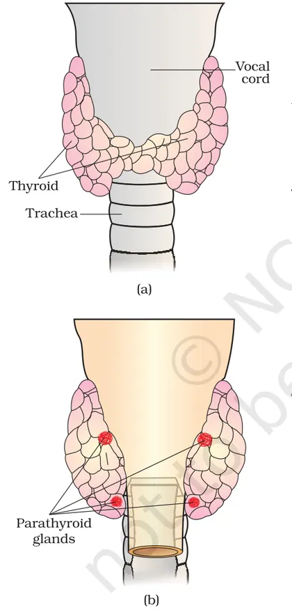
Location & structure: A butterfly-shaped endocrine gland in the neck, anterior to the trachea. Microscopic units are follicles lined by follicular epithelial cells filled with colloid (thyroglobulin). Parafollicular (C) cells lie between follicles.
Major hormones:
- Thyroxine (T4) — main secreted form (prohormone).
- Triiodothyronine (T3) — active form (mostly produced by peripheral deiodination of T4).
- Calcitonin — from C cells; lowers blood calcium slightly by inhibiting bone resorption.
Synthesis of T3/T4 (stepwise):
- Iodide uptake (iodide trapping) into follicular cell from blood via Na+/I- symporter.
- Oxidation of I- to I0 and iodination of tyrosyl residues on thyroglobulin in the colloid → mono- and di-iodotyrosine (MIT, DIT).
- Coupling reactions on thyroglobulin: MIT + DIT → T3; DIT + DIT → T4.
- Endocytosis of iodinated thyroglobulin, proteolysis in lysosomes → release of T3 and T4 into blood.
- Transport: >99% of T3/T4 bind plasma proteins (thyroxine-binding globulin, albumin); free fractions (fT3/fT4) are biologically active.
Physiological actions:
- Increase basal metabolic rate (↑O2 consumption, ↑heat production) by stimulating mitochondrial activity and gene expression of metabolic enzymes.
- Promote growth and neural development (crucial in fetal and neonatal life).
- ↑Heart rate and contractility, ↑lipid and carbohydrate metabolism, affect protein turnover.
Regulation (hypothalamic–pituitary–thyroid axis):
- Hypothalamus secretes TRH → stimulates anterior pituitary to secrete TSH → stimulates thyroid to produce T3/T4.
- Negative feedback: ↑free T3/T4 → ↓TSH and ↓TRH. TSH also stimulates thyroid growth (excess TSH → goitre).
Disorders (common features):
- Hypothyroidism: fatigue, cold intolerance, weight gain, bradycardia. In infants → cretinism (mental retardation, stunted growth). Adults → myxedema.
- Hyperthyroidism: weight loss, heat intolerance, tremor, tachycardia; Graves' disease: autoantibodies stimulating TSH receptor → goitre, exophthalmos.
- Goitre: enlargement of thyroid due to iodine deficiency or TSH overstimulation.
Normal lab ranges (approximate): TSH ~ 0.4–4.0 mIU/L; Free T4 ~ 0.8–1.8 ng/dL; Free T3 ~ 2.3–4.2 pg/mL (laboratory-dependent).
Parathyroid Glands
Location & structure: Usually four small glands on the posterior surface of the thyroid. Two main cell types: chief (principal) cells produce parathyroid hormone (PTH); oxyphil cells of unclear endocrine role.
PTH: the principal hormone regulating blood calcium. Secreted in response to low plasma Ca2+ (sensed directly by Ca2+-sensing receptors on chief cells).
Main actions of PTH (net effect: ↑serum Ca2+):
- Bone: stimulates osteoclast activity indirectly (via osteoblast signalling) → bone resorption → release of Ca2+ and PO4 3-.
- Kidney: ↑reabsorption of Ca2+ in distal tubule, ↓reabsorption of phosphate in proximal tubule (phosphaturia), and stimulates 1α-hydroxylase → conversion of 25(OH)D to active 1,25(OH)2D (calcitriol).
- Intestine (via calcitriol): ↑absorption of Ca2+ and phosphate.
Regulation: PTH secretion is tightly and inversely related to serum ionized Ca2+. Small decreases in Ca2+ cause marked increases in PTH. Rising Ca2+ suppresses PTH secretion.
Disorders:
- Hyperparathyroidism: excess PTH → hypercalcemia → kidney stones, bone pain/osteoporosis, polyuria, neuropsychiatric symptoms.
- Hypoparathyroidism: low PTH → hypocalcemia → tetany, muscle cramps, paresthesias; often follows thyroid/parathyroid surgery.
Interaction between thyroid and parathyroid: calcitonin (thyroid) and PTH (parathyroid) have opposite short-term effects on bone Ca2+ handling; long-term calcium homeostasis is dominated by PTH and vitamin D.
Clinical relevance and screening: newborn TSH/T4 screening prevents cretinism; iodized salt prevents endemic goitre; levothyroxine (synthetic T4) treats hypothyroidism; antithyroid drugs, radioactive iodine, or surgery treat hyperthyroidism. PTH and serum Ca2+/phosphate measurements guide diagnosis of parathyroid disorders.
- Endemic goitre in regions of iodine deficiency — enlarged thyroid due to chronic TSH stimulation.
- Graves' disease — autoimmune hyperthyroidism: weight loss, heat intolerance, palpitations, exophthalmos.
- Congenital hypothyroidism (neonatal screening) — if untreated causes cretinism (stunted growth, intellectual disability).
- Primary hyperparathyroidism — kidney stones and bone demineralization due to chronically high PTH and hypercalcemia.
- Post-thyroidectomy hypoparathyroidism — accidental removal/damage to parathyroids → hypocalcemic tetany.
- Use of levothyroxine (L‑T4) replacement therapy for hypothyroid patients; dose titrated by TSH.
- Chemical formulas: Thyroxine (T4) ≈ C15H11I4NO4; Triiodothyronine (T3) ≈ C15H12I3NO4 (T3 has one fewer iodine than T4).
- Coupling reactions on thyroglobulin: MIT + DIT → T3; DIT + DIT → T4 (where MIT = monoiodotyrosine, DIT = diiodotyrosine).
- Peripheral activation: T4 --(5'-deiodinase)--> T3 + I- (T3 is the more active form).
- Vitamin D activation in kidney: 25(OH)D --(1α-hydroxylase stimulated by PTH)--> 1,25(OH)2D (calcitriol) → ↑intestinal Ca2+ absorption.
- Regulatory relationships: ↑(free T3/T4) → ↓TSH (negative feedback); ↓serum Ca2+ → ↑PTH secretion (inverse relationship).
- Physiological ranges (approx.): serum Ca2+ (total) ≈ 8.5–10.5 mg/dL; TSH ≈ 0.4–4.0 mIU/L (lab-dependent).
Adrenal Glands
Adrenal Glands
The adrenal (suprarenal) glands are paired endocrine glands located on the superior pole of each kidney. Each gland has two structurally and functionally distinct parts: the outer adrenal cortex and the inner adrenal medulla. They secrete steroid and catecholamine hormones that regulate metabolism, salt–water balance, stress responses and some secondary sexual characteristics.
Structure & Zones
- Adrenal cortex (derived from mesoderm) — three concentric zones:
- Zona glomerulosa: produces mineralocorticoids (mainly aldosterone).
- Zona fasciculata: produces glucocorticoids (mainly cortisol).
- Zona reticularis: produces adrenal androgens (e.g., DHEA).
- Adrenal medulla (derived from neural crest) — chromaffin cells that secrete catecholamines: epinephrine (adrenaline) and norepinephrine (noradrenaline).
Major Hormones & Functions
- Aldosterone (mineralocorticoid): increases Na+ reabsorption and K+ secretion in renal distal tubules and collecting ducts → increases blood volume and blood pressure. Stimulated by angiotensin II, high plasma K+, low Na+.
- Cortisol (glucocorticoid): increases gluconeogenesis, mobilizes amino acids and fatty acids, has anti-inflammatory and immunosuppressive effects, helps body adapt to stress. Secretion controlled by hypothalamic CRH → anterior pituitary ACTH → adrenal cortex (negative feedback by cortisol).
- Adrenal androgens (e.g., DHEA): weak androgens converted to stronger androgens/estrogens in peripheral tissues; more important in females for libido and pubic/axillary hair development.
- Adrenaline and Noradrenaline: increase heart rate, cardiac output, bronchial dilation, glycogenolysis, lipolysis, and divert blood to muscles (fight-or-flight). Medullary secretion is under sympathetic nervous system control (splanchnic nerves) and rapid (seconds).
Regulation
- Renin–Angiotensin–Aldosterone System (RAAS): decreased renal perfusion → renin release → converts angiotensinogen to angiotensin I → ACE converts angiotensin I to angiotensin II → stimulates aldosterone release.
- HPA axis: stress or diurnal rhythm → hypothalamic CRH → pituitary ACTH → cortisol secretion; cortisol exerts negative feedback on CRH and ACTH.
- Sympathetic activation: preganglionic fibres stimulate chromaffin cells to release catecholamines immediately during stress.
Clinical Correlates
- Addison's disease (primary adrenal insufficiency): low cortisol and aldosterone → fatigue, hypotension, hyperkalemia, hyponatremia, hyperpigmentation (due to raised ACTH).
- Cushing's syndrome: chronic high cortisol (from pituitary ACTH excess or adrenal tumor) → central obesity, moon face, muscle wasting, hyperglycemia, immunosuppression.
- Conn's syndrome (primary hyperaldosteronism): aldosterone-secreting tumor → hypertension, hypokalemia.
- Pheochromocytoma: catecholamine-secreting tumor of medulla → episodic hypertension, palpitations, sweating, headache.
Takeaway: Adrenal glands integrate short-term (catecholamines) and long-term (steroids) responses to maintain homeostasis under normal and stressful conditions.
- Fight-or-flight response: a sudden threat (e.g., seeing a snake) triggers sympathetic nerves → adrenal medulla releases adrenaline → heart rate and breathing increase, pupils dilate, glucose is mobilized for immediate energy.
- Prolonged academic stress: chronic activation of the HPA axis raises cortisol levels → increased blood glucose, possible suppression of immune responses and impaired wound healing.
- Dehydration or blood loss: reduced kidney perfusion increases renin release → RAAS activation → higher aldosterone to conserve sodium and water, raising blood pressure.
- Pheochromocytoma clinical episode: episodic headaches, sweating, palpitations due to intermittent high catecholamine secretion.
- RAAS pathway (reaction chain): angiotensinogen --(renin)--> angiotensin I --(ACE)--> angiotensin II → stimulates aldosterone secretion
- HPA axis (signal chain): stress/diurnal → CRH (hypothalamus) → ACTH (pituitary) → cortisol (adrenal cortex) → negative feedback on CRH & ACTH
- Blood pressure relation (physiological): BP ≈ Cardiac Output (CO) × Total Peripheral Resistance (TPR)
- Plasma osmolarity (basic relation): Osmolarity ≈ total solute (osmoles) / plasma volume (litres) — relevant when discussing ADH and indirect effects of aldosterone/cortisol on fluid balance
Adrenal Gland
Definition and location: The adrenal (suprarenal) glands are paired endocrine glands located on the superior pole of each kidney. Each gland has two structurally and functionally distinct parts: an outer cortex and an inner medulla.
Structure and embryonic origin: The adrenal cortex is derived from mesoderm and is divided into three zones from outside to inside: zona glomerulosa, zona fasciculata, and zona reticularis. The adrenal medulla is derived from neural crest (ectoderm) and consists of chromaffin cells.
Hormones produced and their actions:
- Cortex (steroid hormones, lipid-soluble):
- Zona glomerulosa: mineralocorticoids (mainly aldosterone) — regulate sodium and potassium balance, blood volume and blood pressure by increasing renal Na+ reabsorption and K+ secretion.
- Zona fasciculata: glucocorticoids (mainly cortisol in humans) — regulate carbohydrate, protein and fat metabolism, increase blood glucose (gluconeogenesis), have anti-inflammatory and immunosuppressive effects, and help in stress adaptation.
- Zona reticularis: adrenal androgens (eg, DHEA) — weak androgens that can be converted to stronger sex steroids in peripheral tissues.
- Medulla (catecholamines, water-soluble):
- Chromaffin cells secrete epinephrine (adrenaline) and norepinephrine (noradrenaline) — responsible for the rapid fight-or-flight response: increase heart rate, contractility, bronchodilation, glycogenolysis, lipolysis, and vasoconstriction in some vascular beds.
Regulation:
- Cortisol secretion is primarily regulated by the hypothalamic-pituitary-adrenal (HPA) axis: hypothalamus releases CRH (corticotropin-releasing hormone) → anterior pituitary releases ACTH → ACTH stimulates cortisol secretion from zona fasciculata. Cortisol exerts negative feedback on CRH and ACTH.
- Aldosterone is mainly regulated by the renin-angiotensin-aldosterone system (RAAS) and by plasma K+; ACTH has a minor stimulatory role.
- Medullary catecholamines are released in response to sympathetic preganglionic stimulation (acetylcholine) and circulating stress signals.
Physiological roles and importance:
- Stress response: cortisol and catecholamines enable the body to respond to physical and emotional stress.
- Metabolic regulation: cortisol promotes gluconeogenesis, protein catabolism and lipolysis to increase blood glucose and fuel availability.
- Fluid-electrolyte balance and blood pressure: aldosterone conserves sodium and water, increasing blood volume and pressure.
- Acute cardiovascular adjustments: epinephrine increases cardiac output and redistributes blood flow.
Common disorders:
- Cushing's syndrome (excess cortisol): features include central obesity, moon face, buffalo hump, muscle wasting, hyperglycemia, hypertension.
- Addison's disease (primary adrenal insufficiency): deficiency of cortisol and aldosterone; symptoms include fatigue, weight loss, hypotension, hyperpigmentation (from high ACTH), hyponatremia, hyperkalemia.
- Conn's syndrome (primary hyperaldosteronism): excess aldosterone → hypertension, hypokalemia, muscle weakness.
- Pheochromocytoma: catecholamine-secreting tumor of medulla → episodic hypertension, palpitations, sweating, headaches.
Clinical notes: Measurement of serum cortisol, plasma ACTH, renin activity, aldosterone levels and urinary catecholamines/metanephrines are used to diagnose adrenal disorders. Imaging (CT/MRI) locates tumors or hyperplasia. Treatment may include hormone replacement, surgery, or drugs that block hormone synthesis.
- Exam stress: During exams, the HPA axis increases cortisol secretion. Short-term cortisol elevation mobilizes glucose and supports alertness, but chronic elevation can cause sleep problems and mood changes.
- Fight-or-flight: When someone sees a threat, sympathetic activation triggers adrenal medulla release of epinephrine and norepinephrine, causing a rapid rise in heart rate and blood pressure and dilation of bronchioles — enabling immediate physical action.
- Dehydration and low blood pressure: Reduced renal perfusion increases renin release, activating the RAAS cascade and increasing aldosterone secretion to conserve sodium and water, restoring blood volume and pressure.
- Pheochromocytoma clinical scenario: A patient with episodic headaches, sweating, and very high blood pressure may have a catecholamine-secreting tumor of the adrenal medulla; diagnosis uses plasma free metanephrines and imaging.
- Steady-state hormone concentration (simple model): C_ss = Secretion rate / Clearance rate
- First-order elimination half-life: t1/2 = ln(2) / k_e, where k_e is the elimination rate constant
- Rate of change of hormone concentration: dC/dt = Secretion(t) - Clearance(t)
- Renin-angiotensin cascade (reaction form): angiotensinogen --(renin)--> Angiotensin I --(ACE)--> Angiotensin II --> stimulates aldosterone secretion
- Mean arterial pressure approximation: MAP ≈ Cardiac output × Total peripheral resistance (MAP ≈ CO × TPR). Aldosterone increases blood volume → increases CO and can raise MAP.
Pancreas and Regulation of Blood Glucose
Overview
The pancreas is a mixed gland with exocrine (digestive enzymes) and endocrine components. The endocrine portion consists of pancreatic islets (islets of Langerhans) that regulate blood glucose by secreting hormones—primarily insulin from beta (β) cells and glucagon from alpha (α) cells.
Structure and cells
- Islets contain β cells (insulin), α cells (glucagon), and δ cells (somatostatin).
- β cells sense blood glucose and secrete insulin; α cells secrete glucagon during low glucose.
Insulin: synthesis and action
Insulin is synthesized as preproinsulin → proinsulin → insulin + C‑peptide. Mechanism of glucose‑stimulated insulin release in β cells: glucose uptake → phosphorylation by glucokinase → increased ATP → closure of K+ATP channels → membrane depolarization → opening of voltage‑gated Ca2+ channels → Ca2+ influx → exocytosis of insulin vesicles.
Major actions of insulin (anabolic hormone):
- Muscle: increases glucose uptake (GLUT4 translocation), glycogen synthesis, protein synthesis.
- Liver: stimulates glycogenesis, inhibits glycogenolysis and gluconeogenesis; promotes lipogenesis.
- Adipose tissue: increases glucose uptake and triglyceride synthesis; inhibits lipolysis.
Glucagon: synthesis and action
Glucagon is secreted by α cells when blood glucose is low. It acts mainly on the liver via a GPCR → adenylate cyclase → ↑cAMP → PKA activation. Effects (catabolic): stimulates glycogenolysis (glycogen → glucose), stimulates gluconeogenesis, promotes lipolysis and ketogenesis.
Homeostatic regulation
Blood glucose is maintained by a negative feedback system: rise in blood glucose → ↑insulin, ↓glucagon → storage and utilization of glucose → glucose level falls toward normal. Fall in blood glucose → ↑glucagon, ↓insulin → mobilization of glucose, synthesis of new glucose.
Key metabolic pathways involved
- Glycolysis: glucose → pyruvate (energy production).
- Glycogenesis: glucose → glycogen (storage, promoted by insulin).
- Glycogenolysis: glycogen → glucose-1-phosphate → glucose (mobilization, promoted by glucagon/epinephrine).
- Gluconeogenesis: noncarbohydrates → glucose (promoted by glucagon).
Physiological ranges and clinical points
- Normal fasting blood glucose: about 70–100 mg/dL (3.9–5.6 mmol/L).
- Postprandial (2 h) normal: <140 mg/dL (<7.8 mmol/L).
- Hypoglycemia: <70 mg/dL; symptoms include sweating, tremor, confusion; can result from excess insulin.
- Diabetes mellitus: chronic hyperglycemia. Type 1: autoimmune destruction of β cells → insulin deficiency. Type 2: insulin resistance ± relative insulin deficiency.
Cell signaling highlights
Insulin receptor is a receptor tyrosine kinase that triggers phosphorylation cascades → metabolic and gene regulation effects. Insulin promotes translocation of GLUT4 vesicles to the cell membrane in muscle and adipose tissue, increasing glucose uptake.
Clinical and physiological examples
After a carbohydrate-rich meal, blood glucose rises, triggering insulin release: liver and muscle store glucose as glycogen and adipose stores triglycerides. During fasting or between meals, glucagon maintains blood glucose by glycogenolysis and gluconeogenesis. In prolonged starvation, ketone bodies increase as fat is used for energy.
Summary
Pancreatic hormones insulin and glucagon work antagonistically to maintain blood glucose within a narrow range by balancing uptake, storage, and production of glucose. Disruption leads to hypo- or hyperglycemia and metabolic disease such as diabetes.
- After a meal: Increased blood glucose → insulin secretion → increased GLUT4 translocation in muscle and adipose → glucose uptake and glycogen synthesis.
- Fasting between meals: Falling blood glucose → glucagon secretion → liver glycogenolysis and gluconeogenesis → blood glucose maintained.
- Exercise: Muscle contractions also increase GLUT4 translocation independently of insulin, increasing glucose uptake.
- Insulin overdose (or missed meal after injecting insulin) can cause acute hypoglycemia: sweating, tremor, confusion; treated with oral glucose or glucagon injection if unconscious.
- Type 2 diabetes: insulin resistance leads to high fasting and postprandial glucose; managed by lifestyle change, oral drugs, sometimes insulin.
- Conversion: glucose (mmol/L) = glucose (mg/dL) ÷ 18
- Glycogenesis (net step): (glucose)n + UDP‑glucose → (glucose)n+1 + UDP (catalyzed by glycogen synthase)
- Glycogenolysis (phosphorolysis): glycogen(n) + Pi → glycogen(n‑1) + glucose‑1‑phosphate (glycogen phosphorylase)
- Simplified insulin/glucagon regulatory relation: ↑blood glucose → ↑insulin, ↓glucagon; ↓blood glucose → ↑glucagon, ↓insulin
- Proinsulin processing: preproinsulin → proinsulin → insulin + C‑peptide
Pancreas (Endocrine Function) and Diabetes
Overview
The pancreas has both exocrine and endocrine parts. The endocrine portion consists of the islets of Langerhans — clusters of cells that secrete hormones directly into the blood to regulate blood glucose and metabolism.
Cell types and hormones
- β (beta) cells: secrete insulin — lowers blood glucose by promoting uptake and storage of glucose.
- α (alpha) cells: secrete glucagon — raises blood glucose by stimulating glycogenolysis and gluconeogenesis in the liver.
- δ (delta) cells: secrete somatostatin — inhibits secretion of insulin and glucagon (paracrine regulator).
- PP cells (F cells): secrete pancreatic polypeptide — influences pancreatic secretions and appetite.
Mechanism of insulin secretion (simplified)
When blood glucose rises (e.g., after a meal):
- Glucose enters β-cells via GLUT transporters and is metabolized to generate ATP.
- Increased ATP/ADP ratio closes ATP-sensitive K+ channels → membrane depolarization.
- Voltage-gated Ca2+ channels open → Ca2+ influx triggers exocytosis of insulin-containing vesicles.
Major actions of insulin
- Increases glucose uptake in muscle/adipose (GLUT4 translocation).
- Stimulates glycogenesis (glycogen synthesis) in liver and muscle.
- Promotes glycolysis and lipogenesis; inhibits gluconeogenesis and glycogenolysis.
- Enhances amino acid uptake and protein synthesis; inhibits protein breakdown.
Glucose homeostasis (feedback loop)
Insulin and glucagon act antagonistically to keep blood glucose within a narrow range. After a meal, insulin dominates to store excess glucose. During fasting, glucagon maintains blood glucose by releasing stored glucose and stimulating new glucose production.
Diabetes mellitus — types and causes
- Type 1 diabetes (insulin-dependent): autoimmune destruction of β-cells → absolute insulin deficiency. Common onset in children/young adults. Requires insulin replacement.
- Type 2 diabetes (non–insulin-dependent): insulin resistance in target tissues plus relative insulin deficiency. Strongly associated with obesity, sedentary lifestyle, and genetics. Managed with lifestyle changes, oral drugs, and sometimes insulin.
- Gestational diabetes: glucose intolerance first recognized during pregnancy due to hormonal insulin resistance; usually monitored and managed to protect mother and fetus.
- Other types: genetic defects, pancreatic disease, drug-induced, endocrine disorders.
Clinical features & complications
Common acute symptoms: polyuria (frequent urination), polydipsia (excessive thirst), polyphagia (increased hunger), unexplained weight loss, fatigue, blurred vision. Severe hyperglycemia can cause diabetic ketoacidosis (DKA) in type 1. Long-term complications: microvascular (retinopathy, nephropathy, neuropathy) and macrovascular (cardiovascular disease) damage.
Diagnosis (important CBSE values)
- Fasting plasma glucose: normal 70–100 mg/dL (3.9–5.6 mmol/L). Diabetes if ≥ 126 mg/dL (≥ 7.0 mmol/L).
- 2-hour plasma glucose during 75 g OGTT: diabetes if ≥ 200 mg/dL (≥ 11.1 mmol/L).
- Random plasma glucose ≥ 200 mg/dL (11.1 mmol/L) with symptoms = diabetes.
- HbA1c >= 6.5% indicates diabetes (also used to monitor long-term control).
Management (overview)
- Type 1: insulin therapy (multiple daily injections or pump), carbohydrate counting, regular monitoring.
- Type 2: lifestyle modification (diet, exercise, weight loss), oral antidiabetic drugs (e.g., metformin, sulfonylureas), sometimes insulin or other injectables (GLP-1 analogues, SGLT2 inhibitors).
- Monitoring: self blood glucose testing, HbA1c every few months to assess average control.
Summary (CBSE focus)
Endocrine pancreas (islets of Langerhans) maintains blood glucose via insulin and glucagon. Diabetes is the result of impaired insulin production or action, diagnosed using blood glucose/HbA1c criteria and managed by medicines and lifestyle to prevent complications.
- A 12-year-old child with sudden weight loss, frequent urination and excessive thirst — suspected Type 1 diabetes; diagnosis confirmed by high fasting glucose and low/absent C-peptide.
- A 50-year-old obese adult with persistent high blood sugar levels controlled initially by diet and metformin — typical Type 2 diabetes with insulin resistance.
- Pregnant woman screened at 24–28 weeks shows elevated 2-hour OGTT value — diagnosed with gestational diabetes and managed with diet/exercise or insulin if needed.
- Diabetic ketoacidosis (DKA): in Type 1, lack of insulin causes fat breakdown, ketone production, dehydration and acidosis — medical emergency presenting with deep rapid breathing and vomiting.
- Conversion between mg/dL and mmol/L for glucose: mmol/L = mg/dL ÷ 18; mg/dL = mmol/L × 18
- Estimated Average Glucose (eAG) from HbA1c (approx.): eAG (mg/dL) = 28.7 × HbA1c (%) − 46.7
- Diagnosis thresholds: Fasting plasma glucose ≥ 126 mg/dL (≥ 7.0 mmol/L); 2-hour OGTT ≥ 200 mg/dL (≥ 11.1 mmol/L); HbA1c ≥ 6.5%
Pineal, Thymus and Other Endocrine Tissues
Overview: Besides the major endocrine glands (pituitary, thyroid, adrenals, pancreas, gonads), several organs/tissues produce hormones that help regulate physiology. Important ones studied in Class 11 are the pineal gland and thymus; other tissues with endocrine functions include the heart, kidney, gastrointestinal tract, adipose tissue, skin, liver and placenta.
Pineal Gland
- Location & structure: Small, cone-shaped endocrine structure in the epithalamus near the midline of the brain.
- Main hormone: Melatonin (derived from tryptophan → serotonin → melatonin).
- Functions:
- Regulates sleep–wake cycle (circadian rhythm): melatonin is high at night and low in daylight.
- Influences seasonal reproductive cycles in some animals (photoperiodism).
- Has antioxidant and modulatory effects on mood and immune function.
- Regulation: Light perceived by retina → signal to suprachiasmatic nucleus (SCN) → pineal via sympathetic pathway; light suppresses melatonin secretion, darkness stimulates it.
- Clinical relevance / examples: Melatonin supplements are used for jet lag and some sleep disorders; altered melatonin patterns are linked with seasonal affective disorder (SAD) and certain sleep problems.
Thymus
- Location & structure: Bilobed organ in the anterior mediastinum, most active in infancy and childhood; involutes after puberty and largely replaced by adipose tissue in adults.
- Hormones: Thymosin, thymopoietin and related peptides.
- Functions:
- Essential for maturation, differentiation and education of T-lymphocytes (cell-mediated immunity).
- Helps establish self-tolerance and functional adaptive immune system during early life.
- Clinical relevance: Thymic disorders or early removal can impair T-cell development; thymic involution occurs with age and stress. Thymus abnormalities are associated with immune defects (e.g., DiGeorge syndrome involves thymic hypoplasia).
Other Endocrine Tissues (brief summary by organ)
- Heart — Atria produce atrial natriuretic peptide (ANP): reduces blood volume and blood pressure by promoting natriuresis and inhibiting renin/aldosterone.
- Kidney — Produces erythropoietin (EPO) to stimulate RBC production in response to hypoxia; juxtaglomerular cells secrete renin (start of renin–angiotensin system).
- Gastrointestinal tract — Endocrine cells secrete gastrin, secretin, cholecystokinin (CCK), gastric inhibitory peptide (GIP) etc., which regulate digestion, enzyme secretion and appetite.
- Adipose tissue — Secretes leptin (regulates appetite and energy balance), adiponectin (metabolic effects) and other cytokines.
- Skin — Produces cholecalciferol (vitamin D3) precursor under UV light; converted in liver/kidney to active calcitriol (hormone regulating Ca2+ homeostasis).
- Liver — Produces insulin-like growth factor 1 (IGF-1) in response to growth hormone; modifies many hormones (activation/inactivation).
- Placenta — Endocrine organ in pregnancy secreting hCG, progesterone, estrogens and human placental lactogen (hPL) to maintain pregnancy and fetal development.
Key concepts & integration
- Many tissues have secondary endocrine roles — paracrine or systemic — and interact with classical endocrine axes.
- Homeostasis is often maintained by feedback loops: for example, ANP opposes the renin–angiotensin–aldosterone system (RAAS) to regulate blood pressure.
- Developmental timing matters: thymus active in early life for immune education; pineal melatonin provides time cues for circadian systems.
Quick clinical/real-life notes
- Melatonin supplements: used short-term for jet lag and some insomnia cases.
- Thymic involution: normal with age; premature loss (congenital) causes immune deficiency.
- EPO therapies: used to treat anemia (e.g., in chronic kidney disease), but misuse can appear in sport doping.
Tip for study: Know each tissue: its hormone(s), main physiological role, how secretion is regulated, and one clinical application/implication.
- Jet lag: rapid travel across time zones disrupts light–dark cues; melatonin taken at the appropriate time can help reset the sleep–wake cycle.
- Seasonal affective disorder (SAD): altered melatonin rhythms and reduced daylight in winter are associated with mood changes; light therapy helps.
- Newborn thymectomy (experimental or congenital thymic aplasia) leads to poor T-cell development and increased susceptibility to infections.
- Chronic kidney disease causes reduced EPO production → anemia; treated with recombinant EPO.
- High blood volume stimulates ANP release from the heart, causing increased sodium excretion and lowered blood pressure.
- Vitamin D synthesis: insufficient sunlight reduces skin production of cholecalciferol → risk of rickets/osteomalacia without dietary supplementation or fortified foods.
- Tryptophan → (tryptophan hydroxylase) → 5‑hydroxytryptophan → serotonin → N‑acetylserotonin → melatonin
- Renin pathway (schematic): angiotensinogen --(renin)--> angiotensin I --(ACE)--> angiotensin II → stimulates aldosterone → ↑Na+ retention, ↑BP
- Vitamin D activation: 7‑dehydrocholesterol --(UV)--> cholecalciferol (vitamin D3) --(liver)--> calcidiol --(kidney)--> calcitriol (active hormone)
- Hormone action (generic scheme): Hormone + Receptor ⇌ Hormone–Receptor complex → intracellular signaling cascade → physiological response
- EPO regulation (qualitative relation): blood O2 tension ↓ ⇒ kidney EPO secretion ↑ ⇒ bone marrow RBC production ↑ ⇒ blood O2 carrying capacity ↑
Reproductive Endocrinology
Overview
Reproductive endocrinology studies hormones and endocrine control of sexual development, gametogenesis and reproduction. The central regulatory axis is the hypothalamo–pituitary–gonadal (HPG) axis: hypothalamus (GnRH) → anterior pituitary (FSH, LH) → gonads (testes or ovaries) producing sex steroids (testosterone, oestrogens, progesterone) and gametes.
HPG axis and feedback
Gonadotropin-releasing hormone (GnRH) is secreted in pulses from the hypothalamus. Pulsatile GnRH stimulates release of follicle-stimulating hormone (FSH) and luteinizing hormone (LH) from the anterior pituitary. FSH and LH act on gonads to produce sex steroids and support gametogenesis. Sex steroids exert negative and (in specific phases) positive feedback on hypothalamus and pituitary to regulate the cycle.
Female reproductive endocrinology — ovarian and menstrual cycle
The ovarian cycle has two main phases: follicular (pre-ovulatory) and luteal (post-ovulatory). Key hormonal events across a typical 28-day cycle:
- Early follicular phase: FSH stimulates follicle growth; follicles produce oestradiol (E2).
- Late follicular/midcycle: Rising E2 exerts positive feedback on the hypothalamus/pituitary, causing an LH surge — this LH surge triggers ovulation (release of the oocyte) and completion of meiosis I.
- Luteal phase: The ruptured follicle becomes corpus luteum, which secretes progesterone (and some oestradiol). Progesterone maintains endometrium and exerts negative feedback on GnRH/LH/FSH. If pregnancy does not occur, corpus luteum degenerates → progesterone falls → menstruation.
- Pulsatility: GnRH pulse frequency and amplitude determine relative LH and FSH secretion.
- Feedback: Sex steroids usually negative-feedback the axis, but sustained high oestradiol midcycle produces a short positive feedback that causes the LH surge and ovulation.
- Hormone mimicry: hCG mimics LH; many drugs act by agonizing or antagonizing GnRH, LH, FSH, or sex-steroid receptors.
Pregnancy and placenta
If fertilisation and implantation occur, the trophoblast secretes human chorionic gonadotropin (hCG), which mimics LH and maintains the corpus luteum (maintains progesterone production) until the placenta takes over steroid synthesis. Placental oestrogens and progesterone support pregnancy. hCG is the basis of pregnancy tests.
Male reproductive endocrinology
In males, LH stimulates Leydig cells to produce testosterone; FSH acts on Sertoli cells to support spermatogenesis and produce inhibin. Testosterone (and inhibin) provide negative feedback on hypothalamus and pituitary to regulate GnRH, LH and FSH levels. Continuous (rather than cyclic) hormone levels maintain ongoing spermatogenesis.
Sexual differentiation and puberty
Genetic sex (SRY on Y chromosome) drives testis development; fetal testes produce testosterone and anti-Müllerian hormone (AMH) to promote male internal/external genital differentiation. Puberty begins when increased pulsatile GnRH secretion elevates LH/FSH and sex steroids, producing secondary sexual characteristics and gametogenesis.
Clinical relevance
Knowledge of reproductive endocrinology underlies: oral contraceptives (combined estrogen–progestin pills suppress ovulation by negative feedback), ovulation tests (detect LH surge), fertility treatments (clomiphene, controlled ovarian stimulation, IVF), hormonal contraception (progestin-only methods), and management of disorders such as polycystic ovary syndrome (PCOS), amenorrhea, hypogonadism, and use of GnRH analogs in hormone-dependent cancers.
Key principles to remember
- Oral contraceptives (combined oestrogen–progestin pills) suppress GnRH/LH/FSH via negative feedback to prevent the LH surge and ovulation.
- Pregnancy test detects hCG produced by the developing placenta; hCG maintains corpus luteum and progesterone secretion in early pregnancy.
- In vitro fertilisation (IVF) uses controlled ovarian hyperstimulation (FSH analogues) and an LH analogue or hCG to trigger ovulation before egg retrieval.
- Polycystic ovary syndrome (PCOS): excess androgens, anovulation and irregular menses — a disorder of endocrine regulation of the ovaries.
- Use of exogenous anabolic steroids elevates blood testosterone → suppresses GnRH/LH/FSH → testicular atrophy and reduced spermatogenesis.
- GnRH (pulsatile) ↑ → LH ↑ + FSH ↑ → Gonadal steroids (Testosterone / Oestradiol / Progesterone) ↑
- High sex steroid (baseline) → negative feedback → ↓ GnRH, ↓ LH, ↓ FSH
- High oestradiol (sustained midcycle) → positive feedback → LH surge → Ovulation
- FSH + Testosterone (local at testis) → Spermatogenesis (Sertoli cell support)
- hCG ≈ LH activity → maintains corpus luteum → sustained progesterone secretion in early pregnancy
Hormonal Regulation of Growth, Development and Metabolism
Overview: Hormones are chemical messengers produced by endocrine glands (animals) or specific tissues (plants) that regulate growth, development and metabolism. They act at low concentrations, bind to specific receptors and change cellular activities by altering gene expression or enzyme activities. Regulation often occurs through feedback loops to maintain homeostasis.
Mechanisms of action:
- Lipid‑soluble hormones (steroid hormones, thyroid hormones): diffuse across cell membranes, bind intracellular receptors, and directly modulate gene transcription (slow but long‑lasting effects).
- Water‑soluble hormones (peptides, catecholamines): bind cell‑surface receptors, activate second‑messenger cascades (cAMP, IP3/Ca2+, kinase cascades) producing rapid but often short‑lived responses.
- Regulation: negative feedback (most common) and positive feedback adjust hormone levels. Receptor down‑ or up‑regulation also modulates sensitivity.
Animal (human) hormones important for growth, development and metabolism:
- Growth Hormone (GH, somatotropin) — secreted by anterior pituitary; stimulates IGF‑1 (insulin‑like growth factor) production in liver; promotes protein synthesis, cell division, bone and muscle growth. Excess in childhood → gigantism; deficiency → pituitary dwarfism; excess in adult → acromegaly.
- Thyroid hormones (T3, T4) — increase basal metabolic rate, stimulate protein synthesis and nervous system development (critical in childhood). Hypothyroidism in children → cretinism (stunted growth, mental retardation); in adults → myxedema; hyperthyroidism → weight loss, heat intolerance, nervousness.
- Insulin and Glucagon (pancreas) — key regulators of carbohydrate, lipid and protein metabolism. Insulin promotes glucose uptake and anabolic processes; glucagon promotes glycogenolysis and gluconeogenesis. Dysregulation → diabetes mellitus (type 1: insulin deficiency; type 2: insulin resistance).
- Adrenal corticosteroids (cortisol) — glucocorticoids increase gluconeogenesis, protein catabolism, and help respond to stress. Chronic excess → Cushing's syndrome; deficiency → Addison's disease.
- Sex hormones (estrogens, androgens, progesterone) — drive sexual differentiation, growth spurts at puberty, and influence bone growth and metabolism.
- Catecholamines (epinephrine, norepinephrine) — increase metabolic rate transiently during stress (fight/flight): increase heart rate, mobilize glucose and fats).
Hormonal control of developmental processes: Hormones determine timing and pattern of development. Examples include GH and thyroid hormone for body and nervous system maturation, sex steroids for pubertal changes, and cortisol for fetal lung maturation.
Plant hormones (phytohormones) regulating growth and development:
- Auxins (IAA) — promote cell elongation, apical dominance, phototropism and root initiation. High auxin at shaded side causes cell elongation and bending toward light.
- Gibberellins (GAs) — promote stem elongation, seed germination and fruit development (bolting in plants).
- Cytokinins — promote cell division, delay leaf senescence and act antagonistically with auxin in organogenesis.
- Abscisic acid (ABA) — induces seed dormancy, closes stomata during water stress, promotes cold/drought tolerance.
- Ethylene — gaseous hormone that promotes fruit ripening, leaf abscission and senescence.
Integration and cross‑talk: Growth and metabolism are controlled by interplay among hormones. Example: insulin and GH both promote growth but through different pathways; thyroid hormones increase sensitivity to catecholamines; in plants, auxin and cytokinin ratios determine root vs shoot development.
Clinical and practical relevance: Diagnosing endocrine disorders (blood hormone assays), therapeutic hormone replacement (insulin, thyroxine), use of plant hormones in agriculture (auxins for rooting, gibberellins to increase fruit size, ethylene to ripen fruit), and herbicides that mimic auxin (e.g., 2,4‑D).
- Pituitary dwarfism (GH deficiency) causing reduced growth in children; treated by GH therapy.
- Gigantism (GH excess before growth plate closure) and acromegaly (GH excess in adults) with characteristic overgrowth.
- Hypothyroidism in infants (cretinism) leading to stunted growth and mental retardation; early thyroxine replacement prevents defects.
- Diabetes mellitus: Type 1 (autoimmune destruction of pancreatic β-cells → insulin deficiency); Type 2 (insulin resistance) → disturbed glucose metabolism.
- Cushing's syndrome (excess cortisol) causing central obesity, muscle wasting and hyperglycaemia; Addison's disease (cortisol deficiency) causing fatigue and hypotension.
- Plant phototropism: auxin redistributed to shaded side of stem → cell elongation there → bending toward light.
- Percentage growth (%) = ((W2 − W1) / W1) × 100, where W1 and W2 are weights at times t1 and t2.
- Specific growth rate (SGR) = (ln W2 − ln W1) / (t2 − t1) (often expressed per day).
- Basal metabolic rate (approximate scaling law): BMR ∝ M^0.75 (Kleiber's law), where M = body mass.
- First‑order hormone elimination: t1/2 = ln(2) / k, where k is the elimination rate constant.
- Fraction of receptors occupied (simple ligand‑receptor equilibrium) = [L] / (Kd + [L]), where [L] is ligand concentration and Kd is dissociation constant.
Regulation of Hormone Secretion
Overview
Hormone secretion is tightly regulated so the body maintains internal stability (homeostasis) and responds appropriately to changes. Regulation occurs by three main kinds of control: humoral, neural and hormonal, plus modulation by rhythms and receptor dynamics. Most control systems use negative feedback; positive feedback is used in special situations.
Main mechanisms
- Humoral regulation: Hormone release in direct response to changing blood levels of ions, nutrients or other molecules. Example: low blood Ca2+ stimulates parathyroid hormone (PTH) secretion; high blood glucose stimulates insulin.
- Neural regulation: Nerve impulses stimulate endocrine cells to release hormones (rapid). Example: sympathetic stimulation of adrenal medulla → epinephrine/norepinephrine release during stress.
- Hormonal regulation (tropic control): One endocrine gland secretes hormones that control another gland’s secretion. Example: hypothalamus releasing TRH → anterior pituitary releases TSH → thyroid releases T3/T4.
Feedback control
- Negative feedback (most common): Hormone’s effects (or end-product levels) inhibit further secretion. Example: increased T3/T4 inhibits TRH and TSH release; high blood glucose + insulin reduce further insulin requirement.
- Positive feedback (rare): Hormone action increases its own secretion until an external event stops it. Example: oxytocin during childbirth — uterine contractions → oxytocin release → stronger contractions until delivery.
Temporal patterns and modulation
- Pulsatile secretion: Many hormones are secreted in bursts (e.g., GnRH), preventing receptor desensitization and maintaining responsiveness.
- Circadian and ultradian rhythms: Daily patterns affect secretion (e.g., cortisol peaks in early morning; melatonin peaks at night).
- Receptor regulation: Up-regulation (more receptors → increased sensitivity) or down-regulation (fewer receptors → decreased sensitivity) alters tissue response to hormones. Chronic high hormone levels often cause down-regulation.
- Permissiveness, synergism and antagonism: Some hormones permit the action of others (e.g., thyroid hormones increase responsiveness to catecholamines), some act together for amplified effect (synergism), and some oppose each other (antagonism, e.g., insulin vs glucagon).
Control loop concept (simple)
Most endocrine control can be viewed as a control loop: a sensor detects a variable → an integrating center computes error relative to setpoint → endocrine gland (effector) secretes hormone → hormone alters the variable → feedback signal modifies further secretion.
Clinical and physiological relevance
- Dysregulation leads to disease: hypothyroidism (insufficient thyroid hormone), Cushing’s syndrome (excess cortisol), diabetes mellitus (impaired insulin production/action).
- Pharmacological hormones can mimic or block normal control (e.g., glucocorticoid therapy can suppress HPA axis via negative feedback).
Summary: Hormone secretion is regulated by humoral, neural and hormonal signals, fine-tuned by feedback (mostly negative), rhythmic secretion patterns and receptor dynamics to produce appropriate physiological responses.
- Insulin and glucagon: After a carbohydrate-rich meal blood glucose rises → pancreatic β-cells secrete insulin (humoral regulation) to lower glucose; when glucose falls, α-cells secrete glucagon to increase blood glucose. Negative feedback maintains glucose homeostasis.
- Thyroid axis (hormonal feedback): Hypothalamus secretes TRH → anterior pituitary secretes TSH → thyroid secretes T3/T4. Rising T3/T4 levels inhibit TRH and TSH (negative feedback).
- Adrenal medulla (neural control): Sympathetic nerve impulses during 'fight-or-flight' stimulate adrenal medulla to rapidly secrete epinephrine and norepinephrine.
- Oxytocin during childbirth (positive feedback): Uterine contractions stimulate oxytocin release; oxytocin increases contraction strength until delivery stops the loop.
- PTH and calcium (humoral regulation + negative feedback): Low plasma Ca2+ stimulates PTH secretion → PTH raises Ca2+; increased Ca2+ inhibits further PTH release.
- \[First-order elimination (hormone clearance): C(t) = C0 * e^{-k t}\]\[where C(t) is concentration at time t\]\[C0 initial concentration\]\[k elimination rate constant.\]
- \[Half-life: t_{1/2} = ln(2)/k.\]
- Dose–response (Hill equation, receptor binding cooperativity): Response = (R_max * [H]^n) / (K_d^n + [H]^n), where [H] = hormone concentration, R_max = maximal response, K_d = concentration at half-max, n = Hill coefficient.
- Simple feedback-control relation (qualitative): Secretion rate ≈ k * (Setpoint − Measured value).
- Receptor-ligand equilibrium: Fraction bound = [H] / ([H] + K_d) (for non-cooperative binding).
Calcium Metabolism and Bone Homeostasis
Overview: Calcium (Ca2+) is essential for skeletal structure and many physiological processes (muscle contraction, blood clotting, neurotransmission, enzyme cofactors). The body maintains plasma Ca2+ within a narrow range (~8.5–10.5 mg/dL) by coordinated actions of bone, intestine, kidney and hormones (parathyroid hormone, calcitonin, active vitamin D).
Major compartments and balance:
- About 99% of body Ca is in bone as hydroxyapatite (structural reservoir); ~1% is in extracellular fluid and soft tissues (ionized Ca2+, protein-bound, complexed).
- Calcium balance = dietary intake + bone release − (urinary excretion + fecal loss). Homeostasis adjusts bone turnover, intestinal absorption and renal excretion.
Key hormones and actions:
- Parathyroid hormone (PTH) — secreted by parathyroid glands when plasma Ca2+ falls. Effects: ↑ bone resorption (activates osteoclasts indirectly), ↑ renal reabsorption of Ca2+ (reduces urinary Ca loss), ↑ phosphate excretion (phosphaturia), and ↑ activation of vitamin D in kidney (↑ 1,25-(OH)2 vitamin D = calcitriol). Net effect: increase plasma Ca2+.
- Calcitonin — secreted by thyroid C cells when Ca2+ is high. Effects: inhibits osteoclast activity → ↓ bone resorption and lowers plasma Ca2+ (important in some species and in acute regulation).
- Vitamin D (calcitriol, 1,25-(OH)2 D3) — produced from skin (7-dehydrocholesterol → cholecalciferol) and activated in liver (25-OH D) and kidney (1,25-(OH)2 D). Effects: ↑ intestinal absorption of Ca2+ and PO4 3−, helps bone mineralization (and in some conditions supports resorption to mobilize Ca).
Bone remodeling (dynamic process):
- Remodeling cycle phases: activation → resorption (osteoclasts digest mineral & matrix) → reversal → formation (osteoblasts deposit osteoid) → mineralization.
- Osteoclasts dissolve mineral (hydroxyapatite) and degrade organic matrix; osteoblasts synthesize collagen and promote mineral deposition.
- Remodeling is regulated by mechanical stress, systemic hormones (PTH, calcitonin, vitamin D, sex steroids), and local factors (cytokines, growth factors).
Feedback control (simplified):
- Fall in plasma Ca2+ → ↑ PTH → ↑ bone resorption, ↑ renal Ca reabsorption, ↑ calcitriol production → ↑ intestinal Ca absorption → plasma Ca2+ rises → PTH secretion suppressed.
- Rise in plasma Ca2+ → ↑ calcitonin (and reduced PTH) → decreased bone resorption → plasma Ca2+ falls to set point.
Clinical relevance / disorders:
- Hypocalcemia (low Ca2+) → tetany (muscle spasms), paresthesias; causes include hypoparathyroidism, vitamin D deficiency.
- Hypercalcemia (high Ca2+) → polyuria, kidney stones, constipation, fatigue; causes include hyperparathyroidism, malignancy.
- Rickets (children) / osteomalacia (adults): defective mineralization due to vitamin D deficiency → soft bones, deformities.
- Osteoporosis: decreased bone mass and increased fracture risk from an imbalance favoring resorption over formation (age, hormonal loss, inactivity).
Integration — organs working together:
- Bone: reservoir that releases or stores Ca2+ via remodeling.
- Kidney: controls Ca2+ excretion/reabsorption and activates vitamin D (1-alpha hydroxylase stimulated by PTH).
- Intestine: site of dietary Ca2+ absorption enhanced by calcitriol.
Key points students should remember:
- PTH raises plasma Ca2+, decreases phosphate reabsorption, and stimulates calcitriol production.
- Calcitonin lowers plasma Ca2+ by inhibiting osteoclasts.
- Vitamin D increases Ca2+ (and phosphate) absorption from gut; essential for bone mineralization.
- Bone remodeling maintains bone strength and mineral homeostasis; imbalance leads to metabolic bone disease.
- Rickets in children due to vitamin D deficiency — bowed legs, delayed tooth eruption; prevented by sunlight exposure and dietary vitamin D/calcium (milk, fortified foods).
- Hypoparathyroidism after thyroid surgery — low PTH → hypocalcemia → muscle tetany and tingling; treated with calcium supplements and active vitamin D.
- Primary hyperparathyroidism (parathyroid adenoma) — high PTH → hypercalcemia → kidney stones, abdominal pain, bone pain (increased bone resorption).
- Osteoporosis after menopause — decreased oestrogen leads to increased bone resorption relative to formation; managed by dietary calcium, vitamin D, weight-bearing exercise and sometimes bisphosphonates.
- Hydroxyapatite dissolution (bone mineral): Ca10(PO4)6(OH)2 ↔ 10 Ca2+ + 6 PO43− + 2 OH−
- Vitamin D activation pathway (simplified): 7-dehydrocholesterol (skin) --UV--> Cholecalciferol (Vit D3) --liver (25-hydroxylase)--> 25‑OH D3 --kidney (1α-hydroxylase, ↑ by PTH)--> 1,25-(OH)2 D3 (calcitriol, active form)
- Approximate normal plasma calcium range: 8.5–10.5 mg/dL (total Ca), ionized Ca ≈ 4.4–5.4 mg/dL (varies with pH and albumin)
- Simple Ca balance concept: Net Ca change = (Dietary Ca absorption) − (Urinary Ca excretion + Fecal Ca loss) ± Bone mineral exchange
Endocrine Disorders and Clinical Correlates
Overview
The endocrine system consists of glands that secrete hormones into the bloodstream to regulate metabolism, growth, fluid balance and reproduction. Hormones work by binding to receptors on/in target cells. Most hormone levels are regulated by negative feedback loops (e.g., hypothalamus → pituitary → peripheral gland).
Basic mechanism and clinical relevance
- Hormone excess or deficiency (or receptor resistance) produces characteristic clinical syndromes.
- Diagnosis uses history, clinical signs, biochemical tests (blood hormone levels, metabolites), imaging and stimulation/suppression tests.
- Treatment: replace deficient hormones, block excess production, treat underlying tumour/autoimmunity, metabolic control (e.g., insulin).
Major glands and principal disorders (with clinical correlates)
Pituitary gland
- Growth hormone (GH): deficiency in childhood → pituitary dwarfism (short stature); excess before epiphyseal closure → gigantism (increased height); excess after closure → acromegaly (enlarged hands, facial bones, prognathism, diabetes).
- Posterior pituitary (ADH): deficiency → diabetes insipidus (polyuria, polydipsia, low urine specific gravity); inappropriate ADH secretion (SIADH) → water retention and hyponatremia.
- Clinical tests: serum GH/IGF-1, oral glucose suppression test for acromegaly, water-deprivation test for diabetes insipidus.
Thyroid gland
- Hypothyroidism (low T3/T4): causes include Hashimoto's (autoimmune), iodine deficiency. Signs: fatigue, cold intolerance, weight gain, slowed mental development in neonates → cretinism (stunted growth, mental retardation). Adults with severe chronic hypothyroidism → myxoedema (facial puffiness, dry skin).
- Hyperthyroidism (high T3/T4): common cause Graves' disease (autoimmune). Signs: weight loss, heat intolerance, tremor, palpitations, exophthalmos (in Graves').
- Goitre: enlargement of the thyroid due to overstimulation (e.g., iodine deficiency or TSH stimulation).
- Clinical tests: serum TSH (most sensitive), free T4, T3, anti-thyroid antibodies; radioiodine uptake in hyperthyroidism.
Parathyroid glands
- Hyperparathyroidism (↑PTH): causes hypercalcemia → bone pain, kidney stones, abdominal groans, psychiatric overtones (memory problems).
- Hypoparathyroidism (↓PTH): hypocalcemia → muscle cramps, tetany, paresthesias.
- Clinical tests: serum calcium, phosphate (usually inverse to Ca), and PTH levels.
Adrenal glands
- Addison's disease (primary adrenal insufficiency): autoimmune destruction → low cortisol and aldosterone → weakness, weight loss, hyperpigmentation, hypotension, hyponatremia, hyperkalemia.
- Cushing's syndrome (excess cortisol): causes include pituitary ACTH-secreting adenoma (Cushing disease), exogenous steroids. Signs: central obesity, moon face, purple striae, muscle wasting, hyperglycemia, immunosuppression.
- Pheochromocytoma (catecholamine‑secreting tumour): episodic hypertension, headache, sweating, palpitations.
- Clinical tests: serum/urine cortisol, dexamethasone suppression test, plasma/urine catecholamines and metanephrines, ACTH levels.
Pancreas (endocrine)
- Diabetes mellitus (DM): chronic hyperglycemia from insulin deficiency (Type 1, autoimmune destruction of beta cells) or insulin resistance with relative insulin deficiency (Type 2).
- Type 1: usually childhood/young adult onset, polyuria, polydipsia, weight loss, risk of diabetic ketoacidosis.
- Type 2: associated with obesity, sedentary lifestyle; complications include neuropathy, retinopathy, nephropathy, cardiovascular disease.
- Clinical tests: fasting plasma glucose, oral glucose tolerance test (OGTT), HbA1c, urine glucose/ketones.
- Treatment: insulin (Type 1), diet/exercise and oral hypoglycemics then insulin if needed (Type 2).
Gonads and reproductive hormones
- Disorders include polycystic ovarian syndrome (PCOS): hyperandrogenism, irregular menses, insulin resistance, infertility.
- Primary amenorrhea, delayed puberty, or virilization can reflect endocrine causes (gonadal failure, pituitary or hypothalamic dysfunction).
Integration and feedback examples
Most endocrine disorders are best understood by looking at feedback patterns. Example: in primary hypothyroidism (thyroid gland failure) low T4/T3 causes high TSH (pituitary tries to compensate). In secondary hypothyroidism (pituitary failure) both TSH and T4 are low.
General diagnostic principles
- Measure hormone and downstream markers (e.g., insulin and blood glucose).
- Use stimulation or suppression tests to distinguish primary from secondary disorders.
- Imaging (ultrasound, MRI) to detect gland enlargement or tumours.
Treatment principles
Replace deficient hormone (levothyroxine for hypothyroidism, insulin for Type 1 DM, hydrocortisone for Addison's), reduce excess hormone (antithyroid drugs, surgery, radiotherapy), manage metabolic consequences (diet, antihypertensives) and treat causes (autoimmunity, tumours).
- A 9-year-old child shows very slow growth and delayed bone age; blood tests show low IGF-1 and low GH response on stimulation test → pituitary GH deficiency (pituitary dwarfism).
- A 35-year-old with weight loss, heat intolerance, palpitations and bulging eyes; tests show suppressed TSH and elevated free T4 → Graves' disease (hyperthyroidism) treated with antithyroid drugs or radioiodine.
- A 50-year-old obese patient with polyuria and polydipsia; fasting plasma glucose 140 mg/dL and HbA1c 7.2% → Type 2 diabetes mellitus; managed with diet, exercise, oral hypoglycemics and later insulin if needed.
- A patient with chronic fatigue, hyperpigmentation, low blood pressure, hyponatremia and low morning cortisol → primary adrenal insufficiency (Addison's disease); treated with corticosteroid and mineralocorticoid replacement.
- A young adult with large hands, coarse facial features, and high IGF-1 despite closed growth plates → acromegaly from a GH-secreting pituitary adenoma.
- BMI = weight (kg) / [height (m)]^2 (used to assess obesity, a risk factor for Type 2 DM)
- Glucose conversion: glucose (mmol/L) = glucose (mg/dL) ÷ 18
- Diabetes diagnostic thresholds: fasting plasma glucose ≥ 126 mg/dL (≥ 7.0 mmol/L); 2‑hour OGTT ≥ 200 mg/dL (≥ 11.1 mmol/L); HbA1c ≥ 6.5%
- Typical TSH reference (laboratory-dependent): ~0.4–4.0 mIU/L (used with free T4 to classify thyroid disorders)
- Serum calcium normal range (typical): ~8.5–10.2 mg/dL — values above/below suggest hyper- or hypoparathyroidism respectively
Chemical Coordination in Plants
Definition and scope: Chemical coordination in plants means regulation and integration of growth, development and responses to the environment by chemical messengers called phytohormones (plant hormones) and other signalling molecules. Unlike animals, plants use a small set of hormones produced at specific sites, transported to targets, and acting at very low concentrations to change cell physiology and gene expression.
Main plant hormones (overview):
- Auxins (e.g. Indole-3-acetic acid, IAA) — synthesized mainly in shoot apices and young leaves. Functions: cell elongation, apical dominance, phototropism and gravitropism, root initiation (used in rooting powders), and inhibition of abscission.
- Gibberellins (GAs, e.g. GA3) — promote stem elongation, seed germination (break dormancy), mobilize food reserves, and stimulate flowering/bolting in some plants.
- Cytokinins (e.g. zeatin) — synthesized in roots; promote cell division, stimulate shoot formation, delay leaf senescence, and work antagonistically with auxin in organogenesis.
- Abscisic acid (ABA) — produced in mature leaves and seeds; induces seed dormancy, inhibits germination, closes stomata under water stress and mediates stress responses.
- Ethylene (C2H4) — a gaseous hormone produced in ripening fruits and stressed tissues; promotes fruit ripening, leaf abscission and senescence.
- Other regulators — brassinosteroids, jasmonates, salicylic acid and systemin (defence/signalling roles) and synthetic regulators (e.g. 2,4-D, a synthetic auxin used as herbicide).
Synthesis and transport: Hormones are synthesized in specific tissues (apices, young leaves, roots, seeds) and are transported via different routes: polar cell-to-cell transport (auxin via carrier proteins like PINs), xylem/phloem long-distance transport (cytokinins, ABA), diffusion (ethylene) or conjugation and release from inactive forms.
Mode of action / signal transduction (general steps):
- Perception: hormone binds to specific receptor (membrane or intracellular).
- Second messengers: changes in Ca2+, cGMP, IP3 or redox status may relay the signal.
- Protein phosphorylation cascade: kinases/phosphatases modify target proteins.
- Transcriptional regulation: transcription factors are activated or repressed, altering gene expression and producing long-term developmental changes.
Key physiological mechanisms (select examples):
- Auxin "acid growth" hypothesis: Auxin stimulates plasma membrane H+-ATPases → cell wall acidification → activation of expansins and wall-loosening enzymes → cell wall loosening and turgor-driven cell elongation.
- Apical dominance: High auxin from shoot apex suppresses lateral bud outgrowth; removal of apex lowers auxin and allows cytokinin action to stimulate lateral buds.
- Tropisms: Phototropism: lateral redistribution of auxin causes differential cell elongation and bending toward light. Gravitropism: auxin redistribution in roots/shoots leads to curvature (note: auxin inhibits elongation in roots but promotes it in shoots).
- Seed dormancy and germination: ABA maintains dormancy; gibberellins stimulate production of hydrolytic enzymes (e.g., alpha-amylase in cereal endosperm) to mobilize food reserves and allow germination.
- Fruit ripening: Ethylene triggers coordinated gene expression leading to softening, colour change and aroma production.
Agricultural and practical applications: rooting powders (auxins), 2,4-D as selective herbicide (synthetic auxin), gibberellins to increase stem elongation or break dormancy, ethylene or its releasers to ripen fruits, cytokinins in micropropagation for shoot induction, ABA-related treatments for stress management and seed storage.
Experimental evidence and classic experiments: Removal of coleoptile tip (Darwin) abolishes phototropic response; applied auxin restores bending. Application of gibberellin to dormant seeds triggers germination; application of ABA induces dormancy. These provide causal links between hormones and plant responses.
Summary: Chemical coordination in plants is achieved by a small set of hormones whose concentrations, sites of synthesis, transport routes and interactions (synergistic or antagonistic) regulate virtually every aspect of plant life — from cell division and elongation to stress responses, dormancy and reproduction. Understanding these hormones and their mechanisms is essential for both basic plant biology and practical crop management.
- Apical dominance: Removing the shoot tip (source of auxin) leads to lateral bud outgrowth; used in pruning to encourage bushier growth.
- Rooting powders: Gardeners use IAA or its synthetic analogues to promote root formation on cuttings.
- Fruit ripening: Ethylene gas is used commercially to ripen bananas and tomatoes uniformly.
- Seed dormancy/germination: High ABA keeps seeds dormant; gibberellin treatments break dormancy and stimulate alpha-amylase production in cereal seeds.
- Use of 2,4-D: A synthetic auxin used as an herbicide to selectively kill broadleaf weeds.
- Delay of senescence: Cytokinins are used to prolong the shelf life of cut flowers by delaying leaf yellowing.
- Indole-3-acetic acid (IAA): C10H9NO2 (major natural auxin)
- Gibberellic acid (GA3): C19H22O6 (representative gibberellin)
- Abscisic acid (ABA): C15H20O4
- Ethylene: C2H4 (gaseous hormone)
- Zeatin (a natural cytokinin): C10H13N5O (representative cytokinin)
- 2,4-D (2,4-dichlorophenoxyacetic acid; synthetic auxin/herbicide): C8H6Cl2O3
Neuroendocrine Integration and Feedback Mechanisms
What it is
Neuroendocrine integration is the coordination between the nervous system and the endocrine system: specialized neurons (neurosecretory cells) convert electrical signals into hormone release, allowing the brain to control distant organs via the blood. Feedback mechanisms are regulatory loops (mainly negative feedback, sometimes positive) that keep physiological variables near a set point.
Key components and pathways
- Hypothalamus – contains neurosecretory cells that synthesise releasing/inhibiting hormones (for anterior pituitary) and neurohormones (oxytocin, ADH) transported to the posterior pituitary.
- Hypophyseal (pituitary) system – anterior pituitary secretes trophic hormones (TSH, ACTH, LH, FSH, GH, prolactin) under hypothalamic control via the hypophyseal portal vessels; posterior pituitary releases hypothalamic neurohormones (ADH, oxytocin) directly into blood.
- Neuroendocrine cell action – neuronal input (sensory, circadian, stress) → action potentials in neurosecretory cells → hormone exocytosis into circulation → target tissues respond via receptors.
Feedback mechanisms
- Negative feedback – increased level/effect of a hormone inhibits its further release (primary homeostatic mechanism). Example: high thyroid hormones (T3/T4) suppress TRH and TSH release; high cortisol suppresses CRH and ACTH (long-loop).
- Short-loop feedback – pituitary hormones inhibit hypothalamic releasing hormones (e.g., ACTH inhibiting CRH).
- Positive feedback – hormone effect amplifies its own production until an event completes (less common). Example: oxytocin during childbirth increases uterine contractions, which stimulate more oxytocin release (birth ends the loop).
- Pulsatile and circadian secretion – some neuroendocrine signals are released in pulses (GnRH → LH/FSH pulses) or show daily rhythms (cortisol peaks in early morning).
Functional significance
Neuroendocrine integration allows rapid sensing (neurons) with sustained, body-wide responses (hormones). Feedback loops provide stability (homeostasis), allow graded responses, prevent hormone overproduction, and create time-structured signalling (pulses, rhythms).
Illustrative sequence: HPA axis (stress)
Stress → hypothalamus releases CRH → anterior pituitary releases ACTH → adrenal cortex secretes cortisol. Rising cortisol then suppresses CRH and ACTH (negative feedback). This axis exemplifies neuroendocrine integration and feedback.
- Blood glucose regulation: Meal raises blood glucose → beta cells release insulin (promotes glucose uptake & storage); when glucose falls, alpha cells release glucagon. Insulin and glucagon work via negative feedback to stabilise blood glucose.
- Hypothalamo–pituitary–adrenal (HPA) axis: Stress induces CRH → ACTH → cortisol; cortisol exerts negative feedback on both pituitary and hypothalamus.
- Thyroid axis: Hypothalamus TRH → pituitary TSH → thyroid T3/T4; T3/T4 inhibit TRH and TSH (negative feedback).
- Oxytocin in childbirth: Cervical stretch → hypothalamus/posterior pituitary releases oxytocin → stronger uterine contractions → more stretch and more oxytocin (positive feedback) until delivery.
- ADH (vasopressin) and osmoregulation: Increased plasma osmolality sensed by hypothalamic osmoreceptors → ADH release → water reabsorption in kidneys → plasma osmolality falls, reducing ADH (negative feedback).
- Pulsatile GnRH: Intermittent GnRH pulses from hypothalamus drive pulsatile LH/FSH release; continuous GnRH suppresses gonadotropins (clinical importance).
- Simple mass-balance (steady state): C* = S / k (C* = steady-state hormone concentration, S = secretion rate, k = clearance rate constant)
- Exponential decay (clearance/half-life): C(t) = C0 · e^(−k·t), where k = ln(2) / t1/2 and t1/2 is hormone half-life
- Stimulus–response (qualitative): Response ∝ [Hormone] × receptor sensitivity (i.e., Response = α · H · R, where α is proportionality constant, H = hormone concentration, R = receptor availability/sensitivity)
- Percent inhibition in negative feedback (conceptual): %inhibition = (1 − (new secretion / basal secretion)) × 100
Plant Tropisms and Responses
Definition: Tropisms are directional growth responses of plants in which the direction of the stimulus determines the direction of growth (towards = positive, away = negative). Plant responses also include non-directional movements (nastic movements) and rapid turgor-driven responses.
Major types of tropisms:
- Phototropism – growth response to light. Shoots typically show positive phototropism (bend towards light); mediated mainly by blue-light photoreceptors (phototropins).
- Gravitropism (Geotropism) – response to gravity. Roots show positive gravitropism (grow downward); shoots show negative gravitropism (grow upward). Gravity sensing involves statoliths (starch grains) in columella cells.
- Hydrotropism – growth toward moisture (roots growing to higher water potential).
- Thigmotropism – growth response to touch/contact (e.g., tendrils coiling around supports).
- Chemotropism – growth response to chemicals (e.g., pollen tube growth toward ovule signals).
- Thermotropism – response to temperature gradients (less common).
Mechanism (general):
- Tropisms are achieved by differential cell elongation on opposite sides of an organ. The side with faster cell elongation becomes the convex side, causing bending.
- Cholodny–Went hypothesis: Asymmetric distribution of the plant hormone auxin (IAA) causes differential growth. A stimulus redistributes auxin to one side; in shoots, higher auxin concentration promotes cell elongation on the shaded side (causing bending toward light). In roots, high auxin inhibits elongation, so redistribution produces opposite bending.
- Acid growth hypothesis: Auxin stimulates H+-ATPases in the plasma membrane, lowering apoplastic pH. Acidification activates expansins and cell-wall loosening enzymes → cell-wall extensibility increases and cells elongate when turgor is maintained.
Key experimental evidence:
- Darwin’s coleoptile experiments: tip senses light; bending requires an intact tip.
- Boysen-Jensen: separation of tip from the growing region by a permeable barrier allowed bending (chemical signal diffusion), impermeable barrier prevented bending.
- Went’s agar block experiment: agar placed on shaded side of coleoptile tip acquired a substance (auxin) that caused bending when applied to decapitated coleoptiles.
Hormonal control & differences:
- Auxins (IAA) — central in tropisms: redistribute asymmetrically to cause differential growth. Promote elongation in shoots, inhibit elongation in roots (dose- and tissue-dependent).
- Other hormones: Cytokinins (cell division, interaction with auxin in organogenesis), gibberellins (stem elongation), ethylene (inhibits elongation, promotes triple response and can mediate touch responses), abscisic acid (stress responses), and peptides/other signals in tropic contexts.
- Photoreceptors: Phototropins (blue light) mediate phototropism; phytochromes (red/far-red) mediate other light responses like shade avoidance and germination.
Nastic movements and rapid responses (contrast with tropisms): Nastic movements are non-directional responses dependent on the structure, not stimulus direction. Examples: nyctinasty (leaf folding in legumes at night), thigmonasty (Mimosa pudica leaf folding on touch), rapid trap closure in Dionaea (Venus flytrap) caused by action potentials and rapid turgor changes.
Physiological basis of rapid movements: Rapid movements often depend on fast changes in cell turgor driven by ion fluxes (K+, Cl–) across membranes, producing water movement and rapid volume changes in motor cells (e.g., pulvini in legumes). Electrical signals (action potentials) can coordinate these changes.
Summary: Tropisms are directional growth responses mainly controlled by asymmetric auxin distribution and differential cell elongation. They allow plants to orient shoots toward light and roots toward water and minerals. Nastic and rapid turgor-mediated movements complement growth-mediated tropisms for behavior and protection.
- Phototropism: Coleoptiles bending toward unilateral light; sunflower stems reorienting leaves toward the sun (heliotropism).
- Gravitropism: Roots growing downward (positive gravitropism) and shoots growing upward (negative gravitropism); statolith sedimentation in root cap cells.
- Hydrotropism: Root tips growing toward a localized water source in soil.
- Thigmotropism: Tendrils of pea plants coiling around supports; climbing plants using touch to climb.
- Chemotropism: Pollen tube growing toward ovule chemo-attractants during fertilization.
- Nastic/rapid response: Mimosa pudica folding leaves on touch (thigmonasty); Venus flytrap snapping shut (rapid turgor and action potentials).
- Relative Growth Rate (RGR) = (ln W2 - ln W1) / (t2 - t1), where W1 and W2 are plant size (mass/length) at times t1 and t2.
- Water potential (ψ) = osmotic potential (ψs) + pressure potential (ψp). Water movement toward higher (less negative) ψ drives hydrotropic responses.
- Average linear growth rate = (L2 - L1) / (t2 - t1), where L1 and L2 are lengths at times t1 and t2 (useful to quantify differential elongation during tropic bending).
Comparative and Minor Endocrine Organs
Overview
In addition to the principal endocrine glands (pituitary, thyroid, adrenal, pancreas, gonads), many organs have secondary or minor endocrine functions. These organs secrete hormones or hormonelike factors that help regulate local and systemic physiology. Comparative endocrinology examines how endocrine structures and hormones vary across animal groups (vertebrates vs invertebrates) and how analogous systems accomplish similar regulatory tasks.
Minor endocrine organs and their major secretions (with functions)
- Heart: Atrial natriuretic peptide (ANP/ANF) — reduces blood volume and blood pressure by increasing sodium and water excretion and by inhibiting renin–angiotensin secretion.
- Kidney: Erythropoietin (EPO) — stimulates red blood cell production; also converts vitamin D to its active form (calcitriol) and releases renin (RAAS activation for blood pressure).
- Liver: Angiotensinogen (RAAS precursor), insulin-like growth factors (IGFs) — mediate growth effects of GH; thrombopoietin — regulates platelet production.
- Gastrointestinal tract: Enteric hormones (gastrin, secretin, cholecystokinin, GIP) — regulate digestion, enzyme secretion and motility.
- Adipose tissue: Leptin, adiponectin, resistin — regulate appetite, energy balance and insulin sensitivity.
- Pineal gland: Melatonin — controls circadian rhythms and reproductive seasonality in some animals.
- Thymus: Thymosins and thymopoietin — involved in T‑cell maturation and immune system development.
- Skin: Precursor conversion of 7‑dehydrocholesterol to cholecalciferol (vitamin D3) under UV; active vitamin D regulates calcium metabolism.
- Placenta (in mammals): hCG, progesterone, estrogen, human placental lactogen — maintain pregnancy and modify maternal metabolism.
Comparative aspects (vertebrates vs invertebrates)
- Vertebrates: Well‑differentiated endocrine glands (thyroid, adrenals, pituitary). Many organs (heart, kidney, liver) have clear endocrine roles as above. Fish have additional specific structures (e.g., corpuscles of Stannius in teleosts producing stanniocalcin for calcium homeostasis).
- Invertebrates (especially insects): Endocrine control is mainly neuroendocrine. Key glands include the prothoracic glands (produce ecdysteroids/ecdysone for moulting), corpora allata (produce juvenile hormone (JH) that determines larval vs adult development), and the corpora cardiaca (store/release neurohormones). Hormonal coordination is crucial for metamorphosis and diapause.
Physiological significance and clinical relevance
Minor endocrine organs provide rapid and fine tuning of homeostasis. Clinical examples: decreased EPO → anemia of chronic disease; altered ANP/renin → hypertension; leptin resistance → obesity; vitamin D deficiency → rickets/osteomalacia; placental hCG → basis of pregnancy tests.
Integration and feedback
Minor endocrine hormones often act within feedback loops or synergize with major endocrine axes (e.g., liver IGFs mediate effects of pituitary GH; kidney renin activates angiotensin II which interacts with adrenal aldosterone and cardiac ANP). Understanding these interactions is essential for physiology and medicine.
Note: Diagrams that compare hormone sources across organs and graphs of hormone levels over time (circadian, developmental or dose–response) are especially useful for visual learning.
- Erythropoietin (EPO) from kidney is used clinically to treat certain types of anemia (e.g., anemia of chronic kidney disease).
- Atrial natriuretic peptide (ANP) increases when blood volume rises; its measurement and function are relevant in heart failure management.
- Placental hCG is the hormone detected by pregnancy test kits; it supports early pregnancy by maintaining corpus luteum.
- Melatonin from the pineal gland regulates sleep–wake cycles; melatonin supplements are used for jet lag and certain sleep disorders.
- Leptin produced by adipose tissue signals satiety; leptin deficiency or resistance is implicated in some forms of obesity.
- Vitamin D synthesis in the skin (UV conversion) followed by liver/kidney activation is essential for calcium homeostasis; deficiency causes rickets.
- \[First‑order elimination (pharmacokinetics): C(t) = C0 · e^{−kt}\]\[where C(t) is concentration at time t\]\[C0 is initial concentration and k is elimination rate constant.\]
- Half‑life: t1/2 = ln(2) / k (for first‑order elimination).
- Steady‑state concentration for continuous infusion: Css = Rate of infusion / Clearance (Css = R0 / CL).
- Dose–response (Hill equation, receptor binding): Response = (Emax · [H]^n) / (EC50^n + [H]^n), where [H] is hormone concentration, Emax is maximal effect, EC50 is concentration for half‑maximal effect and n is Hill coefficient.
- Clearance relation: CL = Rate of elimination / Plasma concentration.
Practical and Experimental Aspects
Overview
Practical and experimental aspects of Chemical Coordination and Integration cover laboratory methods and simple investigations used to demonstrate how hormones and chemical regulators work in animals and plants, how to measure their effects, and how to interpret data. Emphasis is on designing controlled experiments, collecting quantitative data, plotting results, understanding dose–response relationships, and appreciating safety and ethical constraints.
Common objectives of experiments
- Demonstrate physiological effects of hormones (e.g., change in heart rate, blood glucose, growth, or tropic responses).
- Show dose–response relationships and determine relative potency.
- Compare treated vs control samples and calculate percentage change or rates.
- Observe feedback patterns (e.g., hormone secretion vs blood parameter) and explain integration.
Typical experimental approaches (animal systems)
- Daphnia or small invertebrates to study cardioactive agents: measure heart rate (beats per minute) before and after adding graded concentrations of adrenaline, caffeine or acetylcholine. Advantages: visible heart, minimal ethical issues.
- Non-invasive human practicals: measure blood glucose (using glucometer/strips) before and after a standard glucose drink to demonstrate insulin action (requires consent and safety precautions). Record glucose vs time.
- Isolated tissue preparations (demonstration level in guided labs): frog/embryonic heart or cardiac strip perfusion to show effect of sympathetic/parasympathetic agents — usually demonstrated by teacher in proper labs with ethics compliance.
Typical experimental approaches (plant systems)
- Phototropism or geotropism experiments: decapitate or cover coleoptile tips and expose to unilateral light; measure curvature over time to infer role of auxins.
- Auxin/Agar block assay (Avena test style): apply auxin to one side of a coleoptile or use agar blocks soaked in IAA to induce elongation and bending.
- Seed germination/growth bioassays: apply gibberellic acid (GA) to seeds or plants and measure stem elongation vs controls.
- Ethylene treatments: demonstrate accelerated fruit ripening (banana/apple/green tomato) by exposing fruits to ethylene or ethylene-generating conditions.
Experimental design and data handling
- Always include controls (untreated) and replicate samples (biological and technical replicates).
- Independent variable: hormone/chemical concentration or time; dependent variable: physiological response (heart rate, curvature angle, glucose level, growth length).
- Record raw data, compute derived values (percentage change, rate, mean ± SD) and plot appropriate graphs (dose–response, time-course).
- Use simple statistics: mean, standard deviation, and, where required, % change = ((final - initial)/initial)*100.
Safety and ethical considerations
- Prefer invertebrate models (Daphnia) or plant systems for class practicums. For human measurements, obtain consent and follow safety rules; avoid exposing students to harmful chemicals or animal harm.
- Dispose of chemical and biological waste as per school guidelines.
Interpretation: integration and feedback
Use experiments to relate observed effects to endocrine principles: hormone specificity (receptors), dose dependency, second-messenger mediated amplification (e.g., cAMP), and negative/positive feedback control (e.g., insulin–glucagon axis, hypothalamo–pituitary axes).
- Daphnia heart-rate assay: count beats per minute before and after adding increasing concentrations of adrenaline; plot heart rate vs adrenaline concentration to get a dose–response curve.
- Blood glucose time-course in humans: measure fasting glucose, then at 30, 60, 120 minutes after a 75 g oral glucose load to show the effect of insulin (requires consent and glucometer).
- Coleoptile phototropism: grow seedlings in dark, expose to unilateral light, measure curvature angle at intervals to show auxin redistribution causes bending.
- Ethylene ripening assay: place unripe fruit with an ethylene source (ripe banana) in a sealed bag and compare ripening rate with control fruits exposed to air.
- Plant growth regulator bioassay: treat seedling sets with graded concentrations of gibberellic acid and measure stem elongation to derive a growth vs concentration relationship.
- Thyroid-related clinical correlation: observe signs of goitre or discuss TSH–T3/T4 feedback; relate to simple blood test results (conceptual, not performed in school).
- Beats per minute (heart rate) = (beats counted / time in seconds) × 60
- Percentage change = ((final value - initial value) / initial value) × 100
- Dilution formula for preparing hormone/solution: C1 × V1 = C2 × V2
- Molarity (M) = moles of solute / liters of solution
- Rate of change = (final measurement - initial measurement) / time interval
- Basic dose–response (qualitative): Response increases with [agonist] and often follows a sigmoidal curve; EC50 = concentration producing 50% of maximal response (can be estimated from plotted data).
Integration and Homeostasis
Integration and homeostasis describe how organisms detect internal and external changes, coordinate responses through nervous and endocrine systems (integration), and maintain stable internal conditions (homeostasis). Homeostasis is dynamic equilibrium around a set point (e.g., 37°C for human core temperature, ~5 mM blood glucose fasting).
Key components of a homeostatic control system:
- Sensor (receptor): detects deviation in a variable (temperature receptors, glucose sensors).
- Integrator (control centre): compares the signal with a set point and decides response (hypothalamus, pancreatic islets).
- Effector: carries out corrective action (sweat glands, blood vessels, liver, muscles, kidneys).
Types of integration:
- Nervous integration: fast, short-lived responses using nerves and neurotransmitters (withdrawal reflex, rapid vasodilation).
- Endocrine (chemical) integration: slower, longer-lasting control by hormones (insulin, ADH, thyroid hormones).
Feedback mechanisms:
- Negative feedback: the most common mechanism. A deviation from set point triggers responses that reduce the deviation (e.g., high blood glucose → insulin release → glucose uptake → blood glucose falls). Negative feedback maintains stability.
- Positive feedback: amplifies a change until an end condition is reached (e.g., oxytocin-mediated uterine contractions during childbirth; blood clotting cascade). Positive feedback is less common and usually self-limiting by an external event.
Examples of homeostatic systems include thermoregulation (sweating, shivering, vasodilation/constriction), blood glucose regulation (insulin and glucagon), osmotic balance and water regulation (ADH and kidney), calcium balance (PTH and calcitonin), and acid–base balance (respiratory and renal compensation).
When integration fails: loss of homeostatic control causes disease — e.g., diabetes mellitus (impaired insulin signalling → hyperglycaemia), dehydration from ADH deficiency, hyper/hypothyroidism affecting metabolic rate.
Takeaway: Integration (coordinated signalling by nerves and hormones) enables effectors to restore internal variables to set points via feedback control, keeping the internal environment stable despite external changes.
- Blood glucose regulation: After a meal blood glucose rises → pancreatic β-cells release insulin → liver/muscle uptake and glycogenesis → blood glucose falls toward set point; glucagon acts when glucose is low.
- Thermoregulation: When core temperature rises, hypothalamus triggers vasodilation and sweating (heat loss); when low, it triggers vasoconstriction and shivering (heat conservation/production).
- Osmoregulation: High plasma osmolarity → hypothalamic osmoreceptors → ADH release from posterior pituitary → kidneys increase water reabsorption → plasma osmolarity decreases.
- Calcium homeostasis: Low blood Ca2+ → parathyroid hormone (PTH) release → bone resorption, increased renal Ca2+ reabsorption, increased active vitamin D → Ca2+ rises.
- Blood pressure regulation (short term): Baroreceptor reflex: decreased arterial pressure → reduced baroreceptor firing → increased sympathetic outflow → heart rate and vasoconstriction increase blood pressure.
- Simple negative-feedback model (first-order correction): dX/dt = -k (X - X_set), where X is the controlled variable, X_set is the set point, and k is a rate constant. Solution shows exponential return to set point.
- Water potential (relevant to osmoregulation): ψ = ψs + ψp (solute potential + pressure potential).
- Osmotic (van't Hoff) relation for dilute solutions: π = C R T (π = osmotic pressure, C = molar concentration, R = gas constant, T = absolute temperature).
- Heat balance for thermoregulation (steady state): M ± R ± C ± K ± E = 0, where M = metabolic heat production, R = radiation heat exchange, C = convection, K = conduction, E = evaporative heat loss. Signs depend on direction of heat flow.
Plant Hormones and Chemical Coordination in Plants
Overview: Plants coordinate growth, development and responses to the environment using chemical messengers called plant hormones (phytohormones). Unlike animal hormones, small amounts act locally or systemically, often through changes in gene expression, ion fluxes and second messengers (Ca2+, cyclic GMP, IP3).
Major plant hormones, sites of synthesis and main roles
- Auxins (e.g., IAA) — synthesized mainly in shoot apical meristems, young leaves and developing seeds. Promote cell elongation (acid-growth hypothesis), apical dominance, vascular differentiation and root initiation. Polar transport (basipetal) via PIN and AUX/LAX proteins creates gradients that direct tropic responses (phototropism, gravitropism).
- Gibberellins (GAs) — produced in young leaves, embryos, and growing tissues. Promote stem elongation, seed germination (stimulate α-amylase in aleurone), and flowering in some plants. Deficiency gives dwarfism; excess causes tall, spindly growth.
- Cytokinins — synthesized in root tips and transported upward. Promote cell division, delay leaf senescence, stimulate shoot formation and affect nutrient mobilization. Action often depends on auxin:cytokinin ratio.
- Abscisic acid (ABA) — synthesized in mature leaves, roots and seeds. Promotes stomatal closure (drought response), induces seed dormancy and inhibits growth. ABA levels increase under stress.
- Ethylene (gaseous) — produced in ripening fruits, senescing tissues and stressed tissues. Promotes fruit ripening, leaf abscission, and triple response in seedlings; interacts with auxin and ABA in abscission and senescence.
- Brassinosteroids — ubiquitous, promote cell expansion and vascular differentiation; similar to steroid hormones in animals (but plant-specific).
- Jasmonates and Salicylic Acid — important in defense signalling (wounding, pathogen resistance) and in reproductive development.
- Florigen (FT protein) — a mobile protein signal from leaves that induces flowering (not a classical small-molecule hormone).
Mechanisms of action
- Perception by specific receptors (membrane or nuclear). Example: many auxin effects involve the TIR1/AFB receptor that targets Aux/IAA repressors for degradation, freeing ARF transcription factors.
- Second messengers (Ca2+, IP3, cGMP) rapidly alter ion channels, enzyme activity and cytoskeleton for quick responses (stomatal movement, tropisms).
- Longer-term effects via changes in gene transcription and protein synthesis (e.g., GA-induced α-amylase in seeds).
Interactions and balance: Plant responses depend on hormone ratios, not single hormones alone (e.g., apical dominance is maintained by high auxin from the shoot apex and low cytokinin in axillary buds; removing the apex lowers auxin and allows cytokinin-driven bud outgrowth). Hormones can be synergistic or antagonistic.
Tropic and nastic movements
- Phototropism: Blue light perceived by phototropins causes asymmetric auxin redistribution; more auxin on shaded side induces cell elongation and bending toward light.
- Gravitropism: Statoliths (amyloplasts) in root cap and coleoptile cells orient auxin flow; in roots high auxin on lower side inhibits elongation (root bends down), while in shoots it promotes elongation (shoot bends up).
- Nastic movements: Rapid movements (e.g., Mimosa pudica folding) mediated by ion fluxes and turgor changes under neuronal-like electrical signalling and chemical mediators.
Chemical coordination vs. nervous coordination: Plants lack nervous systems; instead they use hormones and mobile proteins to transmit information slowly (minutes to days) for developmental coordination, plus fast electrical and hydraulic signals for rapid local responses.
Applications / Practical significance: Synthetic hormones and inhibitors are used in agriculture — rooting powders (IBA, NAA), gibberellins to increase fruit size or break dormancy, ethylene to ripen climacteric fruits, 2,4-D as a selective herbicide, cytokinins to delay senescence.
Summary: Plant hormones are small amounts of chemical regulators produced at specific sites; they act alone or in combination to regulate virtually every aspect of plant life through complex signalling pathways and cross-talk.
- Phototropism in coleoptile: blue light causes auxin redistribution to the shaded side causing curvature toward light.
- Apical dominance: shoot apex auxin suppresses axillary buds; removing apex (decapitation) releases bud growth.
- Fruit ripening in climacteric fruits (banana, tomato, mango): ethylene production surges and triggers ripening and increased respiration.
- Seed dormancy and germination: ABA induces dormancy in seeds; gibberellins (GA) promote germination by inducing α-amylase to mobilize starch (e.g., barley germination).
- Use of synthetic auxins: 2,4-D as a weed killer (selective herbicide), NAA/IBA used to stimulate root formation in cuttings for propagation.
- Dwarf varieties (e.g., 'Green Revolution' wheat/rice) arise from altered gibberellin biosynthesis or response leading to short, sturdy stems.
- Indole-3-acetic acid (IAA, common natural auxin): C10H9NO2
- Gibberellic acid (GA3, a common gibberellin): C19H22O6
- Abscisic acid (ABA): C15H20O4
- Ethylene (simple gaseous hormone): C2H4
- 1-Naphthaleneacetic acid (NAA, synthetic auxin used as rooting agent): C12H10O2
- 2,4-Dichlorophenoxyacetic acid (2,4-D, synthetic auxin herbicide): C8H6Cl2O3
Plant Responses to Stimuli
Overview
Plants perceive external and internal stimuli and respond by directional growth (tropisms), non-directional movements (nastic movements), or physiological changes via chemical regulators (plant hormones). Responses help plants optimize light capture, water/nutrient acquisition, reproduction and survival.
Main categories
- Tropisms – directional growth responses toward or away from a stimulus. Named after stimulus: phototropism (light), geotropism/gravitropism (gravity), hydrotropism (water), chemotropism (chemicals), thigmotropism (touch).
- Nastic movements – non-directional movements dependent on stimulus intensity, not direction (e.g., nyctinasty, seismonasty). Examples: opening/closing of flowers, Mimosa pudica folding on touch.
- Physiological/chemical responses – stomatal movements, abscission, fruit ripening, dormancy and flowering regulated by hormones (auxins, gibberellins, cytokinins, ethylene, abscisic acid) and signaling molecules.
Mechanism of most growth responses
Many directional responses are produced by unequal cell elongation on opposite sides of an organ. Auxin (indole-3-acetic acid, IAA) redistribution is central: e.g., unilateral light causes IAA to migrate to shaded side of coleoptile/shoot, increasing cell elongation there and causing curvature toward light.
Classic experiments
- Darwin: coleoptile tip required for phototropic response.
- Boysen-Jensen: demonstrated a mobile chemical diffuses from tip (blocked by impermeable barriers but not by gelatin).
- Went: quantified auxin activity with agar blocks — foundation for auxin concept.
Examples of hormonal control
Abscisic acid (ABA) promotes stomatal closure & seed dormancy; ethylene promotes fruit ripening and leaf abscission; cytokinins promote cell division; gibberellins promote stem elongation and seed germination; auxins regulate cell elongation, apical dominance and tropisms.
Adaptive significance
Tropisms and nastic movements orient photosynthetic organs, anchor roots, guide pollen tubes to ovules (chemotropism), and enable rapid defensive movements or closing of stomata under stress.
Practical notes for students
When describing a tropic response, state stimulus, organ, direction (+ or –), and mechanism (hormone redistribution → differential growth). For nastic movements, state stimulus intensity and reversible/rapid nature where applicable.
- Sunflower heliotropism: young sunflower buds track the sun (diurnal movement) to maximize light capture.
- Shoot phototropism: A coleoptile bends toward unilateral light due to auxin accumulation on shaded side.
- Root positive geotropism: Roots grow downward; auxin inhibits cell elongation in roots causing curvature toward gravity.
- Pea tendril thigmotropism/thigmonasty: Tendrils wrap around supports upon touch, enabling climbing.
- Mimosa pudica seismonasty: Rapid folding of leaflets when touched due to changes in turgor pressure.
- Pollen tube chemotropism: Pollen tube grows toward ovule following chemical cues from the female tissues.
- Growth rate (linear) = ΔL / Δt (change in length per unit time).
- Differential growth (driving curvature) = g_shaded - g_light, where g = rate of cell elongation on each side; curvature rate (dθ/dt) ∝ (g_shaded - g_light).
- Auxin-gradient rule (qualitative): Differential cell elongation ∝ Δ[IAA] across organ (higher IAA → higher elongation in shoots; in roots high IAA can inhibit elongation).
- Simple stomatal response (qualitative relation): Stomatal aperture ∝ guard cell turgor ∝ water potential and ion content (ABA increases ion efflux → decreases turgor → closes stomata).
Interactions Between Hormones
Definition: Hormonal interactions describe how two or more hormones acting together influence a physiological process. Interactions modify magnitude, direction or quality of the response compared to individual hormone actions.
Why interactions matter: Most physiological events require coordinated action of several hormones (growth, metabolism, reproduction, stress response). Interactions allow fine regulation, integration of signals and appropriate homeostatic responses.
Major types of interactions
- Synergistic (additive/ potentiation): Two hormones produce a greater combined effect than the sum of their separate effects. Mechanisms include converging second-messenger pathways or simultaneous activation of complementary steps in a pathway.
- Permissive: Hormone A is required for full effect of hormone B. Alone A may have little effect, but it enables or increases the responsiveness of the target tissue to B (often by increasing receptor number or signalling components).
- Antagonistic: One hormone opposes the action of another. Antagonism may be direct (opposite physiological effect) or indirect (reducing receptors or downstream signalling of the other hormone).
- Complementary/Integrative: Different hormones act on different target cells or steps to produce a single coordinated outcome (often discussed as part of synergism in textbooks).
Mechanisms of interaction (brief):
- Receptor regulation: up- or down-regulation of hormone receptors (e.g., thyroid hormone increases β-adrenergic receptor expression).
- Second-messenger cross-talk: hormones using same (e.g., cAMP) or interacting signalling cascades can potentiate or inhibit each other.
- Enzyme synthesis: one hormone induces synthesis of enzymes or transporters required for another hormone's action.
- Opposing metabolic pathways: opposing enzymes or transport processes mediate antagonism (e.g., glycogen synthesis vs breakdown).
Physiological and clinical relevance: Understanding interactions explains complex responses — e.g., stress response (glucagon, epinephrine, cortisol), growth (GH needs thyroid hormones and insulin), and guides therapy (combination hormone replacement or blocking agents).
Summary: Hormonal interactions are essential for integrated body functions. Recognizing whether hormones act synergistically, permissively, or antagonistically helps predict net physiological effects and design treatments.
- Synergistic: Epinephrine and glucagon together produce a much greater increase in blood glucose (glycogenolysis and gluconeogenesis) than either alone.
- Permissive: Thyroid hormones increase the number of β-adrenergic receptors in many tissues; without adequate thyroid hormone, catecholamines (adrenaline) show reduced effects.
- Antagonistic: Insulin and glucagon have opposite effects on blood glucose — insulin lowers blood glucose by promoting uptake and glycogen synthesis, while glucagon raises it by promoting glycogenolysis and gluconeogenesis.
- Antagonistic (calcium regulation): Parathyroid hormone (PTH) increases blood Ca2+ by bone resorption and renal reabsorption, whereas calcitonin lowers blood Ca2+ by inhibiting osteoclasts.
- Complementary/integrative: Female reproductive cycle — estrogen promotes proliferation of endometrium while progesterone makes it secretory; both are required sequentially for normal menstrual cycle and implantation.
- Synergistic (conceptual): Effect_total ≈ E1 + E2 (or > E1 + E2 if potentiation occurs)
- Antagonistic (conceptual): Net_effect ≈ E1 - E2 (if effects are directly opposite)
- Permissive (logic): If Permissive hormone present => Hormone B_effective = f(B); If absent => Hormone B_effective ≈ 0 (or much lower)
- Dose–response (general): Response = f([Hormone]) often follows a sigmoidal curve (e.g., receptor-ligand binding / Hill equation) — useful when plotting combined effects.
Techniques, Assays and Practical Applications
Overview
"Techniques, Assays and Practical Applications" covers methods used to detect, quantify and localize hormones and signalling molecules and how these methods are applied in medicine, agriculture and research. Assays can be biological (bioassays) or analytical (immunoassays, chromatographic and spectrophotometric methods). The results are interpreted using calibration curves and basic concentration/dilution calculations.
Main techniques — concise descriptions
- Bioassays: Use a living tissue/organism response as a measure of hormone activity. Example: Avena (oat) coleoptile curvature bioassay for auxin — angle of curvature vs auxin concentration (dose–response).
- Immunoassays (use antibodies):
- Radioimmunoassay (RIA): uses radioactive labelled antigen and antibody competition to quantify hormones (historically important for very low concentrations).
- ELISA (enzyme-linked immunosorbent assay): enzyme label produces a coloured product; absorbance is read and compared to standards — widely used in clinical and lab tests (e.g., pregnancy tests, insulin assays).
- Chromatography: separates components for identification/quantitation. Types: paper/column, thin-layer chromatography (TLC), gas chromatography (GC) and high performance liquid chromatography (HPLC). Often coupled with mass spectrometry (GC–MS, LC–MS) for precise identification.
- Spectrophotometry and fluorometry: measure light absorbance or fluorescence of a reaction product. Used for enzyme assays and ELISA readouts (Beer–Lambert law applies).
- Receptor-binding and radioligand assays: measure binding of labelled ligand/hormone to receptors to estimate receptor number or ligand concentration.
- Immunolocalization / Immunohistochemistry: localize hormones or signalling proteins in tissues using labelled antibodies (colour or fluorescence microscopy).
How assays are interpreted
Most quantitative assays produce a measurable signal (angle, optical density, counts per minute, peak area). A standard (calibration) curve using known concentrations is prepared; the sample signal is matched to the curve to find concentration. Proper controls and dilutions are essential.
Key practical considerations
- Specificity: antibody/technique should discriminate the hormone from similar molecules.
- Sensitivity: lowest detectable concentration (important for hormones present at pico- to nanomolar levels).
- Accuracy and precision: use replicates, blanks and standards.
- Sample preparation: removal of interfering substances, extraction and purification (often by chromatography).
Practical applications — summary
Diagnosis of endocrine disorders, pregnancy testing, monitoring therapeutic drugs (e.g., insulin), agricultural use of plant growth regulators, environmental monitoring of endocrine-disrupting chemicals, and basic research into hormone action and localization.
- Pregnancy test: lateral-flow immunoassay or ELISA detects human chorionic gonadotropin (hCG) in urine. A capture antibody binds hCG; a labelled antibody gives a visible line — result within minutes.
- Insulin assay: ELISA quantifies insulin in blood to monitor diabetes treatment. Patient serum is compared to standards; optical density (OD) is converted to concentration via a standard curve.
- Avena coleoptile bioassay for auxin (IAA): segments of oat coleoptile bend proportionally to auxin concentration. Measure curvature angle versus known IAA concentrations to build a dose–response curve.
- Thyroid hormone measurement (T3/T4): blood sample processed and hormones separated by chromatography or measured by immunoassay; used to diagnose hypothyroidism or hyperthyroidism.
- Agricultural application: gibberellin treatments measured/used to increase grape or apple size; assays ensure correct dose and avoid residues.
- Dilution equation: C1 × V1 = C2 × V2 (to prepare dilutions or calculate sample concentrations after dilution)
- Beer–Lambert law (spectrophotometry): A = ε × l × c, where A = absorbance, ε = molar absorptivity, l = path length (cm), c = concentration (M)
- Standard-curve linear relation: y = m x + c (OD or signal y vs concentration x). Use the regression to compute unknown concentration: x = (y − c)/m
- Percent bound (immunoassays/RIA): % bound = (Bound counts / Total counts) × 100
- Dilution factor = final volume / aliquot volume (used to back-calculate original concentration)
Key Terms and Concepts
Overview: Chemical coordination and integration in animals is controlled mainly by hormones — chemical messengers secreted by endocrine glands — and by the nervous system. Hormones act on specific target cells to regulate metabolism, growth, reproduction and homeostasis. Key concepts include kinds of hormones, modes of action, major endocrine glands and axes, feedback regulation, and examples of physiological control.
- Endocrine vs Exocrine glands: Endocrine glands (e.g., thyroid, adrenal, pituitary) secrete hormones directly into blood; exocrine glands (e.g., salivary, sweat, pancreas acinar cells) release secretions via ducts.
- Hormone: A chemical messenger produced in minute amounts, transported in blood, acting on distant target cells that bear specific receptors. Types: peptide/protein, steroid, and amino-acid derived (amine) hormones.
- Target cell and receptor: Hormones act only on cells with specific receptors. Receptors may be membrane-bound (peptide/amine hormones) or intracellular (steroid and thyroid hormones).
- Mode of action:
- Peptide/amine hormones: bind membrane receptors → activate second messengers (e.g., cAMP, IP3/DAG) → rapid changes in enzyme activity or ion flux.
- Steroid/thyroid hormones: diffuse into cells, bind intracellular receptors → hormone–receptor complex alters gene transcription → slower but longer-lasting effects.
- Second messenger (cAMP) pathway: A common signalling route where hormone binding activates a G protein, stimulating adenylate cyclase to convert ATP to cAMP, which activates protein kinase A and leads to cellular response.
- Hypothalamo–hypophyseal (pituitary) axis: The hypothalamus controls the pituitary. Anterior pituitary (adenohypophysis) secretes GH, TSH, ACTH, FSH, LH, PRL (tropic and non-tropic). Posterior pituitary (neurohypophysis) stores and releases neurohormones (ADH/vasopressin and oxytocin) produced in hypothalamic neurons.
- Feedback regulation:
- Negative feedback: most common — end product inhibits upstream secretion (e.g., high T3/T4 suppress TRH/TSH).
- Positive feedback: amplifying loop seen in e.g., oxytocin during labour (cervical stretch → oxytocin → stronger contractions → more stretch).
- Hormone interactions:
- Synergistic: combined effect > sum of individual effects (e.g., FSH + estrogen on follicle development).
- Permissive: one hormone enables another to act (e.g., thyroid hormones increase responsiveness to epinephrine).
- Antagonistic: hormones with opposite effects (e.g., insulin vs glucagon on blood glucose).
- Neurohormones and neuroendocrine integration: Some neurons secrete hormones (neurohormones) into blood — e.g., ADH, oxytocin, releasing/inhibiting factors from hypothalamus — linking nervous and endocrine systems.
- Half-life and clearance: Hormones have characteristic half-lives (time for concentration to fall by half). Short half-life → rapid onset/offset; long half-life → prolonged action. Half-life affects dosing in therapeutics.
- Major glands and representative hormones:
- Hypothalamus: TRH, CRH, GnRH, GHRH, somatostatin (regulators)
- Pituitary: GH, TSH, ACTH, FSH, LH, PRL, ADH, oxytocin
- Thyroid: T3, T4 (metabolism), calcitonin (lowers blood Ca2+)
- Parathyroid: PTH (raises blood Ca2+)
- Adrenal cortex: cortisol (glucose metabolism, stress), aldosterone (Na+/K+ balance)
- Adrenal medulla: epinephrine/norepinephrine (fight-or-flight)
- Pancreatic islets: insulin (lowers blood glucose), glucagon (raises blood glucose)
- Gonads: estrogen, progesterone, testosterone (reproduction)
- Pineal: melatonin (circadian rhythms)
- Common endocrine disorders:
- Diabetes mellitus (insulin deficiency/resistance) — hyperglycaemia.
- Diabetes insipidus (ADH deficiency or receptor defect) — polyuria, dilute urine.
- Hypothyroidism (low T3/T4) — fatigue, weight gain; hyperthyroidism (high T3/T4) — weight loss, heat intolerance.
- Cushing’s syndrome (excess cortisol) and Addison’s disease (cortisol deficiency).
- Gigantism/acromegaly (GH excess) and dwarfism (GH deficiency).
- Clinical and physiological relevance: Hormones are used therapeutically (insulin, thyroxine, corticosteroids, contraceptive steroids). Understanding feedback and receptor action is essential for diagnosis and treatment of endocrine diseases.
Tip: Focus on how hormones are produced, how they reach and act on target cells, and how feedback loops maintain homeostasis. Memorise major glands, hormone names and principal effects (e.g., insulin lowers blood glucose; glucagon raises it).
- Insulin and diabetes mellitus: After a meal blood glucose rises → pancreatic β-cells secrete insulin → insulin promotes glucose uptake and glycogenesis → blood glucose falls. In type 1 diabetes insulin deficiency leads to persistent hyperglycaemia.
- ADH and diabetes insipidus: ADH promotes water reabsorption in kidney collecting ducts. ADH deficiency or receptor defect causes excretion of large volumes of dilute urine (diabetes insipidus).
- Oxytocin in childbirth and milk ejection: Cervical stretch triggers oxytocin release → uterine contractions increase (positive feedback). Suckling stimulates oxytocin release → milk ejection from mammary alveoli.
- Thyroid hormone and metabolism: Thyroxine (T4) increases basal metabolic rate and is essential for normal growth and development; hypothyroid infants may develop cretinism if untreated.
- Adrenaline (epinephrine) in 'fight-or-flight': Rapid sympathetic release of adrenaline increases heart rate, dilates bronchi and mobilises glucose for quick energy.
- cAMP formation (second messenger pathway): ATP --(adenylate cyclase)--> cAMP + PPi
- Hormone decay (first-order kinetics): t1/2 = ln(2) / k (where k is the elimination rate constant)
- Vitamin D activation sequence: 7-dehydrocholesterol (skin) --UV--> Previtamin D3 --> Cholecalciferol (vitamin D3) --(liver)--> 25-OH D3 --(kidney, stimulated by PTH)--> 1,25-(OH)2 D3 (calcitriol, active form)
- Simplified signalling sequence for many peptide hormones: Hormone + receptor → G-protein activation → adenylate cyclase → ATP → cAMP → PKA activation → target protein phosphorylation → cellular response
- Glycogen metabolism (simplified): - Glycogenesis (stimulated by insulin): Glucose --(glycogen synthase)--> Glycogen - Glycogenolysis (stimulated by glucagon/epinephrine): Glycogen --(glycogen phosphorylase)--> Glucose-1-phosphate → Glucose
Key Concepts
- Hormone
- Chemical messenger secreted by endocrine cells into the blood to regulate physiology and behaviour of distant target cells.
- Endocrine gland
- Ductless gland that secretes hormones directly into the bloodstream.
- Exocrine gland
- Gland that releases its secretions through ducts onto an epithelial surface or into body cavities.
- Hypothalamus
- Region of the brain that integrates neural and endocrine functions, producing releasing and inhibiting hormones and neurohormones.
- Pituitary gland
- Master endocrine gland located below the hypothalamus; anterior (adenohypophysis) secretes tropic hormones, posterior (neurohypophysis) releases ADH and oxytocin made in hypothalamus.
- Thyroid gland
- Endocrine gland in the neck that produces thyroid hormones (T3 and T4) and calcitonin to regulate metabolism and calcium homeostasis.
- Parathyroid gland
- Small endocrine glands on the thyroid that secrete parathyroid hormone (PTH) to raise blood calcium levels.
- Pancreas (Islets of Langerhans)
- Organ with both exocrine and endocrine functions; islets contain alpha and beta cells that secrete glucagon and insulin respectively to regulate blood glucose.
- Adrenal gland
- Paired glands above kidneys with cortex (produces mineralocorticoids, glucocorticoids, and androgens) and medulla (secretes catecholamines like adrenaline).
- Pineal gland
- Small endocrine gland in the brain that secretes melatonin to regulate circadian rhythms and reproductive timing.
- Insulin
- Peptide hormone secreted by pancreatic beta cells that lowers blood glucose by promoting uptake and storage as glycogen and fat.
- Glucagon
- Peptide hormone from pancreatic alpha cells that raises blood glucose by stimulating glycogenolysis and gluconeogenesis in the liver.
- Thyroid hormones (T3/T4)
- Iodinated hormones (triiodothyronine T3 and thyroxine T4) that regulate metabolic rate, growth and development, and thermogenesis.
- Parathyroid hormone (PTH)
- Hormone from parathyroid glands that increases blood calcium and decreases phosphate levels by acting on bone, kidney, and intestine.
- Adrenaline (Epinephrine)
- Catecholamine hormone from adrenal medulla that prepares body for acute stress by increasing heart rate, blood pressure and blood glucose.
- Cortisol
- Glucocorticoid steroid from adrenal cortex involved in long-term stress response; increases blood glucose, protein catabolism and has anti-inflammatory effects.
- Growth Hormone (GH)
- Anterior pituitary peptide hormone that stimulates growth, cell reproduction and metabolism, acting directly and via IGF-1 from liver.
- Prolactin
- Anterior pituitary hormone that promotes milk production (lactation) in mammary glands and affects reproductive behaviour.
- Negative feedback
- Homeostatic control mechanism where the effect of a hormone inhibits its further secretion to maintain internal balance.
- Second messenger (cAMP)
- Intracellular signalling molecule (e.g., cyclic AMP) generated in response to a hormone binding a membrane receptor, amplifying and relaying the signal inside the cell.
End-of-Chapter Trial Paper & Test Questions
Topic-wise questions to test your understanding of every concept in this chapter.
-
Define a hormone and state two key features of hormones. / हार्मोन को परिभाषित कीजिए और हार्मोनों की दो प्रमुख विशेषताएँ बताइए।
Show answer
A hormone is a chemical messenger secreted by endocrine glands into the blood that acts on specific target cells; key features are that they act at very low concentrations with high potency and act specifically only where appropriate receptors are present. / हार्मोन अंतःस्रावी ग्रंथियों द्वारा रक्त में स्रावित एक रासायनिक संदेशवाहक है जो विशिष्ट लक्ष्य कोशिकाओं पर कार्य करता है; प्रमुख विशेषताएँ हैं कि वे अत्यंत कम सांद्रता पर उच्च सामर्थ्य से कार्य करते हैं तथा केवल वहीं विशिष्ट रूप से कार्य करते हैं जहाँ उपयुक्त ग्राही उपस्थित हों।
-
Why do peptide hormones act faster than steroid hormones? / पेप्टाइड हार्मोन स्टेरॉयड हार्मोनों की तुलना में तेज़ी से क्यों कार्य करते हैं?
Show answer
Water-soluble peptide hormones bind cell-surface receptors and trigger rapid second-messenger cascades (e.g., cAMP) that alter existing enzyme activity, whereas lipid-soluble steroid hormones enter the cell and alter gene transcription, producing slower but longer-lasting effects. / जल-घुलनशील पेप्टाइड हार्मोन कोशिका-सतह ग्राहियों से जुड़कर तीव्र द्वितीयक संदेशवाहक श्रृंखला (जैसे cAMP) प्रेरित करते हैं जो विद्यमान एंजाइम क्रिया बदलती है, जबकि वसा-घुलनशील स्टेरॉयड हार्मोन कोशिका में प्रवेश कर जीन अनुलेखन बदलते हैं, जिससे धीमे परंतु दीर्घकालीन प्रभाव बनते हैं।
-
Explain how the hypothalamus links the nervous and endocrine systems. / हाइपोथैलेमस तंत्रिका एवं अंतःस्रावी तंत्र को कैसे जोड़ता है, समझाइए।
Show answer
The hypothalamus secretes releasing and inhibiting hormones that control the anterior pituitary via the portal system, and it also produces the neurohormones ADH and oxytocin that are released from the posterior pituitary, thus integrating neural inputs with endocrine outputs. / हाइपोथैलेमस मोचक एवं संदमक हार्मोन स्रावित करता है जो पोर्टल तंत्र द्वारा अग्र पीयूष ग्रंथि को नियंत्रित करते हैं, तथा यह तंत्रिका हार्मोन ADH एवं ऑक्सीटोसिन भी बनाता है जो पश्च पीयूष से मोचित होते हैं, इस प्रकार तंत्रिका इनपुट को अंतःस्रावी आउटपुट से समाकलित करता है।
-
Describe negative feedback control using the hypothalamus–pituitary–thyroid axis. / हाइपोथैलेमस–पीयूष–थायरॉइड अक्ष का उपयोग करते हुए ऋणात्मक प्रतिपुष्टि नियंत्रण का वर्णन कीजिए।
Show answer
TRH from the hypothalamus stimulates TSH from the pituitary, which stimulates the thyroid to secrete T3/T4; rising levels of T3/T4 then inhibit further release of TRH and TSH, keeping thyroid hormone levels balanced. / हाइपोथैलेमस से TRH पीयूष से TSH को उद्दीपित करता है, जो थायरॉइड को T3/T4 स्रावित करने के लिए उद्दीपित करता है; T3/T4 के बढ़ते स्तर फिर TRH एवं TSH के आगे मोचन को संदमित करते हैं, जिससे थायरॉइड हार्मोन स्तर संतुलित रहता है।
-
Give one example of positive feedback in the endocrine system and explain it. / अंतःस्रावी तंत्र में धनात्मक प्रतिपुष्टि का एक उदाहरण देकर समझाइए।
Show answer
During childbirth, oxytocin causes uterine contractions which stretch the uterus and stimulate even more oxytocin release, strengthening contractions progressively until delivery is complete. / प्रसव के दौरान ऑक्सीटोसिन गर्भाशय संकुचन उत्पन्न करता है जो गर्भाशय को तानकर और अधिक ऑक्सीटोसिन मोचन को उद्दीपित करता है, जिससे प्रसव पूर्ण होने तक संकुचन उत्तरोत्तर मजबूत होते जाते हैं।
-
Differentiate between endocrine, paracrine and autocrine modes of chemical communication. / रासायनिक संचार के अंतःस्रावी, पैराक्राइन एवं ऑटोक्राइन तरीकों में अंतर बताइए।
Show answer
In endocrine signalling the hormone travels through the blood to distant target cells; in paracrine signalling it acts on neighbouring cells; in autocrine signalling the chemical acts on the very same cell that secreted it. / अंतःस्रावी संकेतन में हार्मोन रक्त द्वारा दूरस्थ लक्ष्य कोशिकाओं तक जाता है; पैराक्राइन संकेतन में यह पड़ोसी कोशिकाओं पर कार्य करता है; ऑटोक्राइन संकेतन में रसायन उसी कोशिका पर कार्य करता है जिसने इसे स्रावित किया।
-
Outline the steps of T3 and T4 synthesis in the thyroid follicular cell. / थायरॉइड पुटकीय कोशिका में T3 तथा T4 के संश्लेषण के चरणों की रूपरेखा दीजिए।
Show answer
Iodide is trapped into the cell via the Na+/I- symporter, oxidised and used to iodinate tyrosyl residues on thyroglobulin forming MIT and DIT; coupling gives MIT+DIT→T3 and DIT+DIT→T4; iodinated thyroglobulin is then endocytosed and broken down to release T3 and T4 into the blood. / आयोडाइड Na+/I- सिमपोर्टर द्वारा कोशिका में फँसाया जाता है, ऑक्सीकृत होकर थायरोग्लोब्युलिन पर टायरोसिल अवशेषों के आयोडीनीकरण में प्रयुक्त होकर MIT एवं DIT बनाता है; युग्मन से MIT+DIT→T3 तथा DIT+DIT→T4 बनते हैं; फिर आयोडीनीकृत थायरोग्लोब्युलिन का अंतःकोशिकाग्रहण एवं विघटन होकर T3 तथा T4 रक्त में मोचित होते हैं।
-
State the cause and main symptom of diabetes insipidus, and how it differs from diabetes mellitus. / डायबिटीज इन्सिपिडस का कारण एवं मुख्य लक्षण बताइए, तथा यह डायबिटीज मेलाइटस से कैसे भिन्न है।
Show answer
Diabetes insipidus is caused by ADH deficiency, leading to polyuria (large volumes of dilute urine) and polydipsia; unlike diabetes mellitus (an insulin problem with high blood glucose and glucose in urine), it involves no glucose abnormality and is treated with desmopressin. / डायबिटीज इन्सिपिडस ADH की कमी से होता है, जिससे बहुमूत्रता (अधिक मात्रा में तनु मूत्र) एवं अत्यधिक प्यास होती है; डायबिटीज मेलाइटस (इंसुलिन संबंधी समस्या जिसमें उच्च रक्त ग्लूकोज एवं मूत्र में ग्लूकोज) के विपरीत इसमें कोई ग्लूकोज असामान्यता नहीं होती और इसका उपचार डेस्मोप्रेसिन से किया जाता है।
Related Laws & Principles
Explore allFoundational laws & principles behind this chapter. Each one opens a full page — what it says, why it matters, five practice questions and the mistakes to avoid.