Overview
This chapter introduces 'Human Health and Disease' with definitions of health, disease and well‑being, and explains why understanding disease causation, prevention and control is essential for individuals and public health. It covers major themes: types of diseases (communicable and non‑communicable), agents and modes of transmission of infectious diseases, basics of diagnosis and treatment, and the body’s defence mechanisms (innate and adaptive immunity). The chapter explains vaccination, immunization programmes, antibiotic use and resistance, and the principles behind prevention and control of epidemics. It also describes major non‑infectious conditions such as cancer, cardiovascular diseases, diabetes and lifestyle‑related disorders, and highlights public health measures, sanitation, vector control and national/international health initiatives. By the end, students will understand disease mechanisms, immune responses, practical diagnosis and prevention strategies, and will be able to relate individual choices and public policies to health outcomes.
Learning Objectives
- Define health, disease, infection, pathogen and carrier state with suitable examples.
- Describe major classes of pathogens (bacteria, viruses, fungi, protozoa, helminths) and name at least one disease caused by each.
- Explain modes of transmission of infectious diseases (direct contact, droplet, feco-oral, vector-borne, vertical) with illustrative examples.
- Differentiate between infectious and non-infectious diseases and give two representative examples of each.
- Classify the body's resistance mechanisms into innate and adaptive immunity and list their cellular and molecular components.
- Explain the mechanisms of humoral and cell-mediated immune responses, including primary and secondary responses and the role of memory cells.
- Describe the structure, replication strategy, mode of transmission and pathogenic effects of HIV and outline major prevention strategies.
- Outline the principles, types of vaccines (live-attenuated, killed, subunit, toxoid, conjugate) and the concept of herd immunity and routine immunization.
Topics in this chapter
30 topics · tap a topic title to jump straight to it.
Health and Disease
Definition of health: According to WHO (1948), health is "a state of complete physical, mental and social well‑being and not merely the absence of disease or infirmity." In practice health also includes metabolic and environmental well‑being and the ability to perform daily activities.
Definition of disease: A disease is any condition that impairs normal structure or function of an organism, producing signs and/or symptoms.
Types of disease:
- Infectious (communicable): Caused by pathogens (viruses, bacteria, fungi, protozoa, helminths). These can spread between hosts: e.g., tuberculosis (airborne), cholera (waterborne), malaria (vector‑borne).
- Non‑infectious (non‑communicable): Not spread by pathogens: e.g., diabetes, hypertension, cancer, genetic disorders, nutritional deficiencies.
Mode of transmission (infectious diseases): Direct contact (touch, droplet), airborne, faeco‑oral (contaminated food/water), vector‑borne (mosquito), fomite (inanimate objects).
Stages of infectious disease: Incubation (no symptoms), prodromal (early mild symptoms), illness (peak symptoms), decline (symptoms decrease), convalescence (recovery).
Host–pathogen interaction and defence:
- Innate immunity (non‑specific): Physical barriers (skin, mucous), chemical barriers (stomach acid), cellular responders (phagocytes, NK cells), inflammation, complement system. Rapid but not specific and has no memory.
- Adaptive immunity (specific): Humoral (B cells → antibodies) and cell‑mediated (T helper and cytotoxic T cells). Slower to develop but specific and has immunological memory.
- Types of acquired immunity: Natural active (infection), natural passive (maternal antibodies via placenta/breast milk), artificial active (vaccination), artificial passive (injection of antibodies/antitoxins).
Vaccination and herd immunity: Vaccines stimulate active immunity without causing disease (types: live attenuated, inactivated/killed, subunit/toxoid, conjugate, nucleic acid vaccines). Herd immunity occurs when sufficient proportion of population is immune, reducing spread and protecting unvaccinated individuals.
Prevention and control:
- Primary prevention: Health education, sanitation, vector control, vaccination, safe food/water.
- Secondary prevention: Early detection and screening, prompt treatment, quarantine/isolation when needed.
- Tertiary prevention: Rehabilitation, long‑term care, limiting complications.
Epidemiology — basic terms: incidence, prevalence, epidemic, endemic, pandemic, sporadic, case fatality rate. These quantify disease frequency and help plan control strategies.
Public health measures: Surveillance, vaccination programmes, vector control, sanitation, antibiotic stewardship (to delay resistance), health policies and education.
Class‑12 perspective — link to curriculum: Understand classification of diseases, immune responses (cells and molecules involved), vaccines and their role, modes of transmission, and simple epidemiological calculations (incidence, prevalence, R0 and herd immunity concept).
- Malaria — protozoan (Plasmodium) transmitted by Anopheles mosquitoes; prevention: insecticide‑treated nets, eliminating stagnant water, antimalarial drugs.
- Tuberculosis — bacterial (Mycobacterium tuberculosis), airborne; prevention: BCG vaccination, early detection, treatment with multi‑drug therapy.
- Cholera — bacterial (Vibrio cholerae), waterborne causing severe diarrhoea; prevention: clean water, ORS (oral rehydration solution), sanitation.
- COVID‑19 — viral respiratory disease (SARS‑CoV‑2); example of a pandemic; control: masks, distancing, vaccination, testing and isolation.
- Type 2 diabetes — non‑communicable metabolic disorder related to lifestyle factors (diet, sedentary life); prevention: healthy diet, exercise, weight control.
- Passive immunity — newborns receive maternal IgG through placenta; rabies post‑exposure prophylaxis may include passive antibody (immunoglobulin).
- Incidence rate = (Number of new cases during a specified period / Population at risk during that period) × multiplier (e.g., per 1,000 or 100,000).
- Prevalence = (Total number of existing cases at a given time / Total population at that time) × multiplier.
- Case Fatality Rate (CFR) = (Number of deaths due to disease / Number of diagnosed cases of that disease) × 100%.
- Basic reproduction number (R0) — average number of secondary cases produced by one infected individual in a fully susceptible population. (Conceptual) R0 > 1: outbreak can grow; R0 < 1: outbreak will decline.
- Herd immunity threshold ≈ 1 − 1/R0 (fraction of population that must be immune to stop sustained transmission).
- Simple SIR model (compartmental differential form): dS/dt = −βSI, dI/dt = βSI − γI, dR/dt = γI, where S, I, R are proportions susceptible, infectious, recovered; β = transmission rate, γ = recovery rate.
Causes of Disease
Overview: Diseases arise when normal body function is disturbed. Causes can be broadly classified as infectious (caused by living agents) and non-infectious (caused by genetic, nutritional, environmental, lifestyle, or other factors). Most disease outcomes depend on interactions among the agent (if any), host susceptibility, and the environment.
Main categories and mechanisms:
- Infectious agents (pathogens): Bacteria, viruses, fungi, protozoa and helminths cause disease by invading tissues, multiplying, producing toxins, or triggering damaging immune responses. Modes of transmission include direct contact, droplet, airborne, vehicle (contaminated water/food), vector-borne and vertical (mother-to-child).
- Genetic causes: Inherited mutations or chromosomal abnormalities (single-gene defects, polygenic predispositions, chromosomal disorders) lead to disease by altering protein structure/function or developmental processes.
- Nutritional causes: Deficiencies (e.g., vitamin C, vitamin D, iron) or excesses (e.g., obesity from caloric excess) impair body processes and increase disease risk.
- Environmental and occupational causes: Physical (radiation), chemical (pollutants, heavy metals), and workplace exposures (silica, asbestos) cause toxic, inflammatory or carcinogenic effects.
- Lifestyle and behavioural causes: Smoking, alcohol, poor diet, lack of exercise, and risky sexual behaviour increase risk of chronic diseases, infections and cancers.
- Immune-related causes: Immunodeficiency (congenital or acquired, e.g., HIV/AIDS) increases susceptibility to infections; autoimmune reactions cause tissue damage (e.g., type 1 diabetes, rheumatoid arthritis).
- Iatrogenic causes: Adverse effects of medical treatments (drug side effects, surgical complications) and hospital-acquired infections (nosocomial) can cause disease.
- Psychosomatic and social determinants: Chronic stress, poverty, and poor sanitation contribute to disease risk by altering immunity, access to care, and exposure to hazards.
Chain of infection (conceptual model): agent → reservoir → portal of exit → mode of transmission → portal of entry → susceptible host. Breaking any link helps prevent spread.
Prevention principles: remove or treat the agent (antibiotics, antivirals, antifungals, deworming), reduce exposure (hygiene, sanitation, vector control, vaccines), strengthen host resistance (nutrition, immunization), and modify environment/behaviour (legislation, workplace safety, health education).
- Bacterial: Mycobacterium tuberculosis causes tuberculosis transmitted by airborne droplets — public-health control via case detection, treatment (DOTS), and isolation of infectious cases.
- Viral: Influenza and SARS-CoV-2 (COVID-19) spread by droplets and aerosols; prevented by vaccination, masks, ventilation and social measures.
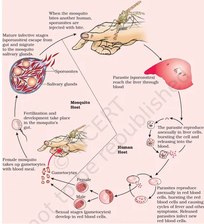
- Protozoal: Plasmodium spp. cause malaria transmitted by Anopheles mosquitoes — prevented by vector control (nets, insecticides) and antimalarial drugs.
- Helminthic: Ascaris lumbricoides causes ascariasis via fecal–oral transmission — prevented by sanitation and deworming programs.
- Fungal: Candida albicans causes thrush (opportunistic) especially in immunocompromised hosts or following antibiotic use.
- Genetic: Sickle cell disease caused by a point mutation in beta-globin gene — inherited, manifests with vaso-occlusion and anemia.
- Incidence rate = (Number of new cases in a specified period / Population at risk during that period) × k (e.g., per 1,000 or 100,000)
- Prevalence = (Number of existing cases at a point in time / Total population at that time) × k
- Attack rate = (Number of people who become ill in an outbreak / Number of persons exposed) × 100%
- Case Fatality Rate (CFR) = (Number of deaths from a disease / Number of confirmed cases of the disease) × 100%
- Mortality rate = (Number of deaths due to a disease in a period / Total population) × k
- Basic reproduction number (R0) — conceptual: average number of secondary cases produced by one infectious case in a fully susceptible population; R0 > 1 → potential for epidemic
Microorganisms in Health and Disease
Overview
Microorganisms (bacteria, viruses, fungi, protozoa and some algae) are microscopic life-forms that interact with humans in two main ways: beneficially (as normal flora, probiotics, decomposers and in biotechnology) and harmfully (as pathogens causing infectious diseases). Understanding their roles, modes of transmission and control measures is central to human health.
Microorganisms that promote health
- Normal flora (commensals): Microbes that colonize skin, gut, mouth and other sites without causing disease under normal conditions. They compete with pathogens and help in digestion and vitamin synthesis (e.g., gut bacteria producing vitamin K and some B vitamins).
- Probiotics and fermentation microbes: Lactobacillus and Bifidobacterium strains restore gut balance; Saccharomyces cerevisiae and starter cultures are used in food fermentation (yogurt, cheese, bread).
- Immune system development: Early microbial exposure helps train the immune response and maintain mucosal immunity.
Microorganisms that cause disease (pathogens)
Pathogens produce disease when they invade tissues, multiply and/or produce toxins. Disease severity depends on pathogen virulence, dose, route of entry and host immunity.
Modes of transmission
- Direct contact (touch, sexual contact) — e.g., herpes, syphilis.
- Indirect contact (fomites) — e.g., common cold spread via doorknobs.
- Droplet and airborne — e.g., influenza, tuberculosis.
- Fecal–oral — e.g., cholera, hepatitis A.
- Vector-borne — e.g., malaria (mosquito), dengue.
- Vertical (mother to child) — e.g., HIV, rubella congenitally.
Host–pathogen interactions
Pathogens adhere to host cells, evade or suppress host defenses, acquire nutrients and multiply. Host responses include innate immunity (barriers, phagocytes, inflammation) and adaptive immunity (antibodies, T cells). Clinical signs result from pathogen damage and immune reactions.
Koch’s postulates (historical criteria)
Robert Koch proposed criteria to link a microbe to a disease: the organism must be present in diseased cases, isolated and grown in pure culture, cause disease when introduced into a healthy host, and be re-isolated. Modern microbiology recognizes limitations (asymptomatic carriers, unculturable organisms) and supplements Koch’s ideas with molecular methods.
Diagnosis, treatment and prevention
- Diagnosis: Microscopy, culture, antigen/antibody tests, PCR and imaging.
- Treatment: Antibacterial (antibiotics), antiviral, antifungal and antiparasitic agents. Supportive care (fluids, oxygen) is often critical.
- Prevention: Vaccination, sanitation, safe water, hand hygiene, vector control, safe food handling and rational antibiotic use.
- Antibiotic resistance: Overuse/misuse selects resistant strains (e.g., MRSA, multidrug-resistant TB) — a major public-health challenge.
Public-health concepts
Incidence and prevalence measure disease frequency; R0 (basic reproduction number) estimates transmission potential of an infectious agent. Outbreak investigation and epidemiological surveillance guide control measures.
Summary
Microorganisms are essential for life and human health but can also cause disease. Prevention (hygiene, vaccination), correct diagnosis and responsible treatment are key to reduce disease burden and limit the spread of resistant strains.
- Gut microbiota: Bacteroides and Firmicutes help digest complex carbohydrates and synthesize vitamins (vitamin K).
- Fermentation: Lactobacillus bulgaricus and Streptococcus thermophilus in yogurt production (beneficial microbes).
- Bacterial disease: Mycobacterium tuberculosis causes tuberculosis (airborne transmission; BCG vaccine used in many countries).
- Viral disease: Influenza virus causes seasonal flu (droplet spread; vaccination and antivirals can reduce severity).
- Water-borne disease: Vibrio cholerae causes cholera (fecal–oral transmission; treated with rehydration and prevented by sanitation).
- Opportunistic infection: Candida albicans causes thrush in immunocompromised individuals when normal flora balance is disturbed.
- Exponential growth (bacteria): N = N0 × 2^(t/g), where N0 = initial number, t = time, g = generation (doubling) time.
- Continuous growth form: N = N0 × e^(µt), where µ = growth rate constant. Relationship: µ = ln(2)/g.
- Doubling time (generation time): g = ln(2)/µ ≈ 0.693/µ.
- Incidence rate: Incidence = (Number of new cases during period) / (Population at risk during period).
- Prevalence: Prevalence = (Total number of cases at a given time) / (Total population at that time).
- Basic reproduction number (R0): R0 = average number of secondary cases produced by one infected individual in a fully susceptible population. (If R0>1, infection can spread; if R0<1, it will decline.)
Modes of Transmission
Definition: Modes of transmission are the ways infectious agents (viruses, bacteria, parasites, fungi) move from a source (infected person, animal or reservoir) to a new host. Understanding modes helps in prevention and control of disease spread.
Major categories:
- Direct transmission – Immediate transfer of pathogen from an infected host to a susceptible host without an intermediate object or organism.
- Direct contact: touch, sexual contact, blood contact (e.g., skin infections, HIV by unprotected sex or needle sharing).
- Droplet: large respiratory droplets produced by coughs/sneezes that travel short distances (≈1–2 m) and deposit on mucous membranes (e.g., influenza, COVID-19 primarily via droplets).
- Vertical (mother-to-child): across placenta, during childbirth or breastfeeding (e.g., HIV, congenital syphilis).
- Indirect transmission – Transfer via an intermediate vehicle, vector, or environmental medium.
- Airborne: small droplet nuclei or particles that remain suspended and travel long distances (e.g., tuberculosis, measles).
- Vehicle-borne: contaminated inanimate objects or substances (fomites: bedding, doorknobs; foodborne: salmonella; waterborne: cholera).
- Vector-borne: living organisms (insects, ticks) that carry and transmit pathogens (e.g., malaria by Anopheles mosquitoes, dengue by Aedes).
- Nosocomial (hospital-acquired) – Infections acquired in healthcare settings often via invasive procedures, contaminated instruments, or cross-transmission by staff (e.g., MRSA, Clostridioides difficile).
- Zoonotic transmission – Pathogens transmitted between animals and humans (direct contact, bites, vector-mediated; e.g., rabies, certain influenza strains, COVID-19 likely originated zoonotically).
Key determinants of transmission:
- Pathogen factors: infectious dose, stability in environment, route of entry.
- Host factors: immune status, behaviour (handwashing, mask use), susceptibility.
- Environmental factors: crowding, ventilation, sanitation, vector presence, seasonality.
Prevention & control principles:
- Interrupt direct transmission: isolation, barrier nursing, condoms, safe injection practices.
- Interrupt indirect transmission: hand hygiene, surface disinfection, safe water/food handling, vector control (nets, insecticides), air filtration and ventilation.
- Vaccination to reduce susceptible pool and lower effective transmission (Rt).
How this links to epidemiology: Modes of transmission determine the shape of outbreaks, appropriate public health interventions and which epidemiological measures (incidence, prevalence, R0) are most informative for control.
- Common cold: Primarily direct droplet and close-contact transmission; also via contaminated hands (fomite).
- Influenza: Droplet transmission, can have short-range airborne spread; prevented by vaccination, masks and ventilation.
- Tuberculosis (TB): Airborne transmission via droplet nuclei that remain suspended; requires prolonged close exposure.
- COVID-19: Primarily droplet and aerosol (airborne) transmission, with fomite and close-contact risks; controlled by masks, ventilation, distancing and vaccination.
- Malaria: Vector-borne transmission by Anopheles mosquitoes; prevented by insecticide-treated nets and vector control.
- Cholera: Waterborne (vehicle-borne) transmission through contaminated water; prevented by clean water, sanitation and oral vaccines.
- Basic reproduction number (R0): R0 = β / γ (in SIR model) where β = transmission rate, γ = recovery rate. Alternative expression: R0 = c × p × d (contact rate × transmission probability per contact × duration of infectiousness).
- Effective reproduction number (Rt): Rt = R0 × S (proportion of population susceptible) — indicates current average secondary cases per infection.
- Attack rate: (Number of new cases during an outbreak / Population at risk) × 100%
- Secondary attack rate: (New cases among contacts of primary cases / Total susceptible contacts) × 100%
- Incidence rate: (New cases in a specified time period / Population at risk during that period) (often per 1,000 or 100,000).
- Prevalence: (Total existing cases at a point in time / Total population) × 100%
Common Infectious Diseases — Examples
What are common infectious diseases? Infectious diseases are disorders caused by pathogenic microorganisms — bacteria, viruses, fungi or parasites — that can be spread, directly or indirectly, from one person to another or from animals or the environment to people. Classifying and understanding examples helps in prevention, diagnosis and control.
Major components to consider
- Causative agent: bacterium (e.g., Salmonella typhi), virus (e.g., Influenza virus), parasite (e.g., Plasmodium spp.), fungus (e.g., Trichophyton spp.).
- Modes of transmission: feco-oral (contaminated water/food), airborne/droplet, direct contact, vector-borne (mosquitoes, ticks), sexual, blood-borne.
- Incubation period: time between exposure and symptoms — important for quarantine and diagnosis.
- Clinical features: range from mild (common cold) to severe (tuberculosis, HIV/AIDS); symptoms guide initial treatment.
- Prevention and control: vaccination, sanitation, safe water, vector control, personal hygiene, safe sex, use of antibiotics/antivirals when indicated and stewardship to prevent resistance.
How the body defends — innate immunity (skin, mucous, phagocytes) and adaptive immunity (antibodies, cell-mediated response). Vaccination trains adaptive immunity to recognise pathogens without causing disease.
Treatment principles — use of specific antimicrobials: antibiotics for bacteria, antivirals for some viral infections, antifungals for fungal infections, antiprotozoal drugs for parasites. Supportive care (e.g., oral rehydration for diarrhoea) is often critical.
Public health role — surveillance, outbreak investigation, vaccination programmes, health education, environmental sanitation and vector control reduce incidence and spread.
Short summary: Knowing typical examples, their agents, transmission modes and control measures helps students apply these concepts to clinical or public-health scenarios and understand CBSE-level questions on human health and disease.
- Cholera — Agent: Vibrio cholerae; Mode: feco-oral (contaminated water/food); Key features: profuse watery diarrhoea, dehydration; Prevention: clean water, sanitation, oral rehydration solution (ORS), vaccination in outbreaks.
- Typhoid — Agent: Salmonella typhi; Mode: feco-oral; Key features: prolonged fever, abdominal pain; Prevention: hygiene, safe food/water, typhoid vaccine; Treatment: appropriate antibiotics.
- Tuberculosis (TB) — Agent: Mycobacterium tuberculosis; Mode: airborne droplets; Key features: persistent cough, weight loss, night sweats; Prevention: BCG vaccine, early detection and DOTS therapy (directly observed treatment).
- Malaria — Agent: Plasmodium spp. (P. falciparum, P. vivax, etc.); Mode: vector-borne (Anopheles mosquitoes); Key features: cyclical fever, chills, anaemia; Prevention: mosquito control, bed nets, chemoprophylaxis; Treatment: antimalarials.
- Dengue — Agent: dengue virus (Flavivirus); Mode: Aedes mosquito bite; Key features: high fever, severe myalgia, haemorrhagic complications in severe cases; Prevention: vector control, avoid stagnant water, supportive care.
- Influenza — Agent: Influenza viruses (A, B); Mode: airborne/droplet; Key features: sudden fever, cough, body ache; Prevention: annual flu vaccine, respiratory hygiene, antivirals in some cases.
- Incidence rate = (Number of new cases in a time period / Population at risk during that period) × multiplier (e.g., 1000 or 100,000). Example: incidence per 1000 = (new cases / population) × 1000.
- Prevalence = (Total number of existing cases at a specific time / Total population at that time) × 100 (or per 1000).
- Attack rate (for outbreaks) = (Number of people who become ill in a group / Number of people at risk in that group) × 100%.
- Case Fatality Rate (CFR) = (Number of deaths due to the disease / Number of diagnosed cases of the disease) × 100%.
- Basic reproduction number (R0) — not a simple algebraic formula here but conceptually: R0 = average number of secondary cases produced by one primary case in a fully susceptible population. If R0 > 1, infection can spread; R0 < 1, it will die out.
Non-infectious Diseases and Lifestyle Disorders
Definition: Non-infectious diseases (also called non-communicable diseases, NCDs) are disorders that are not caused by infectious agents and cannot be transmitted between people. Lifestyle disorders are a subcategory of NCDs whose principal causes are behavioral or environmental risk factors (diet, physical inactivity, tobacco, alcohol, stress).
Main categories:
- Genetic disorders: caused by mutations or chromosomal abnormalities (e.g., sickle cell anemia, hemophilia, Down syndrome).
- Deficiency diseases: due to lack of essential nutrients (e.g., scurvy—vitamin C deficiency; rickets—vitamin D deficiency; iron-deficiency anemia).
- Metabolic/endocrine disorders: e.g., diabetes mellitus (particularly type 2), obesity, thyroid disorders.
- Cardiovascular diseases (CVD): e.g., hypertension, coronary artery disease, myocardial infarction, stroke—often related to atherosclerosis.
- Chronic respiratory diseases: e.g., COPD, chronic bronchitis, emphysema—commonly linked to smoking and pollution.
- Cancer (neoplasia): abnormal cell growth due to genetic and environmental risk factors (smoking, radiation, diet).
- Autoimmune and allergic diseases: e.g., rheumatoid arthritis, type 1 diabetes (autoimmune), asthma (allergic component).
Key mechanisms and examples:
- Atherosclerosis: deposition of cholesterol and lipids in arterial walls → plaque formation → narrowing of lumen → reduced blood flow. Progressive narrowing greatly increases vascular resistance (even small decreases in vessel radius drastically reduce flow).
- Insulin resistance and type 2 diabetes: excess adiposity, sedentary lifestyle and poor diet lead to decreased insulin sensitivity. Chronic hyperglycemia causes vascular damage, neuropathy and increased infection risk.
- Obesity: excess energy intake vs expenditure causes adipose expansion, metabolic changes (inflammation, dyslipidemia) that raise risk of diabetes, hypertension and CVD.
- Carcinogenesis: accumulation of DNA damage and failures in repair/apoptosis give rise to uncontrolled proliferation; lifestyle factors (tobacco, alcohol, diet, UV) increase mutation load.
Clinical features & diagnosis: Symptoms depend on disease (chest pain, breathlessness, polyuria, polydipsia, unexplained weight loss/gain). Diagnosis uses clinical exam, biochemical tests (blood glucose, lipid profile), imaging (ECG, angiography, CT) and histopathology for cancers.
Prevention and management:
- Primary prevention: healthy diet (low saturated fat, adequate fruits/vegetables), regular physical activity, avoiding tobacco and excessive alcohol, maintaining healthy weight.
- Secondary prevention: screening for hypertension, diabetes, hyperlipidemia; early treatment to prevent complications.
- Tertiary care: medications (antihypertensives, statins, insulin/oral hypoglycemics), surgical interventions (angioplasty, bypass, tumor resection), rehabilitation and lifestyle counseling.
Public health perspective: NCDs are the leading cause of mortality worldwide; they are chronic, costly, and largely preventable by population-level policy (tobacco control, healthy food access, active transport).
Connections to CBSE Class 12 syllabus: Understand differences between infectious and non-infectious disease, major examples and mechanisms (e.g., atherosclerosis, insulin resistance), role of lifestyle factors, prevention strategies and the importance of early diagnosis and health education.
- Type 2 diabetes: A 50-year-old sedentary, overweight man develops frequent urination, excessive thirst and high fasting blood glucose; management includes lifestyle modification, metformin and monitoring of HbA1c.
- Myocardial infarction (heart attack): A 55-year-old smoker with high LDL cholesterol and hypertension develops sudden chest pain and shortness of breath due to coronary artery occlusion from an atherosclerotic plaque rupture.
- Obesity leading to metabolic syndrome: Central obesity, raised triglycerides, low HDL, hypertension and insulin resistance in a patient increases risk for CVD and type 2 diabetes.
- Chronic obstructive pulmonary disease (COPD): A long-term smoker develops chronic productive cough and breathlessness due to emphysema and chronic bronchitis.
- Vitamin D deficiency (rickets/osteomalacia): A child with inadequate sunlight exposure and poor diet develops bowed legs and delayed growth.
- Lung cancer: Long-term tobacco use increases mutation burden in bronchial epithelium leading to malignant transformation and progressive respiratory symptoms.
- Body Mass Index (BMI) = weight (kg) / [height (m)]^2 — used to classify underweight, normal, overweight, obesity.
- Waist-to-Hip Ratio = waist circumference / hip circumference — central obesity indicator (higher ratio = higher CVD risk).
- Cardiac Output (CO) = Heart Rate (HR) × Stroke Volume (SV) — CO (L/min) determines blood delivered by heart.
- Mean Arterial Pressure (MAP) ≈ Diastolic BP + 1/3 (Systolic BP − Diastolic BP) — an estimate of average arterial pressure.
- Poiseuille's law (blood flow relation): Q = (π ΔP r^4) / (8 η l) — flow (Q) is proportional to radius^4; small decreases in vessel radius (r) cause large drops in flow and large rises in resistance.
- Conversion for HbA1c to average glucose (approx): Estimated Average Glucose (mg/dL) ≈ 28.7 × HbA1c (%) − 46.7 — used to interpret long-term glucose control.
Defense Mechanisms — Overview
Overview: Defence mechanisms are the body's strategies to prevent infection and eliminate pathogens. They operate at two broad levels: innate (nonspecific) immunity — immediate, general protection present from birth — and adaptive (specific) immunity — slower to develop but highly specific and with memory.
Innate immunity (first and second lines of defence):
- Physical and chemical barriers (first line): skin, mucous membranes, tears, saliva, acidic stomach, normal microbiota that compete with pathogens.
- Cellular components (second line): phagocytes (neutrophils, macrophages) that ingest microbes; natural killer (NK) cells that kill infected or cancerous cells.
- Inflammation and fever: local response — vasodilation, increased vascular permeability, leukocyte recruitment — that helps isolate and remove pathogens. Fever raises body temperature to inhibit microbe growth and stimulate immune activity.
- Soluble factors: complement proteins (opsonization, membrane attack complex), interferons (antiviral signalling), acute phase proteins.
Adaptive immunity (third line of defence):
- Humoral immunity: mediated by B lymphocytes that differentiate into plasma cells producing antibodies (immunoglobulins). Antibodies neutralize pathogens, opsonize for phagocytosis, and activate complement.
- Cell-mediated immunity: mediated by T lymphocytes — helper T cells (CD4+) activate B cells and macrophages; cytotoxic T cells (CD8+) kill infected cells.
- Clonal selection and memory: antigen stimulates specific B or T cell clones which proliferate (clonal expansion). Some become long‑lived memory cells producing a faster, stronger response on re-exposure (secondary response).
- Active vs passive immunity: active — host produces its own immune response (natural infection or vaccination); passive — transfer of ready-made antibodies (maternal IgG across placenta, IgA in breast milk, or therapeutic antiserum) giving immediate, short-term protection.
Failures and dysregulation: Allergies/hypersensitivities are overreactions (e.g., IgE-mediated hay fever); autoimmune diseases result from loss of self-tolerance (e.g., rheumatoid arthritis, type I diabetes); immunodeficiencies (congenital or acquired like HIV) reduce ability to fight infections.
Principles of vaccination: Vaccines present antigen in safe form to induce primary adaptive response and memory, so that on real exposure a rapid secondary response prevents disease. Herd immunity depends on sufficient population immunity to reduce transmission.
Key concepts to remember: innate = fast, nonspecific, no memory; adaptive = slow at first, specific, memory. Effective defence usually requires cooperation of both arms.
- Splinter in skin → local inflammation: pain, redness (vasodilation), swelling (increased permeability), pus formation (neutrophil accumulation).
- Breastfeeding transfers secretory IgA in milk → passive mucosal protection for infant against enteric pathogens.
- Tetanus antitoxin (passive immunization) given after a wound provides immediate antibodies to neutralize toxin.
- Measles vaccination (active immunization) induces memory B and T cells so subsequent exposure produces a rapid secondary antibody response preventing disease.
- Hay fever (allergic rhinitis): pollen-specific IgE bound to mast cells causes histamine release on exposure, producing sneezing and itch.
- HIV infection leads to loss of CD4+ T cells → immunodeficiency, increased opportunistic infections.
- Antigen–antibody equilibrium: Antigen (Ag) + Antibody (Ab) ⇌ Ag·Ab complex. Association constant Ka = [Ag·Ab] / ([Ag][Ab]). Dissociation constant Kd = 1/Ka.
- Antibody titer (practical definition): reciprocal of the highest dilution of serum that still gives a detectable reaction (e.g., agglutination or precipitation).
- Vaccine efficacy (%) = ((AR_unvaccinated − AR_vaccinated) / AR_unvaccinated) × 100, where AR = attack rate (incidence) in the group.
Innate Immunity
Definition: Innate immunity is the body’s first line of defence against infectious agents. It is present from birth, acts immediately or within hours of an antigen’s appearance, is non‑specific, and does not generate immunological memory.
Key features:
- Rapid (minutes–hours), non‑specific recognition of threats.
- No memory — same response on repeated exposure.
- Uses physical, chemical, cellular and humoral mechanisms.
Main components:
- Physical and mechanical barriers: Intact skin, mucous membranes, mucus, cilia (respiratory tract), flushing actions (tears, urine).
- Chemical barriers: Lysozyme (tears, saliva), low pH of stomach, antimicrobial peptides (defensins), surfactant proteins in lungs.
- Microbial flora: Commensal bacteria on skin and mucosa that compete with pathogens.
- Cellular defences:
- Neutrophils — first recruited, phagocytose and kill bacteria.
- Macrophages — phagocytosis, antigen presentation, cytokine production.
- Dendritic cells — bridge innate and adaptive systems by presenting antigen.
- Natural killer (NK) cells — kill virus‑infected and tumor cells via perforin/granzymes; recognise ‘missing self’ (low MHC I).
- Eosinophils and mast cells — important against parasites and in allergy/inflammation.
- Humoral factors:
- Complement system (classical, lectin, alternative pathways) — opsonisation, inflammation, membrane attack complex (lysis of some microbes).
- Interferons (especially type I IFNs) — antiviral state in neighbouring cells.
- Acute phase proteins (e.g., C‑reactive protein) — opsonisation and modulation of inflammation.
- Cytokines (e.g., IL‑1, TNF‑α, IL‑6) — mediate inflammation, fever, and leukocyte recruitment.
Recognition mechanism: Innate cells detect conserved molecular patterns on microbes called PAMPs (pathogen‑associated molecular patterns) via PRRs (pattern recognition receptors) such as Toll‑like receptors (TLRs). This triggers phagocytosis, cytokine release and activation of downstream responses.
Inflammation (basic steps): Tissue injury or infection → detection by resident macrophages/dendritic cells → release of cytokines/chemokines → vasodilation and increased vascular permeability → recruitment of neutrophils and monocytes → phagocytosis and killing of microbes → resolution and repair (or chronic inflammation if unresolved).
Outcome: If innate mechanisms reduce pathogen numbers sufficiently, infection is controlled and adaptive immunity may not be strongly engaged. If not, innate responses activate and shape adaptive immunity (T and B cell responses).
Clinical relevance / examples of defects: Defects in innate components cause susceptibility to infections — e.g., chronic granulomatous disease (defect in NADPH oxidase → impaired killing by phagocytes), complement deficiencies (C3 deficiency → recurrent pyogenic infections).
- Cut on the skin: physical barrier broken → local inflammation (redness, heat, swelling) with neutrophil and macrophage recruitment and phagocytosis of invading bacteria.
- Common cold / influenza: infected epithelial cells produce type I interferons, inducing an antiviral state in neighbouring cells and activating NK cells that kill infected cells.
- Complement-mediated lysis: Gram-negative bacteria may be directly lysed by the membrane attack complex (terminal complement pathway), while C3b opsonisation enhances phagocytosis.
- Nasal mucociliary clearance: mucus traps inhaled particles and cilia move them out of the airway, preventing infection.
- Microbial flora on skin/intestine: commensal bacteria occupy niches and limit colonisation by pathogens (colonisation resistance).
- \[Simple first‑order model of innate pathogen clearance: dP/dt = -k P → P(t) = P₀ e^{-k t}\]\[where P = pathogen load\]\[k = clearance rate constant.\]
- Phagocytic clearance proportional model: dP/dt = -k_phago × P × M, where M = number (or activity) of phagocytes.
- Effective reproduction reduction (conceptual): R_eff = R₀ × (1 - E_innate), where E_innate ∈ [0,1] represents fractional reduction in transmission/infectivity produced by innate mechanisms.
Inflammation and Fever
Overview
Inflammation is the local tissue response to injury or infection whose purpose is to eliminate the initial cause of cell injury, clear out damaged cells and tissues, and establish repair. Fever is a systemic response in which the hypothalamic set point for body temperature is raised, usually in response to infection or inflammation.
Acute inflammation: cardinal signs
- Rubor (redness) – due to vasodilation and increased blood flow.
- Calor (heat) – increased blood flow and metabolism at the site.
- Tumor (swelling) – due to increased vascular permeability and accumulation of fluid (exudate).
- Dolor (pain) – sensitization of nerve endings by mediators (eg, bradykinin, prostaglandins).
- Loss of function – from pain and tissue damage.
Key steps and cellular events
- Recognition of injurious agent – by resident cells (macrophages, dendritic cells) via pattern recognition receptors (eg, TLRs) recognizing PAMPs and DAMPs.
- Vasodilation – mediated by histamine, nitric oxide and prostaglandins. Causes redness and heat.
- Increased vascular permeability – plasma proteins and fluid leak into tissues producing exudate and swelling.
- Leukocyte recruitment and migration – steps include margination, rolling (selectins), adhesion (integrins), diapedesis and chemotaxis (eg, C5a, IL-8).
- Phagocytosis and killing – neutrophils and macrophages ingest microbes/debris; killing via reactive oxygen species, nitric oxide, lysosomal enzymes.
- Resolution or outcome – complete resolution (restoration), healing by fibrosis (scar), abscess formation, or progression to chronic inflammation if injurious agent persists.
Mediators of inflammation
Major mediators include histamine, bradykinin, prostaglandins, leukotrienes, nitric oxide, complement fragments (C3a, C5a), cytokines (eg, IL-1, TNF-α) and chemokines. These are produced by resident cells, recruited leukocytes and plasma protein systems (complement, clotting, kinin).
Chronic inflammation
Occurs when the acute response fails to eliminate the cause. Characterized by predominance of macrophages, lymphocytes, plasma cells, tissue destruction, and attempts at repair with fibrosis (eg, tuberculosis, rheumatoid arthritis).
Fever (pyrexia)
Definition and cause
Fever is an elevation of body temperature due to an increase in the hypothalamic thermoregulatory set point. Common triggers are infection (bacteria, viruses, parasites), inflammatory diseases, malignancy and some drugs.
Pyrogens
- Exogenous pyrogens – microbial products such as lipopolysaccharide (LPS).
- Endogenous pyrogens – cytokines produced by host cells in response to infection or injury (eg, IL-1, IL-6, TNF-α). These act via production of prostaglandin E2 (PGE2) in the hypothalamus to raise the temperature set point.
Mechanism
Endogenous pyrogens increase PGE2 in the preoptic area of hypothalamus → hypothalamic set point rises → physiological responses to reach new set point: peripheral vasoconstriction, shivering, increased metabolism, and behavioral changes (seeking warmth). When infection or inflammation resolves, set point returns to normal → vasodilation and sweating reduce temperature.
Patterns of fever
- Continuous (remains above normal with little variation) – eg, typhoid.
- Remittent (fluctuates but never returns to normal) – many bacterial infections.
- Intermittent (normal at intervals) – eg, malaria.
- Relapsing (periods of fever separated by days or weeks) – eg, certain infections like relapsing fever.
Physiological effects, benefits and dangers
Moderate fever enhances immune function (increases leukocyte activity and impairs growth of some pathogens). High fever can cause dehydration, increased metabolic demand, delirium, seizures (especially in young children) and organ dysfunction if prolonged.
Treatment and management
Antipyretics: paracetamol (acetaminophen) and NSAIDs (eg, ibuprofen) lower PGE2 and reduce set point. Supportive measures: adequate hydration, antipyretics, tepid sponging (when appropriate), cooling for very high fevers, and treating the underlying cause (eg, antibiotics for bacterial infection).
Important clinical correlations
- Appendicitis: localized acute inflammation of the appendix with pain, tenderness and systemic signs.
- Rheumatoid arthritis: example of chronic immune-mediated inflammation with joint destruction.
- Sepsis: systemic inflammatory response to infection that can lead to multi-organ dysfunction and shock.
Summary: Inflammation is a complex host response aimed at removing harmful stimuli and starting repair. Fever is a systemic, regulated rise in body temperature driven by pyrogenic mediators. Both are components of the body s defense but may cause harm if excessive or uncontrolled.
- Insect bite producing redness, swelling and itching (acute local inflammation)
- Appendicitis presenting with localized pain, tenderness and fever (acute inflammation with systemic signs)
- Rheumatoid arthritis causing chronic joint swelling, pain and deformity (chronic inflammation)
- Malaria causing intermittent high fevers every 48-72 hours (intermittent fever pattern)
- Common cold or influenza causing low to moderate fever due to cytokine release after viral infection
- Abscess formation after a skin infection where pus accumulates and a fibrous capsule may form
- Celsius to Fahrenheit: F = (9/5) × C + 32
- Fahrenheit to Celsius: C = (5/9) × (F - 32)
- Q10 temperature coefficient (rule of thumb for metabolic rate changes): Rate2 = Rate1 × Q10^((T2 - T1)/10). For biological systems Q10 ≈ 2, meaning roughly a 10°C increase doubles rate.
Adaptive Immunity — Overview
Adaptive immunity (also called acquired or specific immunity) is the arm of the immune system that recognizes specific foreign molecules (antigens), mounts a targeted response, and retains memory for faster, stronger responses on re-exposure. It complements innate immunity and develops after contact with pathogens or vaccines.
Key characteristics
- Specificity: Recognizes specific antigenic determinants (epitopes).
- Memory: Produces a faster and larger response on subsequent exposures (secondary response).
- Self–nonself discrimination: Tolerance to self antigens; reacts to nonself.
- Diversity: Can recognize vast numbers of different antigens.
Main components
- Humoral immunity (B lymphocytes): B cells recognize antigens (often with T help), differentiate into plasma cells that secrete antibodies (immunoglobulins IgM, IgG, IgA, IgE, IgD) and generate memory B cells.
- Cell-mediated immunity (T lymphocytes): T cells act against infected cells, cancer cells and help other immune cells. Major types: helper T cells (CD4+, activate B cells and macrophages), cytotoxic T cells (CD8+, kill infected cells), regulatory T cells (suppress responses).
- Antigen-presenting cells (APCs): Dendritic cells, macrophages and B cells process antigens and present peptide fragments on major histocompatibility complex (MHC) molecules to T cells. MHC I presents to CD8+; MHC II presents to CD4+.
How adaptive immunity works (overview of sequence)
- Antigen entry & capture by APCs.
- Antigen processing and presentation on MHC molecules.
- Activation of naive T cells (helper and cytotoxic) and B cells (with T helper cell assistance for T-dependent antigens).
- Clonal expansion and differentiation: effector cells (plasma cells and cytotoxic T cells) + memory cells.
- Elimination of pathogen and formation of long-lived memory cells.
Clonal selection (concise)
Each lymphocyte bears antigen receptors of a single specificity. Encounter with its specific antigen (plus co-stimulation) causes that clone to proliferate (clonal expansion) and form effector and memory cells. This explains specificity and memory.
Primary vs secondary response
Primary response: first exposure produces a lag phase, initial IgM production followed by IgG; lower magnitude. Secondary response: memory cells produce a rapid, high-magnitude response dominated by high-affinity IgG (or other isotypes), often preventing disease.
Clinical relevance
- Vaccination: deliberate exposure to antigen to create memory without disease.
- Autoimmunity: failure of self–nonself discrimination leads to attacks on self tissues.
- Transplant rejection: cell-mediated recognition of nonself MHC causes graft rejection.
- Immunodeficiency: defects in adaptive components (e.g., B- or T-cell deficiencies) increase susceptibility to infections.
Summary line: Adaptive immunity is a specific, memory-forming immune system involving B- and T-lymphocytes, antigen presentation via MHC, clonal selection, and distinct primary and secondary antibody responses that underpin vaccination and long-term protection.
- Vaccination against measles: exposure to attenuated virus produces memory B and T cells so subsequent natural exposure does not cause disease.
- Secondary response to chickenpox (varicella): after initial infection, memory cells prevent severe disease on re-exposure; shingles can occur when latent virus reactivates if cell-mediated immunity wanes.
- Organ transplant rejection: recipient T cells recognize donor MHC molecules as nonself and destroy graft cells (cell-mediated adaptive response).
- Monoclonal antibody therapy (e.g., anti-TNF drugs for rheumatoid arthritis): lab-produced antibodies target specific immune molecules to modulate adaptive responses.
- Antigen + Antibody ⇌ Antigen–Antibody complex (immune complex) — represents specific binding equilibrium
- Association constant (affinity): Ka = [AB] / ([A][B]) , Dissociation constant: Kd = 1 / Ka — describes antibody-antigen binding strength
- Clonal selection (schematic): Naive B cell + specific antigen → activated B cell → plasma cells (antibody-secreting) + memory B cells
- Primary vs secondary (qualitative): Primary: lag → IgM peak (lower) → IgG rises; Secondary: short lag → rapid high IgG peak
Cells and Organs of the Immune System
Overview
The immune system protects the body from pathogens and damaged cells by coordinated action of specialised cells and organs. Cells of the immune system arise from hematopoietic stem cells in bone marrow and are broadly classified into innate (non-specific, rapid) and adaptive (specific, memory-forming) components. Organs are divided into primary (where immune cells develop) and secondary (where immune responses are initiated).
Primary lymphoid organs
- Bone marrow: Site of hematopoiesis. B lymphocytes mature here in humans; myeloid lineages (neutrophils, monocytes, eosinophils, basophils, mast cells) are also produced.
- Thymus: T lymphocyte maturation and selection (positive and negative selection) occur here. Functional size reduces (involution) after puberty.
Secondary lymphoid organs
- Lymph nodes: Filter lymph, concentrate antigens; site of interactions between antigen-presenting cells, T cells and B cells leading to activation and clonal expansion.
- Spleen: Filters blood-borne antigens; contains white pulp (immune reactions) and red pulp (removal of old RBCs).
- Mucosa-associated lymphoid tissue (MALT): Tonsils, Peyer’s patches, appendix — protect mucosal entry points.
Cells of innate immunity
- Neutrophils: Phagocytic first responders; kill bacteria by phagocytosis and release of reactive oxygen species. Most abundant WBC.
- Macrophages: Phagocytose pathogens and dead cells; present antigen to T cells (as antigen-presenting cells, APCs); secrete cytokines to recruit cells.
- Dendritic cells: Potent APCs that capture antigen in tissues and migrate to lymph nodes to activate naive T cells.
- Natural Killer (NK) cells: Kill virus-infected and tumour cells without prior sensitisation (recognise missing MHC I).
- Eosinophils, basophils, mast cells: Involved in parasitic infections and allergic responses (release histamine, major basic protein, etc.).
Cells of adaptive immunity
- B lymphocytes (B cells): Recognise antigen (often with T-cell help), differentiate into plasma cells that secrete antibodies (humoral immunity). Some become memory B cells.
- T lymphocytes (T cells):
- CD4+ Helper T cells (TH): help B cells and macrophages by cytokines; subtypes (TH1, TH2, TH17) direct type of response.
- CD8+ Cytotoxic T cells (TC): kill infected or altered self-cells presenting antigen on MHC I (cell-mediated immunity).
- Regulatory T cells (Treg): suppress immune responses to maintain self-tolerance and prevent autoimmunity.
- Plasma cells: Antibody factories derived from activated B cells.
- Memory cells: Long-lived B and T cells that provide rapid, stronger secondary responses on re-exposure.
Key interactions and processes
- Antigen presentation: APCs (dendritic cells, macrophages, B cells) process antigen and present peptides on MHC molecules—MHC I for CD8+ T cells; MHC II for CD4+ T cells.
- Clonal selection and expansion: A specific lymphocyte recognising its antigen is activated, proliferates (clonal expansion) and differentiates into effector and memory cells.
- Humoral vs cell-mediated immunity: Humoral—antibodies neutralise extracellular pathogens and toxins. Cell-mediated—cytotoxic T cells and macrophages clear intracellular pathogens and infected cells.
Clinical correlations
- Vaccination: Uses an antigen to generate memory B and T cells so that a faster and higher secondary response protects upon real infection.
- Allergy: Excessive IgE-mediated mast cell degranulation (histamine) causes symptoms like asthma, hay fever.
- Autoimmunity: Failure of self-tolerance (e.g., rheumatoid arthritis, type 1 diabetes) where immune cells attack self-tissues.
- Immunodeficiency: Primary (genetic) or secondary (e.g., HIV infects CD4+ T cells) leads to susceptibility to infections.
Understanding the cells and organs together explains how the body recognises, responds to, remembers and resolves infections—core concepts tested in Class 12 Biology.
- Vaccination with tetanus toxoid: B and T cell memory formation leads to rapid antibody production on exposure to Clostridium tetani.
- Bacterial infection (e.g., a cut causing pus): neutrophils rapidly migrate to the site and phagocytose bacteria; macrophages clean up debris and present antigen.
- Allergic rhinitis (hay fever): exposure to pollen triggers IgE on mast cells causing histamine release, sneezing and watery eyes.
- Parasitic worm infection: eosinophils bind and release toxic proteins to kill multicellular parasites.
- HIV infection: virus targets CD4+ helper T cells, reducing cell-mediated and humoral immune responses, leading to opportunistic infections.
- Spleen removal (splenectomy): increased susceptibility to encapsulated bacteria (e.g., Streptococcus pneumoniae) because splenic clearance and antibody responses are reduced.
- Antigen–antibody equilibrium: Ag + Ab ⇌ Ag·Ab; association constant Ka = [Ag·Ab] / ([Ag][Ab]); dissociation constant Kd = 1 / Ka (lower Kd = higher affinity).
- Antibody titre (practical lab definition): titre = reciprocal of the highest dilution of serum that still gives a positive reaction (used to quantify antibody concentration).
- Clonal expansion (cell doubling): N = N0 × 2^n, where N0 = initial activated lymphocyte count and n = number of cell divisions (gives approximate fold increase during proliferation).
Antigens and Epitopes
Antigen (Ag): Any molecule or molecular fragment that can specifically bind to components of the immune system (antibodies or T‑cell receptors) and may provoke an immune response. Typical antigens are proteins or large polysaccharides; small molecules (haptens) are antigenic only when covalently attached to a carrier.
Epitope (Antigenic Determinant): A specific portion of an antigen that is recognized and bound by an antibody or a T‑cell receptor. One antigen molecule can contain several different epitopes.
Key distinctions
- B‑cell (antibody) epitopes: usually accessible regions on the native 3D surface of the antigen. They can be linear (continuous amino‑acid stretches) or conformational/discontinuous (brought together by protein folding).
- T‑cell epitopes: short linear peptides produced by antigen processing and presented on MHC molecules to T‑cell receptors.
Properties that determine antigenicity/immunogenicity
- Foreignness: more foreign → more antigenic.
- Size: larger molecules (>10 kDa, especially proteins) are better immunogens.
- Chemical complexity and heterogeneity: more complex → better.
- Dose, route and adjuvants: affect the magnitude and type of response.
Antigen–antibody interaction: Binding is non‑covalent (hydrogen bonds, ionic interactions, hydrophobic effects, van der Waals). Multiple binding sites on antibodies (bivalent IgG, pentameric IgM) lead to avidity (overall strength) which depends on individual binding affinity and valency.
Clinical and biological significance
- Vaccine design: selection of protective epitopes to elicit neutralizing antibodies or T‑cell responses.
- Allergy and hypersensitivity: small antigenic determinants (allergens) trigger IgE‑mediated responses.
- Blood transfusion and transplantation: recognition of blood group antigens and HLA epitopes causes rejection or hemolytic reactions.
- Autoimmunity: immune recognition of self‑epitopes leads to disease.
- Diagnostics: assays (ELISA, Western blot) detect antigen–antibody binding using known epitopes.
Additional notes: Haptens (e.g., some drugs like penicillin) become antigenic after binding host protein. Cross‑reactivity occurs when unrelated antigens share similar epitopes. Affinity maturation in B cells increases antibody affinity for an epitope over time after repeated antigen exposure.
- Blood groups A and B: A and B antigens on RBC membranes are carbohydrate epitopes; anti‑A or anti‑B antibodies cause agglutination in incompatible transfusions.
- Rh factor and hemolytic disease of the newborn: Maternal anti‑Rh (D) antibodies target fetal Rh epitopes causing hemolysis.
- Vaccines: Tetanus toxoid provides protein epitopes that stimulate protective IgG antibodies; influenza vaccines include surface protein epitopes (haemagglutinin) to elicit neutralizing antibodies.
- Allergens: Pollen proteins contain epitopes recognized by IgE in allergic individuals causing hay fever.
- Haptens: Penicillin becomes antigenic after forming covalent adducts with serum proteins and can trigger allergic reactions.
- Pathogens: The HIV gp120 protein contains epitopes targeted by broadly neutralizing antibodies; bacterial capsules (e.g., Streptococcus pneumoniae) bear polysaccharide epitopes used in conjugate vaccines.
- Antibody–antigen reversible binding: Ab + Ag ⇌ Ab–Ag
- Affinity constant (association): Ka = [Ab–Ag] / ([Ab][Ag])
- Dissociation constant: Kd = 1 / Ka = ([Ab][Ag]) / [Ab–Ag] (lower Kd → higher affinity)
- Fraction of antigen bound (Langmuir form): Fraction bound = [Ab] / ([Ab] + Kd) (or more generally ligand concentration / (ligand concentration + Kd))
Antibodies — Structure and Function
Overview
Antibodies (immunoglobulins, Igs) are Y-shaped glycoproteins produced by B cells (plasma cells) that specifically bind antigens to neutralize pathogens and mark them for elimination. They are central to humoral immunity and are produced during primary and secondary immune responses.
Basic structure
- Each antibody is composed of four polypeptide chains: two identical heavy (H) chains and two identical light (L) chains linked by disulfide bonds. Typical molecular mass: IgG ≈ 150 kDa (H ≈ 50 kDa, L ≈ 25 kDa).
- Each chain has a variable (V) region at the N-terminus and one or more constant (C) domains at the C-terminus. The variable regions of one heavy + one light chain form an antigen-binding site.
- Complementarity determining regions (CDRs, three in each V domain) form the actual antigen-contact loops — 6 CDRs make one paratope.
- Functionally the antibody splits into: Fab (fragment antigen binding) — contains V and first C domain of H and L chains and binds antigen; Fc (fragment crystallizable) — composed of C domains of heavy chains, mediates effector functions (complement activation, Fc receptor binding).
- Hinge region between CH1 and CH2 gives flexibility. Heavy chains have 3 or 4 constant domains depending on isotype (IgG, IgA, IgD: 3; IgM, IgE: 4).
Major isotypes and key functions
- IgG – monomer, most abundant in serum, crosses placenta (provides neonatal immunity), opsonization, neutralization, complement activation; half-life ≈ 21 days.
- IgM – secreted as a pentamer (with J chain), first antibody produced in primary response, very effective at agglutination and activating complement.
- IgA – in secretions (dimeric in mucosal secretions with secretory component), protects mucosal surfaces (saliva, tears, breast milk).
- IgE – monomer, binds Fc receptors on mast cells and basophils, mediates allergic responses and defense vs helminths.
- IgD – mainly B-cell receptor on naive B cells; role in B-cell activation.
How antibodies work (mechanisms)
- Neutralization: antibody blocks pathogen attachment or toxin activity (e.g., antitoxin antibodies).
- Agglutination/Precipitation: cross-linking of antigens into complexes that are cleared more easily.
- Opsonization: Fc region binds phagocyte Fc receptors enhancing phagocytosis.
- Complement activation: Fc region (mainly IgM and IgG) triggers classical complement pathway leading to lysis or opsonization.
- Antibody-dependent cell-mediated cytotoxicity (ADCC): NK cells recognize Fc-bound antibody and kill the target cell.
Generation of antibody diversity
- V(D)J recombination: random somatic recombination of Variable (V), Diversity (D, heavy chain only), and Joining (J) gene segments during B-cell development (mediated by RAG1/2 enzymes) forms diverse V regions.
- Junctional diversity: nucleotide addition/deletion at joining sites increases variability.
- Somatic hypermutation and affinity maturation: activation-induced cytidine deaminase (AID) introduces point mutations in variable regions of activated B cells in germinal centers; B cells with higher-affinity receptors are selected.
- Class (isotype) switching: AID-mediated recombination changes heavy-chain constant region (e.g., from μ to γ, α, ε) so the same antigen specificity can be expressed with different effector functions (IgM → IgG/IgA/IgE) under cytokine control.
Primary vs secondary immune response
In a primary response, IgM appears first then IgG; in a secondary (memory) response, IgG rises faster and to higher levels and affinity is greater due to memory B cells and affinity maturation.
Clinical and practical notes
- Passive immunity: transfer of antibodies (e.g., maternal IgG via placenta; antivenoms and immunoglobulin therapy) provides immediate short-term protection.
- Vaccination stimulates active immunity and memory B cells that produce high-affinity IgG on re-exposure.
- Monoclonal antibodies (mAbs) are engineered antibodies used as therapeutics (e.g., rituximab for CD20+ B-cell lymphoma, trastuzumab for HER2+ breast cancer) and diagnostics.
- Autoimmune diseases often involve pathogenic autoantibodies (e.g., rheumatoid factor, anti-thyroid antibodies).
Key facts summary
- Antigen-binding site = variable regions of heavy + light chains; 6 CDRs per site.
- Fab = antigen binding; Fc = mediates effector functions.
- IgM = primary response, pentameric; IgG = major serum Ab & memory response; IgA = mucosal; IgE = allergy; IgD = BCR.
- Vaccination: After DPT vaccine, primary response shows IgM then IgG; booster dose gives rapid, high IgG (memory response).
- Passive immunization: Anti-snake venom is a preparation of antibodies given after snake bite to immediately neutralize venom.
- Neonatal immunity: Maternal IgG crosses the placenta to protect the newborn for the first months of life.
- Allergy: Pollen-specific IgE bound to mast cells causes histamine release and allergic symptoms on re-exposure.
- Therapeutic monoclonal antibodies: Trastuzumab (Her2-targeting mAb) binds tumor cell receptors to block growth and recruit immune effectors.
- Antigen–antibody binding (law of mass action): Ka = [Ag·Ab] / ([Ag][Ab]) , where Ka is the association (affinity) constant; Kd = 1/Ka (dissociation constant).
- Antibody titer definition: titer = reciprocal of the highest dilution of serum that gives a positive reaction (e.g., agglutination or precipitation).
- Valency concept (qualitative): Effective binding strength (avidity) increases with valency — multivalent antibodies (IgM pentamer) can bind multiple epitopes increasing overall avidity even if individual affinity is moderate.
Classes of Immunoglobulins
Introduction
Immunoglobulins (Igs) or antibodies are glycoprotein molecules produced by plasma cells in response to antigens. In humans there are five major classes: IgG, IgM, IgA, IgE and IgD. Each class differs in structure, location, function and abundance. These differences determine how the immune system neutralises pathogens, activates complement, mediates allergy and provides passive immunity.
Overview of structure (common features)
- Basic unit: four polypeptide chains — 2 heavy (H) chains + 2 light (L) chains forming a Y-shaped molecule with two antigen-binding (Fab) arms and one Fc region.
- Variable (V) regions at tips bind antigen; constant (C) regions determine class and effector functions.
- Forms: monomers or polymers (dimer, pentamer) joined by J (joining) chain; secretory IgA has an extra secretory component for mucosal transport.
Class-wise details
IgG
- Structure: monomer (~150 kDa).
- Abundance: most abundant in serum (~70–75% of total Ig).
- Functions: opsonisation, neutralisation of toxins and viruses, complement activation (mainly subclasses IgG1 & IgG3), antibody-dependent cell-mediated cytotoxicity (ADCC).
- Special feature: crosses the placenta — provides passive immunity to the fetus; long half-life (~21 days).
- Role in immune response: principal antibody of secondary (memory) response; high affinity after class switching and affinity maturation.
IgM
- Structure: primary form is pentamer in serum (5 monomers + J chain) — very large (~900 kDa).
- Abundance: ~5–10% of serum Ig.
- Functions: very effective at agglutination and at activating the classical complement pathway (one IgM pentamer bound to antigen can efficiently activate complement).
- Special feature: first antibody produced in a primary immune response; appears early but is short-lived (half-life ~5 days).
- Valency: pentameric valency ≈10 antigen-binding sites (steric/functional valency often fewer).
IgA
- Structure: serum IgA is mostly a monomer; secretory IgA (sIgA) in mucous secretions is a dimer joined by J chain and secretory component.
- Abundance: ~10–15% of serum Ig but is the main antibody class in mucosal secretions (saliva, tears, gut, bronchial secretions, breast milk).
- Functions: neutralisation at mucosal surfaces, prevents attachment of pathogens to epithelial cells; protects neonates via breast milk (sIgA).
- Half-life: serum IgA ~6 days (approx.).
IgE
- Structure: monomer.
- Abundance: extremely low in serum (<0.01% of total Ig).
- Functions: binds to high-affinity Fc receptors on mast cells and basophils; cross-linking by allergen triggers degranulation and release of histamine — central to allergic (type I hypersensitivity) reactions and defence against helminths.
- Half-life: short (~2 days in serum; longer when bound to Fc receptors on cells).
IgD
- Structure: monomer; found on surface of naive B cells as B-cell receptor (BCR).
- Abundance: very low in serum.
- Functions: role mainly as membrane receptor involved in B cell activation and signalling; serum function not well defined.
Functional summary
- Primary immune response: IgM is produced first; later class switch leads to IgG, IgA or IgE depending on cytokine signals.
- Secondary (memory) response: rapid and larger production of high-affinity IgG (and other switched classes) due to memory B cells.
- Complement activation: classical pathway activated effectively by IgM and certain IgG subclasses.
- Passive immunity: maternal IgG transferred via placenta; sIgA transferred in breast milk protects infant gut.
Key clinical relevance: measurement of Ig classes helps diagnose immunodeficiencies (e.g., low IgA), allergic disease (elevated IgE), recent infection (high IgM) and past exposure or vaccine response (IgG). Therapeutic monoclonal antibodies are mostly IgG isotypes because of long half-life and effector functions.
- Maternal-fetal passive immunity: IgG crosses the placenta and protects newborns in early life.
- Breastfeeding: secretory IgA in breast milk protects an infant's gut from pathogens (e.g., reduces diarrhoea risk).
- Allergic reactions/asthma: environmental allergen cross-links IgE on mast cells causing histamine release (sneezing, bronchospasm).
- Acute infection diagnosis: detection of pathogen-specific IgM indicates a recent/acute infection (e.g., IgM anti-HAV for acute hepatitis A).
- Vaccine response: after primary vaccination IgM appears first; on booster dose, high titres of IgG appear rapidly (memory response).
- Monoclonal antibody drugs: most therapeutic antibodies (e.g., anti-TNF drugs) are engineered IgG molecules to neutralise targets and recruit immune effectors.
- Approximate molecular weights: IgG ≈ 150 kDa; IgM (pentamer) ≈ 900 kDa; secretory IgA (dimer + secretory component) ≈ 385 kDa.
- Valency (antigen-binding sites): IgG (monomer) = 2; IgA (dimer) ≈ 4; IgM (pentamer) ≈ 10.
- Serum distribution (approx.): IgG ≈ 70–75% of total Ig; IgA ≈ 10–15%; IgM ≈ 5–10%; IgE + IgD <1%.
- Typical serum half-lives (approx.): t1/2(IgG) ≈ 21 days; t1/2(IgM) ≈ 5 days; t1/2(IgA) ≈ 6 days; t1/2(IgE) ≈ 2 days; t1/2(IgD) ≈ 2–3 days.
- Complement activation requirement (qualitative): Classical pathway activated when IgM pentamer or IgG molecules are bound to antigen (C1q binding to Fc region).
B-cell Responses and Antibody Production
B-cell responses — overview
B lymphocytes (B cells) are the main cells of humoral immunity. They recognise antigens through surface B-cell receptors (BCRs; membrane-bound immunoglobulins), become activated, proliferate (clonal expansion) and differentiate into antibody-secreting plasma cells and long-lived memory B cells. The antibodies (immunoglobulins, Ig) produced neutralise pathogens, opsonise them for phagocytosis, activate complement and mediate other effector functions.
Steps of B-cell activation and antibody production
- Antigen recognition: BCR binds specific antigen (native/soluble or on pathogen surface).
- Antigen internalisation and presentation: Antigen–BCR complex is internalised; peptides are presented on MHC class II molecules on the B cell surface.
- T-helper (Th) cell help – T-dependent activation: Helper T cells (usually Th2) with TCR specific for the same antigen/MHC II interact with the B cell and provide co-stimulatory signals (CD40L–CD40) and cytokines (IL-4, IL-5, IL-21). This leads to strong activation, germinal centre reactions and memory formation.
- T-independent activation: Some antigens (e.g., repetitive bacterial polysaccharides) can cross-link many BCRs and activate B cells without T-cell help; mostly IgM responses with limited memory.
- Clonal expansion: Activated B cells proliferate to produce many identical clones (clonal selection principle).
- Germinal centre reactions: In secondary lymphoid organs, B cells undergo somatic hypermutation (in variable-region genes) and affinity maturation, and class (isotype) switching under cytokine influence. High-affinity clones are selected.
- Differentiation: Some B cells become plasma cells (antibody factories), others become memory B cells for faster secondary responses.
Antibody structure and classes
Antibodies are Y-shaped molecules made of two identical heavy chains and two identical light chains. Each arm has a variable (V) region that binds antigen (Fab) and a constant (C) region (Fc) that mediates effector functions. Main human isotypes and typical functions:
- IgM: First antibody in primary response, pentameric (high avidity), good at complement activation.
- IgG: Most abundant in serum, opsonisation, complement activation, crosses placenta (provides neonatal immunity).
- IgA: Dimeric in secretions (saliva, mucus, breast milk); protects mucosal surfaces.
- IgE: Binds mast cells/basophils, mediates allergic responses and protection against parasites.
- IgD: Mostly membrane-bound on naïve B cells (role in activation).
Mechanisms of antibody-mediated protection
- Neutralisation: Antibodies block pathogen attachment/entry or toxin activity.
- Agglutination/precipitation: Cross-linking pathogens or soluble antigens to facilitate clearance.
- Opsonisation: Antibody Fc binds Fc receptors on phagocytes, enhancing phagocytosis.
- Complement activation: Classical pathway activation leading to lysis or opsonisation.
- Antibody-dependent cellular cytotoxicity (ADCC): NK cells recognise antibody-coated targets and kill them.
Primary vs secondary (memory) responses
Primary response: first exposure → lag phase (days), low-to-moderate IgM then IgG levels. Secondary response (upon re-exposure or booster): much shorter lag, faster and larger production of high-affinity IgG (and other switched isotypes) because memory B cells respond rapidly and germinal-centre-derived high-affinity clones are available.
Important molecular processes
- V(D)J recombination: Generates BCR/antibody diversity during B-cell development.
- Somatic hypermutation: Point mutations in V regions during germinal-centre reactions increase affinity.
- Class (isotype) switching: DNA recombination in heavy-chain constant region under cytokine control (e.g., IL-4 promotes switching to IgE; TGF-β can promote IgA).
Clinical and practical relevance
Vaccination mimics primary exposure to create memory B cells so that booster or real infection elicits a rapid, high-affinity secondary response. Monoclonal antibodies (lab-produced) are used as diagnostics and therapeutics. Defects in B-cell development or function (e.g., agammaglobulinemia, common variable immunodeficiency) lead to recurrent infections. Allergies are caused by inappropriate IgE responses.
Note for students: Understand the sequence of events (recognition → activation → clonal expansion → differentiation) and the differences between Ig classes and primary vs secondary responses. Diagrams of antibody structure and antibody–time curves are very helpful.
- Vaccination: First (primary) dose of tetanus vaccine produces a primary IgM followed by IgG response; a booster dose later elicits a stronger, faster IgG-dominated secondary response because of memory B cells.
- Breastfeeding: Secretory IgA in breast milk protects infant gut mucosa from pathogens by preventing attachment and invasion.
- Allergy: Pollen-specific IgE bound to mast cells triggers histamine release on re-exposure, causing allergic symptoms.
- Hemolytic disease of newborn: Maternal IgG against fetal Rh antigen crosses the placenta and can destroy fetal red blood cells if mother is sensitised.
- Monoclonal antibody therapy: Therapeutic antibodies (e.g., anti-PD-1 in cancer, anti-TNF in autoimmune disease) target specific molecules to modulate immune responses.
- Antigen–antibody binding equilibrium: Ag + Ab ⇌ Ag·Ab ; Association constant Ka = [Ag·Ab] / ([Ag][Ab])
- Dissociation constant: Kd = 1 / Ka = koff / kon (lower Kd → higher affinity)
- Antibody decay (first-order approximation): C(t) = C0 · e^(−kt) ; half-life t1/2 = ln(2) / k (e.g., IgG t1/2 ≈ 21 days)
- Titer definition (laboratory): antibody titer = reciprocal of the highest dilution of serum that gives a positive reaction (e.g., agglutination)
T-cell Mediated Immunity
Overview
T-cell mediated immunity (cell-mediated immunity) is the arm of adaptive immunity in which T lymphocytes (T cells) recognise and eliminate infected cells, abnormal cells (tumour), and coordinate other immune cells. Unlike B cells that produce antibodies, T cells act mainly by direct cell-to-cell contact and by secreting cytokines.
Types of T cells and functions
- CD8+ Cytotoxic T lymphocytes (CTL, Tc): recognise antigenic peptides presented on MHC I molecules (on all nucleated cells). They kill virus‑infected cells and tumour cells using perforin/granzymes and Fas–FasL pathways.
- CD4+ Helper T cells (Th): recognise peptides on MHC II (on antigen‑presenting cells). They secrete cytokines and differentiate into subsets: Th1 (cellular immunity, activates macrophages and CTLs), Th2 (humoral immunity, helps B cells), Th17 (inflammation), Tfh (help B cells in germinal centres).
- Regulatory T cells (Treg): maintain self‑tolerance and suppress excessive immune responses.
- Memory T cells: long-lived cells that provide rapid, amplified responses on re‑exposure to antigen.
Antigen processing & presentation — two main pathways
- MHC I pathway: Endogenous antigens (viral or abnormal intracellular proteins) are degraded by proteasomes, peptides transported into ER by TAP, loaded onto MHC I, and presented to CD8+ T cells.
- MHC II pathway: Exogenous antigens taken up by endocytosis are processed in endosomes/lysosomes, loaded onto MHC II in antigen presenting cells (dendritic cells, macrophages, B cells), and presented to CD4+ T cells.
Activation of naive T cells — key steps
- Antigen recognition: T cell receptor (TCR) binds specific peptide–MHC complex.
- Co-stimulation: Second signals (e.g., CD28 on T cell binding B7/CD80–CD86 on APC) are required to avoid anergy.
- Cytokine signals: Cytokines from APCs and local environment direct differentiation (IL-12 → Th1, IL-4 → Th2, etc.).
- Clonal expansion and differentiation: Activated T cells proliferate and become effector and memory cells.
- Effector functions: CTLs kill target cells; Th cells activate macrophages, help B cells and orchestrate the immune response.
Mechanisms of target-cell killing by CD8+ T cells
- Perforin‑granzyme pathway: Perforin forms pores in the target cell membrane; granzymes enter and induce apoptosis.
- Fas–FasL interaction: Binding of FasL on T cell to Fas on target triggers apoptotic signalling.
Biological outcomes and importance
- Elimination of intracellular pathogens (viruses, some bacteria, protozoa).
- Anti-tumour immunity by recognising tumour‑specific or tumour‑associated antigens.
- Role in transplant rejection (host T cells recognise donor MHC/peptides).
- Contribution to delayed-type hypersensitivity (Type IV hypersensitivity) — e.g., tuberculin reaction.
- Memory T cells provide faster and stronger secondary responses — principle behind many vaccines.
Regulation and clinical relevance
- Autoimmunity: Faulty regulation of T cells can cause diseases like type 1 diabetes, rheumatoid arthritis.
- Immunodeficiency: HIV infects CD4+ T cells causing loss of helper function and opportunistic infections.
- Immunotherapy: Cancer therapies (e.g., checkpoint inhibitors, CAR‑T cells) harness or modify T cell responses.
Summary (concise)
T-cell mediated immunity is essential for defence against intracellular pathogens and abnormal cells, relies on antigen presentation via MHC molecules, requires antigen recognition plus co-stimulation for activation, and produces effector and memory T cells that kill targets directly or regulate other immune cells.
- Viral infection (e.g., influenza): infected cells present viral peptides on MHC I; CD8+ CTLs recognise and kill these cells, limiting spread.
- Tuberculosis skin test (Mantoux test): injection of tuberculin evokes a Type IV hypersensitivity driven by Th1 cells and macrophage activation.
- HIV infection: virus specifically targets CD4+ T cells, leading to progressive loss of helper T cell function and opportunistic infections.
- Organ transplant rejection: host T cells recognise donor MHC and peptides, mounting a cell-mediated response that can destroy the graft.
- Cancer immunotherapy (CAR‑T cells): patient T cells are engineered to recognise tumour antigens and then administered to kill cancer cells.
- Clonal expansion (cell divisions): N = N0 × 2^n (N0 = initial activated T cells, n = number of cell divisions)
- CD4:CD8 ratio (clinical reference): typically ~2:1 in healthy humans (values vary by lab).
- Antigen recognition condition (qualitative): TCR specificity + peptide–MHC complex + co-stimulation → T cell activation
- Kinetics concept (primary vs secondary response): magnitude_secondary >> magnitude_primary; time_secondary < time_primary (no numeric universal constant — principle only)
Antigen Processing and Presentation
Definition & significance: Antigen processing and presentation is the cellular mechanism by which protein antigens are degraded into short peptide fragments and displayed on the cell surface bound to Major Histocompatibility Complex (MHC) molecules so that T lymphocytes can recognize them. This process links innate detection of foreign material to activation of adaptive immunity (T cells), and is essential for fighting infections, forming immunological memory, and for vaccine responses.
Main components:
- Antigen-presenting cells (APCs): Dendritic cells, macrophages, B cells (professional APCs). Nearly all nucleated cells can present via MHC I.
- MHC molecules: MHC class I (expressed on nearly all nucleated cells) and MHC class II (expressed mainly on professional APCs).
- T cells: CD8+ T cells recognize peptides on MHC I; CD4+ T helper cells recognize peptides on MHC II.
Two classical pathways:
1. Endogenous (MHC I) pathway — presents intracellular antigens (e.g., viral proteins, mutated proteins):
- Cytosolic proteins (normal or viral) are ubiquitinated and degraded by the proteasome into peptides.
- Peptides are transported into the endoplasmic reticulum (ER) by TAP (Transporter associated with Antigen Processing).
- In the ER, peptides are loaded onto newly synthesized MHC class I molecules; peptide–MHC I complexes are transported to the cell surface in vesicles.
- Displayed peptide–MHC I is scanned by CD8+ (cytotoxic) T cells; recognition can trigger killing of the infected cell.
2. Exogenous (MHC II) pathway — presents extracellular antigens (e.g., bacteria, soluble proteins):
- APCs take up extracellular antigen by phagocytosis, endocytosis or receptor-mediated uptake into endosomes/lysosomes.
- Proteases in the endosome/lysosome degrade antigen into peptides.
- MHC class II α and β chains are synthesized in the ER with an invariant chain (Ii) blocking the peptide-binding groove to prevent premature loading.
- MHC II–Ii complexes traffic to endosomal compartments where Ii is degraded, leaving CLIP, and HLA-DM (in humans) facilitates exchange of CLIP for antigenic peptides.
- Peptide–MHC II complexes are transported to the cell surface for recognition by CD4+ helper T cells.
Other important concepts:
- Cross-presentation: Some dendritic cells can present extracellular antigens on MHC I (useful for initiating CD8+ responses to viruses that do not infect DCs).
- Co-stimulation and cytokines: For full T cell activation, peptide–MHC recognition must be accompanied by co-stimulatory signals (e.g., B7 on APC binding CD28 on T cell) and cytokines; without co-stimulation, T cells may become anergic.
- Superantigens: Certain bacterial toxins (e.g., Staphylococcal enterotoxins) bind outside the peptide groove and link MHC II to TCRs nonspecifically, causing massive T cell activation and cytokine storm.
- Clinical relevance: Vaccines depend on APC uptake and presentation to generate helper and cytotoxic T cell responses; transplant rejection is driven by recognition of foreign MHC; defects in processing (e.g., TAP deficiency, Bare Lymphocyte Syndrome) cause immune deficiency.
Step-by-step simplified flow (summary):
- Antigen uptake → Processing into peptides → Loading on MHC (I or II) → Transport to cell surface → Recognition by appropriate T cell → T cell activation → Effector response.
Tips to remember: MHC I = Intracellular (endogenous), presents to CD8+; MHC II = Extracellular (exogenous), presents to CD4+. Proteasome & TAP are key for MHC I; endosomal proteases, invariant chain and HLA-DM are key for MHC II.
Illustrative diagram ideas (see graphs section): Show proteasome/TAP/ER route on one side and phagolysosome/MHC II route on the other; depict T cell receptor contact with peptide–MHC complex and co-stimulatory molecules between APC and T cell.
- Viral infection: A virus infects a body cell. Viral proteins produced in the cytosol are degraded by the proteasome, peptides are loaded onto MHC I via TAP, and the infected cell presents viral peptides to CD8+ T cells which then kill the infected cell.
- Bacterial infection: Macrophages phagocytose bacteria, degrade their proteins in lysosomes, load peptides on MHC II, and present them to CD4+ helper T cells; activated helper T cells then stimulate macrophages and B cells to clear the infection.
- Vaccine action: Protein or inactivated vaccines are taken up by dendritic cells; antigen processing and presentation on MHC II activate CD4+ T cells, which help B cells produce high-affinity antibodies and form memory cells.
- Transplant rejection: Donor MHC molecules differ from the recipient's. Recipient T cells recognize foreign MHC–peptide complexes on graft cells or donor APCs, leading to an immune attack on the transplant.
- Superantigen example: Certain bacterial toxins (e.g., toxic shock syndrome toxin) bind MHC II and TCR outside the peptide-binding site, causing non-specific massive T cell activation and high cytokine release.
- Endogenous pathway (schematic): Cytosolic protein --(ubiquitination)--> Proteasome --(peptides)--> TAP transport --> ER + MHC I --> peptide–MHC I complex (surface)
- Exogenous pathway (schematic): Extracellular antigen --(endocytosis/phagocytosis)--> Endosome/Lysosome --(proteolysis)--> peptides + MHC II (Ii removed) --> peptide–MHC II complex (surface)
- T cell activation (conceptual): APC(peptide–MHC) + TCR recognition + Co-stimulation (e.g., B7–CD28) + Cytokine signals --> T cell activation/expansion
- Binding equilibrium (basic ligand–receptor concept useful for affinity discussion): Kd = [P][L]/[PL] (lower Kd = higher affinity of peptide for MHC/TCR)
Complement System
What is the complement system?
The complement system is a set of >30 plasma and membrane-associated proteins that form an enzymatic cascade and bridge innate and adaptive immunity. When activated, complements tag pathogens, recruit inflammatory cells, and directly lyse target cell membranes.
Major functions
- Opsonization: C3b coats microbes to enhance phagocytosis.
- Inflammation and chemotaxis: small fragments (C3a, C5a) act as anaphylatoxins and chemoattractants.
- Direct lysis: formation of the membrane attack complex (MAC, C5b–C9) creates pores in microbial membranes.
- Immune complex clearance: complement helps remove antigen–antibody complexes and apoptotic cells.
Activation pathways (overview)
- Classical pathway — initiated by C1 binding to antigen–antibody complexes (IgM or IgG). Produces C3 convertase C4b2a.
- Lectin pathway — initiated by mannose-binding lectin (MBL) binding to microbial carbohydrates; similar downstream events to classical, producing C4b2a.
- Alternative pathway — initiated directly on pathogen surfaces by spontaneous hydrolysis of C3 (tickover) and stabilized by properdin; produces C3 convertase C3bBb.
Core cascade (simplified)
All pathways converge at C3 cleavage: C3 → C3a (inflammatory mediator) + C3b (opsonin). C3b participates in forming C5 convertase, which cleaves C5 → C5a (potent chemoattractant) + C5b (initiates MAC assembly). C5b recruits C6, C7, C8 and multiple C9 molecules to form the pore-forming MAC (C5b-9).
Regulation
- Plasma and membrane regulators prevent host damage: C1 inhibitor (C1INH) blocks C1 activation; Factor H and Factor I inactivate C3b; CD55 (DAF) disrupts convertases; CD59 prevents MAC formation on host cells.
Clinical relevance
- Deficiencies: MAC component deficiencies (C5–C9) increase susceptibility to Neisseria infections. C1 inhibitor deficiency causes hereditary angioedema (excess bradykinin-mediated edema).
- Autoimmunity: defective complement-mediated clearance of immune complexes can contribute to SLE.
- Pathogen evasion: encapsulated bacteria (e.g., Streptococcus pneumoniae) resist complement; some microbes express proteins that bind Factor H to evade opsonization.
Summary
The complement system is a rapid, amplifying cascade that helps recognize and eliminate microbes, enhances inflammation and phagocytosis, and must be tightly regulated to avoid host tissue damage.
- CH50 assay — measures classical pathway function by quantifying serum’s ability to lyse antibody-coated sheep red blood cells; used to detect complement deficiencies.
- Hereditary angioedema — caused by C1 inhibitor (C1INH) deficiency; unregulated activation leads to episodic, non-pruritic edema.
- Recurrent Neisseria infections — people with terminal complement (C5–C9) deficiencies (impaired MAC formation) have increased risk of meningococcal disease.
- Paroxysmal nocturnal hemoglobinuria (PNH) — deficiency of GPI-anchored complement regulators (CD55/CD59) on RBCs causes complement-mediated intravascular hemolysis.
- Vaccination and opsonization — effective antibody generation plus complement activation increases clearance of pathogens (e.g., response to pneumococcal vaccine enhanced by complement-mediated opsonization).
- C3 → C3a + C3b (cleavage by C3 convertase)
- Classical/Lectin pathway: Ag–Ab (IgM/IgG) or MBL → C1 (or MASPs) → C4 → C4a + C4b; C2 → C2a + C2b; C4b2a = C3 convertase
- Alternative pathway: C3 (tickover) → C3(H2O) or C3b + Factor B → C3bB; Factor D cleaves → C3bBb = C3 convertase (stabilized by properdin)
- C5 convertases: Classical/lectin: C4b2a3b; Alternative: C3bBbC3b → C5 → C5a + C5b
- MAC assembly: C5b + C6 + C7 + C8 + (multiple) C9 → C5b–9 (pore formation → cell lysis)
- Regulation examples: C1INH blocks C1r/C1s; Factor I + cofactors cleaves C3b → iC3b (inactive opsonin)
Hypersensitivity and Allergy
Overview
Hypersensitivity is an exaggerated or inappropriate immune response that causes host tissue damage. "Allergy" is commonly used for IgE‑mediated (Type I) hypersensitivity but in medicine hypersensitivity includes four types (I–IV) based on mechanism, time course and mediators.
Classification and mechanisms
- Type I — Immediate (IgE mediated)
Mechanism: First exposure to an allergen leads to Th2 activation, IL‑4/IL‑13 driven class switching of B cells to produce IgE. IgE binds FcεRI receptors on mast cells and basophils (sensitisation). On re‑exposure the allergen cross‑links IgE on these cells causing degranulation and release of histamine, tryptase, heparin, leukotrienes and prostaglandins. Effects: vasodilation, increased vascular permeability, bronchoconstriction, mucus secretion, itch. - Type II — Antibody mediated (cytotoxic)
Mechanism: IgG or IgM directed against antigens on cell surfaces or extracellular matrix. Binding leads to complement activation, opsonisation and phagocytosis or antibody dependent cellular cytotoxicity (ADCC). Examples: transfusion reactions (ABO incompatibility), autoimmune hemolytic anemia, some forms of pemphigus. - Type III — Immune complex mediated
Mechanism: Soluble antigen binds antibody forming immune complexes that deposit in vessel walls or tissues. Complexes activate complement, attract neutrophils and cause enzyme‑mediated tissue injury. Time course: hours to days. Examples: serum sickness, post‑streptococcal glomerulonephritis, some manifestations of SLE. - Type IV — Delayed (cell mediated)
Mechanism: T lymphocytes (Th1, CD8+ Tc cells) recognize antigen and release cytokines (eg, IFN‑γ, TNF) that activate macrophages or effect direct cytotoxicity. Reaction occurs 48–72 hours after exposure. Examples: tuberculin (Mantoux) test, contact dermatitis (poison ivy, nickel), graft rejection.
Clinical features and diagnosis
- Type I: rhinitis, conjunctivitis, urticaria (hives), bronchial asthma, anaphylaxis. Tests: skin prick test, specific serum IgE (RAST/ImmunoCAP).
- Type II: hemolysis, thrombocytopenia, tissue‑specific autoimmune disease. Tests: direct/indirect Coombs test, antigen/antibody detection.
- Type III: fever, rash, arthritis, nephritis. Tests: complement levels (often low), immune complex assays.
- Type IV: localized redness, induration. Tests: patch test (contact dermatitis), tuberculin skin test.
Treatment and prevention
- Type I: avoid allergen, antihistamines, inhaled/oral corticosteroids, bronchodilators for asthma, epinephrine for anaphylaxis, allergen immunotherapy (desensitisation).
- Type II/III/IV: remove offending antigen/drug, immunosuppressive therapy (steroids, disease‑modifying drugs), plasmapheresis in severe antibody/complex mediated disease.
Key points
- Allergy usually means Type I hypersensitivity (IgE). Hypersensitivity is a broader term including Types I–IV.
- Time course differs: immediate (minutes) for Type I, immune complex reactions within hours–days, delayed cell mediated in 48–72 hours.
- Different diagnostic tests and treatments are used for each type; correct classification guides management.
- Pollen‑induced allergic rhinitis (hay fever) — seasonal sneezing, watery eyes (Type I)
- Peanut allergy causing anaphylaxis — sudden bronchospasm, hypotension — emergency epinephrine required (Type I)
- Acute hemolytic transfusion reaction when incompatible blood is given — fever, hemoglobinuria (Type II)
- Serum sickness after administration of foreign antiserum — fever, rash, joint pain due to immune complex deposition (Type III)
- Contact dermatitis from nickel jewelry or poison ivy — delayed redness and blistering at contact site after 48–72 hours (Type IV)
- Positive Mantoux test (tuberculin) indicating cell‑mediated hypersensitivity to Mycobacterium tuberculosis antigens (Type IV)
- Antigen–antibody binding equilibrium (affinity): Ka = [Ag·Ab] / ([Ag] × [Ab])
- Dissociation constant: Kd = 1 / Ka (lower Kd = higher affinity)
- Simplified reaction path for Type I: Allergen + IgE(bound to FcεRI on mast cell) → cross‑linking → degranulation → histamine + leukotrienes → bronchoconstriction, vasodilation
- Classical complement activation (simplified): Ag‑Ab complex + C1q → C3 convertase → C5b‑9 (MAC) → cell lysis (relevant in Type II/III)
Autoimmune Diseases
Definition: Autoimmune diseases are conditions in which the immune system mistakenly recognises the body's own cells, tissues or organs as foreign and mounts an immune response against them, causing chronic damage and dysfunction.
Basic mechanism: The immune system normally distinguishes self from non-self by central and peripheral tolerance mechanisms. Autoimmunity occurs when tolerance fails, producing autoreactive T cells and/or autoantibodies. Key processes include:
- Loss of self-tolerance — defects in thymic selection (central tolerance) or peripheral regulatory mechanisms (e.g., Treg cells).
- Molecular mimicry — foreign antigens (often microbial) resemble self-antigens, triggering cross-reactive immune responses.
- Epitope spreading — initial immune response expands to target additional self-epitopes.
- Immune complex deposition — antigen–antibody complexes deposit in tissues activating complement and inflammation (typical of systemic autoimmunity).
- Cell-mediated cytotoxicity — autoreactive CD8+ T cells directly kill target cells (common in organ-specific autoimmunity).
Classification:
- Organ-specific autoimmune diseases — immune attack is directed at a single organ or cell type (examples: Type 1 diabetes pancreas β-cells, Hashimoto's thyroiditis).
- Systemic autoimmune diseases — multiple organs and systems are involved (examples: Systemic lupus erythematosus, rheumatoid arthritis).
Pathogenesis (summary): Genetic predisposition (HLA alleles), environmental triggers (infections, drugs, toxins), hormonal factors (higher prevalence in females), and immune dysregulation combine to break tolerance. Autoantibodies and autoreactive T cells cause inflammation through complement activation, Fc receptor–mediated phagocytosis, cytokine release and direct cytotoxicity, leading to tissue injury and clinical disease.
Clinical features & diagnosis: Symptoms vary by disease but commonly include chronic inflammation, fatigue, organ-specific dysfunction (e.g., hypothyroidism, hyperglycaemia, joint pain). Diagnosis uses serology (autoantibodies such as ANA, anti-dsDNA, anti-TPO, anti-GAD, RF, anti-CCP), functional tests (thyroid function tests, blood glucose), imaging and sometimes biopsy. Important diagnostic concepts include test sensitivity, specificity and antibody titre interpretation.
Treatment principles: There is no universal cure. Treatments aim to reduce immune-mediated damage and manage symptoms:
- Immunosuppressive drugs (corticosteroids, methotrexate, azathioprine).
- Targeted biologicals (TNF inhibitors, anti-CD20 — rituximab) for some conditions.
- Plasmapheresis to remove pathogenic autoantibodies in selected acute cases.
- Replacement therapy when organ function is lost (insulin for type 1 diabetes, thyroid hormone for hypothyroidism).
- Supportive care: physiotherapy, pain control, infection prevention.
Prevention & prognosis: True prevention is difficult. Early diagnosis and therapy reduce irreversible damage. Prognosis varies: some diseases can be controlled long-term; others cause progressive disability or life-threatening complications if untreated.
Summary (Class 12 focus): Autoimmune diseases arise from a breakdown of immune tolerance leading to autoantibodies and autoreactive T cells. They are classified as organ-specific or systemic and are diagnosed by clinical features and specific laboratory tests. Management uses immunosuppression, biologic agents and organ-specific replacement therapies.
- Type 1 diabetes mellitus — autoimmune destruction of pancreatic β-cells leading to insulin deficiency.
- Hashimoto's thyroiditis — autoimmune destruction of thyroid causing hypothyroidism; anti-thyroid peroxidase (anti-TPO) antibodies often present.
- Graves' disease — autoantibodies (TSI) stimulate thyroid → hyperthyroidism and goitre.
- Rheumatoid arthritis — chronic autoimmune inflammation of synovial joints; rheumatoid factor (RF) and anti-CCP antibodies may be positive.
- Systemic lupus erythematosus (SLE) — multisystem disease with autoantibodies (ANA, anti-dsDNA), immune complex deposition and varied manifestations (skin, kidney, joints).
- Multiple sclerosis — autoreactive T cells attack myelin in CNS causing demyelination and neurological deficits.
- Sensitivity = True Positives / (True Positives + False Negatives) — fraction of diseased correctly identified.
- Specificity = True Negatives / (True Negatives + False Positives) — fraction of healthy correctly identified.
- Positive Predictive Value (PPV) = True Positives / (True Positives + False Positives).
- Antibody titre (practical definition) = reciprocal of the highest serum dilution that gives a positive reaction (e.g., if 1:160 is last positive, titre = 160).
Immunodeficiency Disorders
Definition: Immunodeficiency disorders are conditions in which one or more components of the immune system are absent or dysfunctional, leading to increased susceptibility to infections, poor response to vaccines and, in some cases, autoimmunity or malignancy.
Classification
- Primary (congenital) immunodeficiencies: Caused by inherited genetic defects that directly affect immune cells or molecules. Usually present in infancy or childhood. Examples: Severe Combined Immunodeficiency (SCID), X‑linked agammaglobulinemia, DiGeorge syndrome, Chronic Granulomatous Disease (CGD).
- Secondary (acquired) immunodeficiencies: Caused by external factors that impair immune function. Can occur at any age. Examples: HIV/AIDS, malnutrition, chemotherapy, long‑term corticosteroid use, diabetes mellitus.
Basic Pathophysiology
- Defects in B cells cause impaired antibody production → recurrent bacterial infections, especially of the respiratory tract.
- Defects in T cells affect cell‑mediated immunity → severe viral, fungal, and opportunistic infections.
- Combined B‑ and T‑cell defects (as in SCID) result in severe, life‑threatening infections early in life.
- Defects in phagocytes (neutrophils, macrophages) lead to poor killing of ingested microbes → formation of granulomas, recurrent abscesses (e.g., CGD).
- Complement deficiencies predispose to specific bacterial infections and impaired immune complex clearance.
Clinical Features
- Recurrent, severe or unusual infections (opportunistic organisms).
- Poor wound healing, chronic diarrhea, failure to thrive (in children).
- Persistent thrush, pneumonias, meningitis, skin abscesses.
- Family history of early deaths from infection suggests inherited immunodeficiency.
Diagnosis (Common Tests)
- Complete blood count with differential (look for lymphopenia, neutropenia).
- Absolute lymphocyte and neutrophil counts (see formulas provided).
- Serum immunoglobulin levels (IgG, IgM, IgA, IgE).
- Flow cytometry to quantify T, B, and NK cells (CD4+, CD8+, CD19+, CD56+).
- Functional assays: nitroblue tetrazolium (NBT) or dihydrorhodamine (DHR) for neutrophil oxidative burst (CGD); vaccine response assessment for humoral function.
- Specific tests for HIV (ELISA, rapid tests, PCR for viral RNA) in suspected secondary immunodeficiency.
Treatment and Management
- Treat infections promptly with appropriate antimicrobials.
- Replacement therapy: intravenous or subcutaneous immunoglobulin (IVIG/SCIG) for antibody deficiencies.
- Hematopoietic stem cell (bone marrow) transplantation for severe congenital disorders (e.g., SCID).
- Antiviral therapy (antiretroviral therapy, ART) for HIV/AIDS to restore immune function.
- Gene therapy is emerging for some genetic immunodeficiencies (e.g., ADA‑SCID).
- Avoid live attenuated vaccines in severe immunodeficiency; ensure household contacts are immunized.
Prevention & Prognosis
- Early diagnosis improves outcomes — screening (newborn) and family genetic counseling for inherited disorders.
- Good nutrition, infection control, prophylactic antimicrobials in select cases.
- Prognosis varies: from manageable chronic disease (with IVIG or ART) to life‑threatening (untreated SCID).
Key Points for Class 12 (CBSE)
- Know the difference between primary and secondary immunodeficiency with examples.
- Understand the impact of T‑cell loss (e.g., HIV depleting CD4+ cells) and B‑cell/antibody deficiencies.
- Recognize clinical clues: recurrent/opportunistic infections, family history, poor vaccine response.
- Severe Combined Immunodeficiency (SCID) — absence or severe dysfunction of both T and B lymphocytes; infants present with recurrent severe infections; treatment: bone marrow transplant or gene therapy.
- DiGeorge syndrome — 22q11.2 deletion causing thymic aplasia; T‑cell deficiency; associated with congenital heart defects and hypocalcaemia.
- X‑linked agammaglobulinemia (Bruton) — defect in B cell development; very low/absent immunoglobulins; recurrent bacterial infections; treated with IVIG.
- Chronic Granulomatous Disease (CGD) — defect in phagocyte oxidative burst (NADPH oxidase); recurrent catalase‑positive bacterial and fungal infections; diagnosed by NBT/DHR tests.
- AIDS (caused by HIV) — virus infects and destroys CD4+ T cells leading to progressive secondary immunodeficiency and opportunistic infections; treated with combination antiretroviral therapy (ART).
- Iatrogenic immunodeficiency — long‑term corticosteroids, chemotherapy or immunosuppressive drugs (after organ transplant) causing increased infection risk.
- Absolute Lymphocyte Count (ALC) = Total WBC count × (% lymphocytes) / 100
- Absolute Neutrophil Count (ANC) = Total WBC count × (% neutrophils + % bands) / 100
- Absolute CD4 Count = Total lymphocyte count × (% CD4+ cells from flow cytometry) / 100
- Fold change in antibody titre = titre_after / titre_before (used when assessing vaccine response)
HIV and AIDS
Definition: Human Immunodeficiency Virus (HIV) is a retrovirus that infects cells of the immune system, primarily CD4+ T lymphocytes. Acquired Immunodeficiency Syndrome (AIDS) is the advanced clinical stage of HIV infection marked by severe immune deficiency and opportunistic infections.
Causative agent and types: HIV is an enveloped RNA retrovirus (family Retroviridae). Two main types: HIV‑1 (globally common, more virulent) and HIV‑2 (mainly West Africa, slower progression).
Structure (brief): spherical, envelope with glycoproteins gp120 and gp41, capsid containing two copies of +ssRNA, and key enzymes: reverse transcriptase (RT), integrase, and protease.
Replication cycle (key steps):
- Attachment: gp120 binds CD4 receptor and a co‑receptor (CCR5 or CXCR4) on host cells.
- Fusion and entry: viral envelope fuses with host membrane; core enters cytoplasm.
- Reverse transcription: viral RNA → DNA by RT (error‑prone).
- Integration: viral DNA (provirus) integrates into host genome via integrase.
- Transcription/translation: host machinery produces viral RNA and proteins.
- Assembly and budding: immature virions bud from membrane; protease cleaves Gag/Pol to mature infectious virions.
Pathogenesis: HIV infects and destroys CD4+ T cells and impairs immune function. Early infection shows high viremia and acute symptoms. A clinically latent phase follows with progressive CD4 decline. When CD4 count falls below critical levels (e.g., <200 cells/µL) or opportunistic infections/cancers appear, AIDS is diagnosed.
Clinical stages: acute/primary infection (flu‑like illness), clinical latency (months–years, often asymptomatic), symptomatic HIV infection (persistent generalized lymphadenopathy, weight loss), and AIDS (opportunistic infections like Pneumocystis pneumonia, TB, candidiasis; malignancies such as Kaposi's sarcoma).
Diagnosis: screening by antibody/antigen tests (rapid tests, ELISA; 4th generation tests detect p24 antigen + antibodies). Confirmatory testing (Western blot historically; now algorithmic confirmatory assays). Nucleic acid tests (PCR) detect viral RNA—used for early detection (window period) and viral load measurement.
Treatment (principles): Antiretroviral therapy (ART) — combination therapy using drugs from different classes to suppress viral replication, restore/maintain immune function, and prevent resistance. Major drug classes: NRTIs, NNRTIs, protease inhibitors, integrase strand transfer inhibitors (INSTIs), fusion/entry inhibitors. Early and lifelong ART is standard of care; treatment reduces viral load to undetectable levels and greatly lowers transmission risk ("U=U": undetectable = untransmittable).
Prevention: safe sex (condoms), harm reduction for injecting drug users (needle exchange), screening of blood products, prevention of mother‑to‑child transmission (PMTCT: maternal ART, infant prophylaxis, safer delivery and feeding practices), pre‑exposure prophylaxis (PrEP), post‑exposure prophylaxis (PEP), education and stigma reduction.
Public health and social aspects: HIV carries significant stigma; counseling, confidentiality, and community support are essential. Regular testing, contact tracing in public‑health contexts, and equitable access to ART are critical to control the epidemic.
Key points for students:
- Understand viral structure and the reason for high mutation rate (error‑prone reverse transcriptase) leading to drug resistance and immune escape.
- Know modes of transmission: unprotected sexual contact, contaminated blood products/needles, mother‑to‑child (in utero, intrapartum, breast‑feeding).
- Recognize diagnostic tests and the concept of window period and viral load/CD4 monitoring.
- Mother‑to‑child transmission: Without treatment, an HIV‑positive mother can transmit the virus during pregnancy, delivery, or breastfeeding. With PMTCT (maternal ART, infant prophylaxis), transmission risk falls to <5% in many programs.
- Needle‑sharing among intravenous drug users: sharing contaminated needles can transmit HIV rapidly within a drug‑using network; needle‑exchange programs reduce such transmission.
- Contaminated blood transfusion (historical example): Before routine blood screening was implemented, transfusion was a notable source of HIV spread; modern screening has made this route extremely rare.
- Antiretroviral treatment success: A patient on effective ART typically shows a rapid fall in plasma viral load to undetectable levels and a gradual rise in CD4 count, reducing opportunistic infections and transmission risk.
- Sensitivity = TP / (TP + FN) (ability of a test to detect true positives)
- Specificity = TN / (TN + FP) (ability of a test to detect true negatives)
- Positive Predictive Value (PPV) = TP / (TP + FP); Negative Predictive Value (NPV) = TN / (TN + FN)
- \[Exponential growth/decay (useful to model viral load or CD4 count changes): N(t) = N0 × e^{rt}\]\[where r > 0 for growth\]\[r < 0 for decay\]\[Doubling time t_d = ln(2)/r and half‑life t_{1/2} = ln(2)/|r|.\]
- Simplified R0 conceptual relation: R0 ≈ β × c × D, where β = transmission probability per contact, c = mean contact rate, D = duration of infectiousness (useful in epidemiology to think about control measures).
Vaccination and Immunization
Definition
Vaccination is the administration of a vaccine (antigenic material) to stimulate an individual's immune system to develop adaptive immunity to a pathogen. Immunization is the process by which a person becomes protected against a disease through vaccination or through recovery from the disease.
Basic principles
- Vaccines present a safe form of antigen so the immune system can form memory cells without causing the disease.
- On first exposure (primary response) B cells produce mainly IgM, later IgG; memory B and T cells form. On re-exposure (secondary/booster) response is faster, stronger and dominated by IgG.
- Active immunity: host produces own antibodies and memory (long-term). Passive immunity: preformed antibodies given (short-term).
Types of vaccines
- Live attenuated: weakened pathogen (e.g., measles, mumps, rubella). Strong, long-lasting immunity but not for immunocompromised.
- Inactivated (killed): whole killed organism (e.g., inactivated polio vaccine). Safer but may need boosters.
- Subunit/conjugate: purified antigens or polysaccharides linked to a carrier (e.g., HepB, Hib).
- Toxoid: inactivated toxins (e.g., tetanus, diphtheria).
- Recombinant, DNA, mRNA and viral vector: use genetic technology to express antigen (e.g., HepB recombinant; COVID-19 mRNA and viral vector vaccines).
Mechanism (overview)
- Antigen uptake by antigen-presenting cells (APCs) → processing and presentation on MHC molecules.
- Activation of helper T cells (CD4+) → cytokine signals to B cells and cytotoxic T cells.
- B cell activation, proliferation, class switching (IgM → IgG/IgA), affinity maturation and formation of memory B cells and plasma cells.
- On re-exposure, memory cells mount a rapid secondary response with higher-affinity antibodies.
Key public-health concepts
- Herd immunity: when a large fraction of a population is immune, transmission chains are interrupted and unvaccinated individuals are protected indirectly.
- Cold chain: many vaccines require controlled temperature storage from manufacture to administration to retain potency.
- Booster doses restore waning immunity by eliciting a secondary response.
Safety, contraindications and side effects
- Most vaccines cause mild local or systemic side effects (fever, soreness). Serious reactions are rare.
- Live attenuated vaccines are usually contraindicated in severely immunocompromised persons and during pregnancy.
- Vaccine safety is monitored by post-marketing surveillance to detect adverse events.
Class 12 level emphasis
Understand the difference between vaccination and immunization, types of vaccines, the immune mechanisms (primary vs secondary response, role of memory cells), herd immunity concept and simple formulas for herd threshold and vaccine efficacy.
- Smallpox: global eradication achieved by intensive vaccination campaigns (live vaccinia vaccine).
- Polio: near-eradication through oral (OPV) and inactivated polio vaccines (IPV); high coverage prevented paralytic cases.
- Measles: outbreaks occur when vaccine coverage falls below herd-immunity threshold (measles R0 high).
- Tetanus: toxoid vaccine protects by inducing antitoxin antibodies; passive immunization with tetanus immunoglobulin given after contaminated wounds if not immunized.
- COVID-19: multiple vaccine platforms used (mRNA, viral vector, inactivated) — illustrates rapid vaccine development and need for boosters against variants.
- Herd-immunity threshold: p_c = 1 - 1/R0 (where R0 is the basic reproduction number; p_c is fraction of population that must be immune).
- Vaccine efficacy (approximate): VE = (AR_unvaccinated - AR_vaccinated) / AR_unvaccinated × 100% (AR = attack rate).
- Alternate VE using relative risk: VE = (1 - RR) × 100% where RR = AR_vaccinated / AR_unvaccinated.
Diagnosis of Infectious Diseases
Diagnosis of infectious diseases means identifying the causative agent and the state of infection in a patient so appropriate treatment and public‑health action can be taken. Diagnosis combines clinical assessment (history, symptoms, signs) with laboratory methods that detect the pathogen itself, its components (antigen, nucleic acid), or the host response (antibodies).
Major steps in laboratory diagnosis:
- Specimen collection and transport: choose correct specimen (blood, urine, sputum, CSF, swabs), collect aseptically, and transport under recommended conditions to preserve viability/antigens/RNA.
- Direct detection: microscopic examination and staining (e.g., Gram stain, Ziehl–Neelsen for Mycobacterium tuberculosis, Giemsa for malaria parasites) to see organisms directly.
- Culture and isolation: grow bacteria/fungi on selective media for identification and antibiotic susceptibility. Culture is gold standard for many bacterial infections but may take time.
- Antigen detection and rapid tests: lateral flow assays and ELISA detect pathogen proteins (e.g., dengue NS1, malaria RDTs, rapid antigen tests for SARS‑CoV‑2). They are fast but vary in sensitivity.
- Nucleic acid based tests (NAATs): PCR, RT‑PCR and real‑time PCR detect pathogen DNA/RNA with high sensitivity and specificity. Useful for early detection and quantitation (viral load). Quantitative PCR uses Ct (cycle threshold) values to infer starting amount.
- Serology (antibody tests): detect host antibodies (IgM, IgG) against the pathogen (ELISA, agglutination). Useful for retrospective diagnosis or when pathogen is hard to isolate. Note: antibodies appear after a window period.
- Imaging and ancillary tests: chest X‑ray, CT, and other tests can support diagnosis (e.g., TB, pneumonia) but do not identify the microbe.
Interpretation and confirmation:
- Screening tests (fast, high sensitivity) are followed by confirmatory tests (high specificity) to avoid false positives.
- Consider false negatives (due to low pathogen load or early infection) and false positives (cross‑reactivity, contamination).
- Combine laboratory results with clinical context, epidemiology and exposure history for accurate diagnosis.
Advantages and limitations (summary):
- Microscopy: rapid, cheap but requires skilled personnel and may lack sensitivity.
- Culture: definitive and allows susceptibility testing but time‑consuming.
- Antigen tests & rapid tests: quick and field‑usable but sensitivity varies.
- PCR/NAAT: very sensitive and specific, detects early infection, but needs lab infrastructure and has risk of contamination.
- Serology: good for past exposure and immune status but limited in early infection due to window period.
Quality indicators and public health use: accurate diagnosis guides therapy, infection control and epidemiological surveillance (case confirmation, outbreak investigation, monitoring treatment response).
- Tuberculosis: Sputum smear microscopy (Ziehl–Neelsen), culture on Lowenstein–Jensen medium, and molecular detection by GeneXpert (PCR) which also detects rifampicin resistance.
- COVID‑19: RT‑PCR on nasopharyngeal swab as the standard for early diagnosis; rapid antigen tests for quick screening; chest CT for assessing lung involvement in severe cases.
- Malaria: Peripheral blood film (Giemsa stain) to see Plasmodium species and parasitemia; rapid diagnostic tests detecting parasite antigens for field diagnosis.
- HIV: Screening by ELISA for antibodies, confirmatory Western blot or 2nd/3rd generation algorithms; viral load by PCR for treatment monitoring.
- Dengue: NS1 antigen ELISA in early phase, IgM/IgG serology later; PCR can detect viral RNA in acute phase.
- Sensitivity = TP / (TP + FN) where TP = true positives, FN = false negatives
- Specificity = TN / (TN + FP) where TN = true negatives, FP = false positives
- Positive Predictive Value (PPV) = TP / (TP + FP)
- Negative Predictive Value (NPV) = TN / (TN + FN)
- Accuracy = (TP + TN) / (TP + TN + FP + FN)
- Prevalence = (TP + FN) / Total tested
Chemotherapy and Antimicrobial Agents
Overview
Chemotherapy in the context of infectious disease means using chemical agents to kill or inhibit pathogenic microorganisms. Antimicrobial agents include antibiotics (for bacteria), antivirals, antifungals, antiprotozoals and antihelminthics. The key principle is selective toxicity: harm the pathogen without injuring the host.
Main classes and targets
- Cell wall synthesis inhibitors: β-lactams (penicillins, cephalosporins), glycopeptides (vancomycin) — act on peptidoglycan synthesis.
- Protein synthesis inhibitors: act on bacterial ribosomes (30S or 50S) — aminoglycosides, tetracyclines (30S); macrolides, chloramphenicol (50S).
- Metabolic pathway inhibitors: sulfonamides and trimethoprim inhibit folic acid synthesis required for nucleotides.
- Nucleic acid synthesis inhibitors: quinolones inhibit DNA gyrase; some agents block RNA polymerase.
- Membrane disruptors: polymyxins (alter membrane permeability).
- Antivirals, antifungals, antiprotozoals: target virus-specific enzymes (e.g., reverse transcriptase), fungal ergosterol, protozoan metabolic pathways.
Pharmacological concepts
- Therapeutic index (TI): measure of drug safety (higher is safer).
- Minimum Inhibitory Concentration (MIC): lowest concentration that prevents visible growth. MIC guides dosing.
- Time-dependent vs concentration-dependent killing: some drugs work best if serum levels remain above MIC (time-dependent), others if peak concentrations are high (concentration-dependent).
Resistance mechanisms
- Enzymatic drug inactivation (e.g., β-lactamases).
- Modification of drug target (altered ribosomal binding sites, modified PBPs).
- Reduced permeability or increased efflux pumps.
- Acquisition of resistance genes by horizontal gene transfer.
Clinical considerations and stewardship
Rational use of antimicrobials (right drug, dose, duration) prevents resistance and adverse effects. Combination therapy is used to broaden spectrum, prevent resistance, or obtain synergistic effects (but may cause antagonism). Side effects range from allergic reactions to organ toxicity and disturbance of normal flora (superinfections).
Distinguishing chemotherapy for cancer vs antimicrobials
Both are chemical therapies. Antimicrobials target microorganisms; anticancer chemotherapy targets rapidly dividing host cells and has different toxicity and monitoring requirements.
- Penicillin used for streptococcal infections — inhibits bacterial cell wall synthesis (peptidoglycan).
- Acyclovir for herpes simplex virus — inhibits viral DNA polymerase after phosphorylation by viral kinase.
- Fluconazole for systemic or superficial fungal infections — interferes with ergosterol synthesis in fungal membranes.
- Chloroquine and artemisinin derivatives for malaria; combined therapy (ACT) used to delay resistance.
- Methicillin-resistant Staphylococcus aureus (MRSA) as a real-life example of antibiotic resistance requiring alternative drugs like vancomycin.
- Overuse of antibiotics in livestock leading to increased resistant bacteria circulating in humans (public health example).
- Therapeutic index (TI) = TD50 / ED50 (TD50 = dose toxic to 50% of subjects; ED50 = dose effective in 50%)
- \[First-order elimination: C(t) = C0 × e^{−kt} (C = concentration at time t\]\[C0 = initial concentration\]\[k = elimination rate constant)\]
- Half-life: t1/2 = ln(2) / k
- Clearance relationship: CL = k × Vd (CL = clearance; Vd = volume of distribution)
- Bioavailability (oral vs IV): F = (AUC_po / AUC_iv) × (Dose_iv / Dose_po) (AUC = area under concentration–time curve)
Antimicrobial Resistance
Definition: Antimicrobial resistance (AMR) is the ability of microorganisms (bacteria, viruses, fungi, parasites) to survive exposure to an antimicrobial drug that would normally kill them or stop their growth. In bacteria this is commonly called antibiotic resistance.
Why it matters: AMR makes standard treatments ineffective, leads to persistent infections, increases risk of spread, raises healthcare costs, and causes higher morbidity and mortality.
How resistance arises (conceptual overview):strong
- Genetic changes: Spontaneous mutations in chromosomal genes can reduce drug binding or alter metabolic pathways.
- Horizontal gene transfer: Plasmids, transposons and phages transfer resistance genes (e.g., β‑lactamases) between bacteria, even across species.
- Selection pressure: Use (and misuse) of antimicrobials kills susceptible microbes, allowing resistant ones to survive and multiply.
Common bacterial resistance mechanisms:
- Enzymatic drug inactivation (e.g., β‑lactamases breaking down penicillins)
- Alteration of drug targets (e.g., changes in penicillin-binding proteins in MRSA)
- Reduced drug uptake or increased efflux (pumps that remove drug from the cell)
- Metabolic pathway changes or bypass (alternative enzymes/pathways)
- Biofilm formation that limits drug penetration
Causes / drivers of AMR: Overuse and misuse of antibiotics in humans (unnecessary prescriptions, improper duration), use of antimicrobials in livestock and agriculture, poor infection prevention and control in healthcare, lack of new antibiotics, substandard medicines, and global travel and trade spreading resistant strains.
Consequences: Treatment failure, need for more toxic/expensive drugs, longer hospital stays, higher mortality, increased healthcare burden, and potential loss of modern medical procedures (surgeries, chemotherapy) that rely on effective antimicrobials.
Prevention and control (summary): Rational use of antibiotics (antibiotic stewardship), complete prescribed courses only when indicated, vaccination to reduce infections, good hygiene and infection control (hand washing, sterilization), surveillance of resistance patterns, reduced use in agriculture, research and development of new drugs and diagnostics.
Class 12 relevance: Understand mechanisms (enzyme production, target modification, efflux), the role of selection pressure, examples of resistant pathogens (MRSA, MDR‑TB, ESBL E. coli), and public‑health measures to limit spread.
- MRSA (Methicillin-resistant Staphylococcus aureus): resistance via altered penicillin-binding proteins; causes difficult-to-treat skin and hospital-acquired infections.
- MDR-TB (Multidrug-resistant Mycobacterium tuberculosis): resistant to at least isoniazid and rifampicin; requires longer, more toxic treatment.
- ESBL-producing E. coli and Klebsiella: produce extended-spectrum β-lactamases that inactivate many penicillins and cephalosporins.
- CRE (Carbapenem-resistant Enterobacteriaceae): resistant to carbapenems — last-resort antibiotics — making infections hard to treat.
- Drug-resistant malaria (Plasmodium falciparum): resistance to chloroquine and other antimalarials in some regions, complicating control.
- MIC fold-change = MIC_resistant / MIC_sensitive (MIC = minimum inhibitory concentration, the lowest drug concentration that prevents visible growth).
- \[Selection logistic model (frequency change under selection): p(t) = p0 * e^{s t} / [1 - p0 + p0 * e^{s t}]\]\[where p0 = initial frequency of resistant type\]\[s = selection coefficient\]\[t = time.\]
- Selection coefficient (approximate) from frequencies: s ≈ (1/t) * ln[ (p(t) / (1 - p(t))) / (p0 / (1 - p0)) ] . (Used to quantify fitness advantage of resistant strain.)
- Proportion resistant P = R / (R + S) where R = number (or prevalence) of resistant isolates and S = number of susceptible isolates (useful for surveillance summaries).
Cancer — Basic Concepts
Definition: Cancer is a set of diseases characterised by uncontrolled cell division, loss of normal cell behaviour (such as contact inhibition and differentiation), invasion of surrounding tissues and, in many cases, spread (metastasis) to distant sites.
Key features:
- Uncontrolled proliferation — cells divide without normal regulatory signals.
- Loss of differentiation — cells often become less specialised (anaplasia).
- Local invasion — breakdown of basement membranes and stromal invasion.
- Metastasis — tumour cells enter blood/lymph to form secondary tumours.
- Angiogenesis — tumours stimulate new blood vessel growth to get oxygen and nutrients.
Causes / risk factors: mutations in DNA caused by carcinogens (chemicals like tobacco smoke, physical agents like ionising radiation, biological agents like oncogenic viruses—e.g., HPV), inherited predisposition (BRCA1/2), ageing, chronic inflammation, lifestyle factors (diet, alcohol) and certain hormones.
Molecular basis:
- Proto-oncogenes → oncogenes: gain-of-function mutations or overexpression (e.g., RAS) drive proliferation.
- Tumour suppressor genes: loss-of-function mutations remove growth brakes (e.g., TP53, RB).
- DNA repair genes: defects increase mutation accumulation (e.g., BRCA).
- Hallmarks of cancer (short list): sustained proliferative signalling, evasion of growth suppressors, resistance to cell death, replicative immortality, angiogenesis, invasion and metastasis, genome instability.
Types: Carcinomas (epithelial origin; most common), sarcomas (connective tissue), leukemias/lymphomas (blood and lymphoid tissues), germ cell tumours, etc. Tumours can be benign (localised, encapsulated, non-invasive) or malignant (invasive, metastasising).
Diagnosis and staging: Clinical exam, imaging (X-ray, CT, MRI, PET), cytology/biopsy and histopathology, tumour markers (e.g., PSA, CA-125). Staging commonly uses the TNM system (Tumour size/local extent, Node involvement, Metastasis) and grading describes how differentiated the tumour cells are.
Treatment: surgery (removal), radiotherapy (ionising radiation to kill dividing cells), chemotherapy (cytotoxic drugs), targeted therapy (inhibitors against specific molecular targets), immunotherapy (stimulate immune response against tumour), hormonal therapy (for hormone-sensitive cancers). Often multimodal approaches are used.
Prevention: avoid tobacco, limit alcohol, healthy diet & exercise, vaccination against oncogenic viruses (HPV, Hepatitis B), reduce exposure to carcinogens, regular screening (Pap smear, mammography, colonoscopy) to detect pre-cancerous lesions or early-stage cancer when treatment is most effective.
Prognosis depends on type of cancer, stage at detection, grade, patient age and general health, and available treatments. Early detection greatly improves outcomes.
Practical notes for students: understand differences between benign and malignant growth, the role of mutations and key genes (oncogenes and tumour suppressors), basic diagnostic methods, major treatment types and common prevention strategies.
- Lung cancer linked to long-term tobacco smoking — example of a chemical carcinogen (tobacco smoke) causing mutations in lung epithelial cells; presents with cough, weight loss, metastasis to brain/bone common.
- Cervical cancer caused by persistent infection with high-risk human papillomavirus (HPV). Preventable by HPV vaccination and detectable early by Pap smear.
- Breast cancer associated with inherited BRCA1/BRCA2 mutations — shows how hereditary defects in DNA repair raise lifetime risk; treated with surgery, chemo, hormonal and targeted therapies.
- Skin melanoma caused by excessive UV radiation (sunlight) exposure — shows role of physical carcinogens and importance of early excision to prevent metastasis.
- Chronic hepatitis B infection leading to liver cancer (hepatocellular carcinoma) — example of viral oncogenesis and role of vaccination in prevention.
- Exponential tumour-cell growth: N(t) = N0 * e^(k t), where N(t) is cell number at time t, N0 is initial cell number, k is growth rate constant.
- Doubling time (DT): DT = ln(2) / k. Alternatively: N(t) = N0 * 2^(t / DT).
- Approximate cells per volume: 1 mm^3 of tumour ≈ 10^6 cells → 1 cm^3 (1000 mm^3) ≈ 10^9 cells (useful for estimating detectable tumour burden).
- Tumour volume approximations: spherical V = (4/3)πr^3; ellipsoid/clinical approximation V ≈ (π/6) × Length × Width × Height.
Prevention and Control of Diseases
Introduction: Prevention and control of diseases aims to reduce occurrence, limit spread and minimize consequences of disease in individuals and populations. Public health strategies combine biological, social and environmental measures.
Levels of Prevention
- Primary prevention – actions to prevent disease before it occurs: health education, vaccination (immunization), safe water and sanitation, vector control (e.g., insecticide-treated bed nets), food safety, personal protective measures (masks, condoms), environmental sanitation and legislation (e.g., seat-belt laws).
- Secondary prevention – early detection and prompt treatment to halt progression and reduce transmission: screening programmes (e.g., Pap smear, TB sputum microscopy), case finding, contact tracing, isolation and quarantine, chemoprophylaxis.
- Tertiary prevention – reduce complications and disability in established disease: specific therapy (antibiotics, antivirals), rehabilitation, follow-up care, and social support.
Modes of Control
- Immunization – active and passive immunity to prevent infection and protect herd (e.g., childhood vaccination schedule; passive immunoglobulins for rabies).
- Surveillance and reporting – continuous monitoring of disease incidence so outbreaks are detected early (notifiable disease systems).
- Case management – prompt diagnosis and effective treatment to reduce severity and infectivity (e.g., DOTS for tuberculosis).
- Containment measures – isolation (separating sick persons), quarantine (restricting movement of exposed but asymptomatic persons), travel advisories, contact tracing.
- Environmental control – safe water, waste disposal, food hygiene, vector habitat reduction and insecticide spraying (e.g., draining stagnant water for malaria control).
- Behavioral interventions – health education, promotion of handwashing, safe sex practices, smoking cessation.
- Antimicrobial stewardship – rational use of antibiotics and antivirals to delay antimicrobial resistance.
Concepts and Targets
- Elimination vs. eradication: Elimination = zero incidence of disease in a defined area (e.g., measles eliminated locally when achieved). Eradication = permanent global reduction to zero (smallpox eradicated).
- Herd immunity: when a sufficient fraction of population is immune, disease transmission declines and protects susceptible individuals.
- R0 (basic reproduction number): average number of secondary cases produced by one infected individual in a fully susceptible population. Control strategies aim to reduce the effective reproductive number (Re) below 1.
- Antimicrobial resistance (AMR): overuse/misuse of antimicrobials leads to resistant strains; control requires stewardship, infection prevention and development of new drugs.
Public Health Measures — Realistic Steps
- Vaccination campaigns and maintaining cold chain for vaccine potency.
- Routine surveillance and outbreak investigation teams.
- Provision of clean water, chlorination and sewerage systems.
- Vector control programmes: bed nets, indoor residual spraying, larval source management.
- Health education in schools and communities for behaviour change.
- Access to diagnostics, essential medicines and trained health workforce.
Practical examples (brief): immunize to prevent measles outbreaks; DOTS and contact tracing to control TB; bed nets and antimalarials to lower malaria burden; sanitation and oral rehydration to control cholera; quarantine and vaccination to control epidemics like COVID-19 and smallpox historically.
Key challenges: vaccine hesitancy, weak surveillance, poor sanitation, limited access to healthcare, globalization and travel, zoonotic spillover, and antimicrobial resistance.
Summary: A combined approach — prevention (immunization, sanitation), early detection (surveillance, screening), effective treatment and public education — is essential to prevent and control diseases at individual and population levels.
- Smallpox eradication: global vaccination campaign leading to eradication in 1980 — an example of successful eradication.
- Polio immunization drives: mass immunization and surveillance to eliminate polio from many regions; use of oral polio vaccine and inactivated polio vaccine.
- DOTS for Tuberculosis: supervised treatment to ensure compliance and reduce drug resistance.
- Malaria control: use of insecticide-treated bed nets, indoor residual spraying and antimalarial drugs to reduce morbidity and mortality.
- COVID-19 control measures: combination of masking, social distancing, testing-tracing-isolation, vaccination and travel restrictions to reduce transmission.
- Cholera control: provision of safe water, sanitation, rapid rehydration therapy, and targeted vaccination in outbreaks.
- Incidence = (Number of new cases in a period) / (Population at risk during the period).
- Prevalence = (Total number of cases at a point in time) / (Total population at that time).
- Case Fatality Rate (CFR) = (Number of deaths due to disease) / (Number of diagnosed cases) × 100%.
- Basic reproduction number R0: average secondary cases from one index case in a fully susceptible population (no simple algebraic universal formula; estimated epidemiologically).
- Herd immunity threshold = 1 − 1/R0 (fraction of population that needs immunity to stop sustained transmission).
- Vaccine efficacy (approx.) = (Attack rate in unvaccinated − Attack rate in vaccinated) / (Attack rate in unvaccinated) × 100%.
Water-, Food- and Air-borne Diseases and Control
Overview
Water-, food- and air-borne diseases are infections transmitted mainly by contaminated water, contaminated food, or infectious droplets/aerosols. These diseases are caused by bacteria, viruses, protozoa or toxins and are major public‑health problems because they spread rapidly and can cause outbreaks or epidemics.
Modes of transmission
- Water-borne: Pathogens in drinking or bathing water (faecal contamination, poor sanitation). Example agents: Vibrio cholerae, Salmonella typhi, hepatitis A virus, Giardia.
- Food-borne: Contaminated or improperly handled/ stored food (food poisoning, enteric pathogens, toxins). Example agents: Salmonella spp., Escherichia coli, Staphylococcus aureus enterotoxin, Clostridium botulinum.
- Air-borne / droplet-borne: Respiratory droplets or aerosols from infected persons; droplet nuclei that remain suspended for long periods. Example agents: Mycobacterium tuberculosis (droplet nuclei), influenza virus, measles virus, rhinoviruses, SARS-CoV-2.
Common diseases — brief descriptions
- Cholera (Vibrio cholerae): Acute watery diarrhoea ("rice‑water" stools), severe dehydration. Rapid fluid replacement (ORS) is critical.
- Typhoid (Salmonella typhi): Prolonged fever, abdominal pain; transmitted via faeco-oral route through contaminated water/food.
- Hepatitis A / E: Viral hepatitis transmitted by contaminated food/water causing jaundice and liver inflammation.
- Dysentery (Shigella, Entamoeba histolytica): Bloody diarrhoea with abdominal cramps.
- Food poisoning: Due to bacterial toxins (Staph. aureus, Bacillus cereus) or pathogenic bacteria (Salmonella, E. coli) causing vomiting, diarrhoea and sometimes systemic illness.
- Botulism (Clostridium botulinum): Foodborne neurotoxin causing progressive flaccid paralysis—medical emergency.
- Influenza & common cold: Viral respiratory infections spread by droplets and aerosols.
- Tuberculosis (TB) (Mycobacterium tuberculosis): Air‑borne, chronic respiratory disease; requires long-course antibiotic therapy and public-health control.
- Measles: Highly contagious airborne viral infection with fever and rash; preventable by vaccination.
Clinical features & priorities for treatment
- Diarrhoeal diseases: risk of severe dehydration — priority is rehydration (ORS, IV fluids if severe), followed by antimicrobial therapy when indicated.
- Foodborne toxin illnesses: supportive care; antitoxins for botulism and specialized care when required.
- Airborne infections: isolation/respiratory precautions, antiviral/antibacterial drugs when available, and vaccination for prevention.
Prevention and control — personal and community measures
- Safe water: Source protection, chlorination, filtration, boiling of drinking water, regular testing of public water supplies.
- Sanitation and hygiene: Proper sewage disposal, hand‑washing with soap (especially before eating and after defecation), clean toilets.
- Food safety: Proper cooking, avoiding cross‑contamination, refrigeration, pasteurization, adherence to HACCP principles in food industry, safe storage and handling.
- Vaccination: Hepatitis A, typhoid (Vi), cholera (where indicated), measles, influenza — important population‑level tools.
- Clinical/public health: Early diagnosis, appropriate antibiotics/antivirals, ORS therapy for dehydration, outbreak investigation, contact tracing, quarantine/isolation when needed.
- TB control: BCG vaccination (children), active case finding, directly observed therapy short‑course (DOTS) to ensure treatment completion and prevent drug resistance.
- Respiratory precautions: Covering coughs, masks in outbreaks, ventilation of indoor spaces, avoiding crowding during epidemics.
Role of surveillance and outbreak response
Rapid detection of increases in incidence, laboratory confirmation, identification of contaminated water/food sources, public advisories, closure of implicated food outlets and mass immunization (when appropriate) are key steps in controlling outbreaks.
Key public‑health messages for students
- Always wash hands with soap before eating and after using the toilet.
- Drink safe water — boil or chlorinate if source is uncertain.
- Cook food thoroughly and store at safe temperatures.
- Stay home when contagious; follow vaccination schedules.
- Cholera — Vibrio cholerae infection from contaminated water; causes profuse watery diarrhoea; treated with ORS and sometimes antibiotics; prevented by safe water and sanitation.
- Typhoid — Salmonella typhi transmitted via faeco-oral route through contaminated food/water; characterized by prolonged fever; controlled by vaccination, improved sanitation and antibiotics.
- Hepatitis A — viral infection spread through contaminated food/water causing jaundice; prevented by vaccine and hygiene.
- Food poisoning (Salmonella, Staphylococcus aureus) — causes vomiting and diarrhoea after contaminated food; prevented by proper cooking/refrigeration and hygiene.
- Botulism — Clostridium botulinum toxin in improperly canned foods causing neuroparalysis; emergency treatment includes antitoxin and intensive care.
- Influenza — airborne viral infection causing seasonal epidemics; control by vaccination, antivirals and respiratory hygiene.
- Incidence rate = (Number of new cases during a period / Population at risk during that period) × 1000 (or ×100)
- Prevalence = (Total number of cases (new + pre-existing) at a given time / Total population) × 1000 (or ×100)
- Attack rate (outbreak) = (Number of new cases during an outbreak / Population at risk at start of outbreak) × 100
- Case Fatality Rate (CFR) = (Number of deaths from the disease / Number of diagnosed cases of the disease) × 100
- Mortality rate = (Number of deaths due to disease / Total population) × 1000
- Basic reproduction number (R0) in simple SIR model = β / γ (β = transmission rate, γ = recovery rate)
Vector-borne and Zoonotic Diseases
Definition: Vector-borne diseases are illnesses transmitted to humans by another living organism (a vector) that carries the infectious agent from an infected host to a susceptible host. Common vectors include mosquitoes, ticks, fleas and flies. Zoonotic diseases (zoonoses) are infections that are naturally transmitted between vertebrate animals and humans; transmission may be direct (bite, contact) or indirect (via vectors, contaminated food or environment).
Key distinctions
- Vector-borne: requires an arthropod (vector) for transmission (e.g., malaria, dengue).
- Zoonotic: involves an animal reservoir or source; may or may not require a vector (e.g., rabies is zoonotic and transmitted by animal bite; plague is zoonotic and often vector-borne via fleas).
Important components of transmission cycles
- Pathogen – the microbe (virus, bacterium, protozoan, helminth).
- Reservoir – species that maintain the pathogen in nature (animals, sometimes humans).
- Vector – organism that mechanically or biologically transfers the pathogen (e.g., mosquito). Biological vectors allow pathogen development inside them.
- Host – susceptible human or animal that develops disease.
Typical examples of transmission mechanisms
- Biological transmission: pathogen multiplies/undergoes development in vector (e.g., Plasmodium spp. in Anopheles).
- Mechanical transmission: vector physically carries pathogen from one place to another without development (e.g., houseflies carrying bacteria on their legs).
- Direct zoonotic transmission: animal to human via bite, contact, inhalation, ingestion (e.g., rabies, leptospirosis, brucellosis).
Why these diseases matter
Vector-borne and zoonotic diseases cause significant morbidity and mortality worldwide, show seasonal and ecological patterns, can cause outbreaks when humans encroach on wildlife habitats, and are influenced by climate, land-use changes and human behaviour.
Control and prevention
- Vector control: elimination of breeding sites (source reduction), insecticide spraying, larvicides, biological control (fish, Bacillus thuringiensis israelensis), use of bed nets and repellents.
- Vaccination where available (e.g., rabies pre- and post-exposure prophylaxis; Japanese encephalitis vaccine).
- Surveillance and rapid diagnosis, case management, and public education.
- One Health approach: integrated actions across human health, animal health and environment to prevent spillover and outbreaks.
Class 12 level focus: understand common examples (malaria, dengue, chikungunya, filariasis, Japanese encephalitis, plague, Lyme disease, rabies), the role of vectors/reservoirs, the life cycle concept (e.g., Plasmodium alternates between human and mosquito), and basic epidemiological measures used to describe and control outbreaks.
- Malaria — caused by Plasmodium spp. (P. falciparum, P. vivax, etc.), transmitted by female Anopheles mosquitoes; parasite develops in mosquito (biological vector).
- Dengue — caused by dengue virus (Flavivirus), transmitted by Aedes aegypti/Aedes albopictus; causes periodic epidemics in tropical/subtropical urban areas.
- Chikungunya — viral disease transmitted by Aedes mosquitoes; causes fever and severe joint pain.
- Filariasis — caused by Wuchereria bancrofti and others; transmitted by Culex/Anopheles/Aedes mosquitoes; adult worms inhabit lymphatics resulting in elephantiasis.
- Japanese encephalitis — flavivirus transmitted by Culex mosquitoes; pigs and birds act as amplifying hosts.
- Plague — caused by Yersinia pestis; primarily zoonotic with rodents as reservoirs and fleas as vectors; can cause bubonic, septicemic or pneumonic forms.
- Incidence rate = (Number of new cases in a defined period) / (Population at risk during that period)
- Prevalence = (Total number of cases at a given time) / (Total population)
- Case Fatality Rate (CFR) = (Number of deaths from disease / Number of diagnosed cases of disease) × 100
- Basic reproduction number (R0) — general concept: average number of secondary cases produced by a primary case in a fully susceptible population. If R0>1, infection can spread; if R0<1, it will die out.
- Vectorial capacity (Ross-Macdonald concept): C = (m a^2 p^n) / (-ln p) - m = density of vectors per human - a = human biting rate per vector per day - p = daily survival probability of the vector - n = extrinsic incubation period (days) (This expresses the potential of a vector population to transmit a pathogen.)
- R0 (Ross-Macdonald form) ≈ (ma^2 p^n b c) / (r (−ln p)) - b = probability a vector infects a human per bite - c = probability a vector becomes infected from an infectious human - r = recovery rate of humans (1/infectious period)
Key Concepts
- Health
- A state of complete physical, mental and social well-being, not merely the absence of disease or infirmity.
- Disease
- A condition that impairs normal functioning of the body or mind, often characterized by specific signs and symptoms.
- Pathogen
- An organism or agent (bacteria, virus, fungus, protozoan, helminth) that causes disease in a host.
- Infectious disease
- A disease caused by pathogens that can be transmitted from one individual to another, directly or indirectly.
- Non-infectious disease
- A disease not caused by pathogens and not transmissible between people; may be genetic, nutritional, metabolic or degenerative.
- Zoonosis
- An infectious disease that can be transmitted between animals and humans.
- Vector
- An organism, often an arthropod, that transmits a pathogen from one host to another without being affected itself.
- Carrier
- A person or animal harboring a pathogen without showing symptoms, capable of transmitting the infection to others.
- Epidemic
- A sudden increase in the number of cases of a disease above the expected level in a community or region.
- Pandemic
- An epidemic that has spread across multiple countries or continents, affecting a large number of people.
- Immunity
- The ability of the body to resist or eliminate pathogens and their harmful effects through specific and non-specific defenses.
- Innate immunity
- The non-specific, first-line defenses present from birth (physical barriers, phagocytes, inflammatory responses).
- Adaptive (acquired) immunity
- Specific immunity developed after exposure to a particular antigen, involving lymphocytes and memory cells.
- Active immunity
- Immunity developed when an individual's own immune system is stimulated to produce antibodies and memory cells after infection or vaccination.
- Passive immunity
- Short-term immunity produced by the transfer of antibodies from another individual, not by the host's own immune response.
- Vaccination
- Deliberate administration of antigenic material (vaccines) to stimulate adaptive immunity and prevent disease.
- Antigen
- A molecule or molecular fragment, often on pathogens, that is recognized by the immune system and triggers an immune response.
- Antibody
- A protein (immunoglobulin) produced by B cells that specifically binds to an antigen to neutralize or mark it for destruction.
- Antibiotic
- A drug that kills or inhibits the growth of bacteria and is used to treat bacterial infections.
- Antibiotic resistance
- The ability of bacteria to survive and grow in the presence of antibiotics that would normally kill them, often due to genetic changes.
End-of-Chapter Trial Paper & Test Questions
Topic-wise questions to test your understanding of every concept in this chapter.
-
Define health as per the WHO (1948) definition and explain how it differs from merely the absence of disease. / WHO (1948) के अनुसार स्वास्थ्य को परिभाषित करें और बताएं कि यह केवल रोग की अनुपस्थिति से कैसे भिन्न है।
Show answer
According to WHO, health is a state of complete physical, mental and social well-being and not merely the absence of disease or infirmity; it also includes the ability to perform daily activities. / WHO के अनुसार स्वास्थ्य पूर्ण शारीरिक, मानसिक और सामाजिक कल्याण की अवस्था है, न कि केवल रोग या अशक्तता की अनुपस्थिति; इसमें दैनिक कार्य करने की क्षमता भी शामिल है।
-
Distinguish between innate and adaptive immunity with respect to specificity and memory. / विशिष्टता और स्मृति के संदर्भ में सहज (innate) और अर्जित (adaptive) प्रतिरक्षा में अंतर बताएं।
Show answer
Innate immunity is rapid, non-specific and has no memory, using barriers and phagocytes; adaptive immunity is slower, highly specific (B and T cells) and possesses immunological memory via memory cells. / सहज प्रतिरक्षा तीव्र, अविशिष्ट और स्मृतिहीन होती है तथा अवरोधों व भक्षककोशिकाओं का उपयोग करती है; अर्जित प्रतिरक्षा धीमी, अत्यधिक विशिष्ट (B व T कोशिकाएं) होती है और स्मृति कोशिकाओं द्वारा प्रतिरक्षात्मक स्मृति रखती है।
-
Why does the secondary immune response after vaccination protect against disease more effectively than the primary response? / टीकाकरण के बाद द्वितीयक प्रतिरक्षा प्रतिक्रिया प्राथमिक प्रतिक्रिया की तुलना में रोग से अधिक प्रभावी रूप से रक्षा क्यों करती है?
Show answer
Vaccination creates memory B and T cells, so on real exposure the secondary response is faster and produces higher antibody titres, neutralising the pathogen before disease develops. / टीकाकरण स्मृति B व T कोशिकाएं बनाता है, इसलिए वास्तविक संपर्क पर द्वितीयक प्रतिक्रिया तेज होती है और उच्च प्रतिरक्षी टाइटर उत्पन्न करती है, जिससे रोग विकसित होने से पहले रोगाणु निष्क्रिय हो जाता है।
-
If a disease has a basic reproduction number R0 of 4, calculate the herd immunity threshold and interpret it. / यदि किसी रोग का मूल प्रजनन अंक R0 = 4 है, तो झुंड प्रतिरक्षा सीमा (herd immunity threshold) की गणना करें और इसकी व्याख्या करें।
Show answer
Threshold = 1 − 1/R0 = 1 − 1/4 = 0.75, i.e. 75% of the population must be immune to stop sustained transmission. / सीमा = 1 − 1/R0 = 1 − 1/4 = 0.75, अर्थात निरंतर संचरण रोकने के लिए जनसंख्या के 75% को प्रतिरक्षित होना आवश्यक है।
-
Differentiate between natural passive immunity and artificial active immunity, giving one example of each. / प्राकृतिक निष्क्रिय प्रतिरक्षा और कृत्रिम सक्रिय प्रतिरक्षा में अंतर बताएं, प्रत्येक का एक उदाहरण दें।
Show answer
Natural passive immunity is the transfer of ready-made antibodies (e.g., maternal IgG across the placenta), giving immediate short-term protection; artificial active immunity is the host's own response induced by vaccination (e.g., measles vaccine), giving long-lasting protection. / प्राकृतिक निष्क्रिय प्रतिरक्षा तैयार प्रतिरक्षियों का स्थानांतरण है (जैसे अपरा से मातृ IgG), जो तत्काल अल्पकालिक रक्षा देती है; कृत्रिम सक्रिय प्रतिरक्षा टीकाकरण से प्रेरित मेजबान की अपनी प्रतिक्रिया है (जैसे खसरा टीका), जो दीर्घकालिक रक्षा देती है।
-
Malaria and cholera spread by different modes of transmission. Identify each mode and suggest one targeted prevention measure for each. / मलेरिया और हैजा भिन्न संचरण विधियों से फैलते हैं। प्रत्येक विधि की पहचान करें और प्रत्येक के लिए एक लक्षित रोकथाम उपाय सुझाएं।
Show answer
Malaria is vector-borne (Anopheles mosquito), prevented by insecticide-treated nets and eliminating stagnant water; cholera is waterborne (faeco-oral), prevented by clean water and sanitation/ORS. / मलेरिया वाहक-जनित है (एनोफिलीज मच्छर), जिसे कीटनाशक-उपचारित जालियों और रुके पानी को हटाकर रोका जाता है; हैजा जलजनित है (मल-मुख मार्ग), जिसे स्वच्छ जल और स्वच्छता/ORS द्वारा रोका जाता है।
-
Explain how chronic overuse and misuse of antibiotics leads to antibiotic resistance and why this is a public health concern. / एंटीबायोटिक के दीर्घकालिक अति-उपयोग और दुरुपयोग से एंटीबायोटिक प्रतिरोध कैसे उत्पन्न होता है और यह सार्वजनिक स्वास्थ्य की चिंता क्यों है, समझाएं।
Show answer
Overuse selects for resistant strains (e.g., MRSA, MDR-TB) that survive and multiply, so antibiotics become ineffective; this threatens treatment of common infections, requiring antibiotic stewardship to delay resistance. / अति-उपयोग प्रतिरोधी प्रजातियों (जैसे MRSA, MDR-TB) का चयन करता है जो जीवित रहकर बढ़ती हैं, जिससे एंटीबायोटिक अप्रभावी हो जाते हैं; यह सामान्य संक्रमणों के उपचार को खतरे में डालता है, अतः प्रतिरोध को टालने हेतु एंटीबायोटिक प्रबंधन आवश्यक है।
-
Using Poiseuille's law concept, explain why even a small narrowing of an artery due to atherosclerosis greatly reduces blood flow. / पॉइज़्यूली के नियम की अवधारणा का उपयोग करते हुए समझाएं कि एथेरोस्क्लेरोसिस के कारण धमनी का थोड़ा सा संकुचन भी रक्त प्रवाह को बहुत अधिक क्यों घटा देता है।
Show answer
Flow Q is proportional to the fourth power of the radius (Q ∝ r⁴), so a small decrease in vessel radius causes a large drop in flow and a large rise in resistance, severely reducing blood supply. / प्रवाह Q त्रिज्या की चौथी घात के समानुपाती होता है (Q ∝ r⁴), इसलिए वाहिका की त्रिज्या में थोड़ी कमी प्रवाह में बड़ी गिरावट और प्रतिरोध में बड़ी वृद्धि कराती है, जिससे रक्त आपूर्ति गंभीर रूप से घट जाती है।
Related Laws & Principles
Explore allFoundational laws & principles behind this chapter. Each one opens a full page — what it says, why it matters, five practice questions and the mistakes to avoid.