Overview
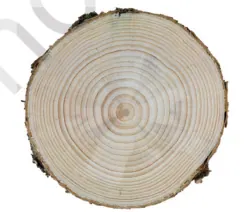
Chapter 'Tissues' (Class 9 Science, NCERT) introduces the concept of tissues as groups of similar cells performing a common function. It covers plant tissues (meristematic and permanent — simple and complex) and animal tissues (epithelial, connective, muscular, and nervous), emphasizing structure–function relationships and cell specialization. The chapter explains how tissues contribute to growth, support, transport, protection, movement and coordination, and it relates microscopic structure to macroscopic functions. Students learn to identify different tissues, describe their location and functions, draw labelled diagrams, and perform simple observational activities with prepared slides or temporary mounts. The chapter also builds foundational ideas for later study of organs and systems.
Learning Objectives
- Define tissue and list its major types in plants and animals.
- Describe meristematic tissue with reference to its structure, types and functions.
- Classify permanent tissues into simple and complex categories and state their key characteristics.
- Differentiate parenchyma, collenchyma and sclerenchyma in terms of structure, function and location.
- Identify the main elements of xylem and phloem and state the role of each in conduction.
- Draw and label neat diagrams of xylem and phloem showing their constituent cells.
- Explain the types of epithelial tissue and correlate their structure with functions and locations in animals.
- Compare connective, muscular and nervous tissues with examples and primary functions.
Topics in this chapter
18 topics · tap a topic title to jump straight to it.
Introduction to Tissues
What is a tissue? A tissue is a group of similar cells that perform a common function. Cells with similar structure and function group together to form tissues; tissues combine to form organs, organs form organ systems and together make an organism.
Why tissues are needed
Single cells can carry out life processes, but multicellular organisms need specialised regions to perform specific tasks efficiently. Grouping cells into tissues allows division of labour, increased efficiency, mechanical support and coordinated function.
Classification (broad)
- Plant tissues: Meristematic (actively dividing) and Permanent (differentiated). Permanent tissues are further classified into Simple (parenchyma, collenchyma, sclerenchyma) and Complex (xylem, phloem).
- Animal tissues: Epithelial, Connective, Muscular and Nervous tissues.
Plant tissues — key types & features
- Meristematic tissue: Small cells, thin walls, dense cytoplasm and prominent nucleus; found at root and shoot tips (apical meristem) and in cambium (lateral meristem). Function: growth by cell division.
- Parenchyma: Living thin-walled cells with intercellular spaces; functions: storage (e.g., potato tuber), photosynthesis (leaf mesophyll), and secretion.
- Collenchyma: Living cells with unevenly thickened primary walls; provide flexible support in regions of new growth (e.g., petiole).
- Sclerenchyma: Thick lignified secondary walls, dead at maturity; provide rigid support (fibres in jute, sclereids in pear grit).
- Xylem (complex): Conducts water and minerals; made of tracheids, vessel elements (dead), xylem parenchyma and fibres.
- Phloem (complex): Transports food (sugars); made of sieve tube elements, companion cells, phloem fibres and phloem parenchyma.
Animal tissues — key types & features
- Epithelial tissue: Tightly packed cells forming coverings and linings (e.g., skin epidermis, lining of gut). Types by shape: squamous, cuboidal, columnar; by arrangement: simple, stratified.
- Connective tissue: Cells scattered in extracellular matrix; includes blood (fluid matrix), bone, cartilage, adipose tissue and tendons. Functions: support, binding, transport, storage.
- Muscle tissue: Composed of elongated cells (fibres) capable of contraction. Types: skeletal (striated, voluntary), cardiac (striated, involuntary, branched), smooth (non-striated, involuntary).
- Nervous tissue: Made of neurons and neuroglia; neurons transmit electrical impulses for coordination and control (brain, spinal cord, nerves).
Key concepts to remember
- Specialisation: Cells become structurally and functionally suited for a task (differentiation).
- Structure–function relationship: Form of tissue relates to its function (e.g., thin squamous epithelium for diffusion; thick sclerenchyma for strength).
- Organisation: Cell → Tissue → Organ → Organ system → Organism.
Tip for study: Learn typical examples and locations (e.g., xylem in stem, muscle in heart, parenchyma in potato) and be able to sketch simple labelled diagrams of tissue types and a plant stem cross-section.
- Parenchyma in potato tuber stores starch (visible as white flesh).
- Collenchyma in the petiole of stems gives flexible support to leaves.
- Sclerenchyma fibres in jute and hemp provide strong fibres used in ropes.
- Xylem vessels in bamboo conduct water from roots to leaves.
- Phloem in sugarcane transports sugars produced in the leaves to storage organs.
- Epithelial tissue: skin epidermis protects the body from microbes and water loss.
- Surface area of a sphere: A = 4πr^2 (useful to compare area available for exchange)
- Volume of a sphere: V = (4/3)πr^3
- Surface-area-to-volume ratio (SA:V) for a sphere: SA/V = 3/r — explains why cells stay small and why tissues (many cells) are needed for efficient exchange
- Qualitative relation (diffusion): diffusion time ∝ distance^2 — indicates why thin layers or many small cells speed up transport
Plant Tissues — Overview
What is a tissue? A tissue is a group of similar cells that perform a common function. In plants, tissues are broadly classified into meristematic (actively dividing) and permanent (differentiated) tissues.
1. Meristematic tissues
Meristematic tissues consist of cells that are small, thin‑walled, with dense cytoplasm and prominent nuclei. These cells divide actively by mitosis and give rise to other tissues. Types of meristems:
- Apical meristem—found at root and shoot tips; responsible for primary (length) growth.
- Intercalary meristem—found at internodes or leaf bases (common in grasses); helps rapid elongation.
- Lateral meristem—includes vascular cambium and cork cambium; responsible for secondary (thickness) growth.
2. Permanent (simple and complex) tissues
Permanent tissues arise from meristematic cells and have lost the ability to divide (or divide rarely). They are of two broad kinds:
Simple permanent tissues (one cell type):
- Parenchyma—most common; thin primary walls, large intercellular spaces, functions: storage (e.g., potato tuber), photosynthesis (chlorenchyma in leaves), and healing/wound repair.
- Collenchyma—elongated cells with unevenly thickened primary walls (pectin/cellulose); provides flexible support (e.g., strands under leaf veins, petioles).
- Sclerenchyma—thick lignified secondary walls, dead at maturity, provides rigid support. Two forms: fibers (e.g., jute, hemp) and sclereids (stone cells in pear).
Complex permanent tissues (more than one cell type working together):
- Xylem—conducts water and dissolved minerals upward; consists of tracheids, vessels, xylem parenchyma and xylem fibers. Vessels and tracheids are dead and lignified. Example: wood is largely secondary xylem.
- Phloem—transports organic nutrients (mainly sugars) both up and down; consists of sieve tube elements, companion cells, phloem parenchyma and phloem fibers. Sieve tube elements are living but lack nucleus.
Other important tissues
Epidermis (outer protective layer) with cuticle, stomata (guard cells) for gas exchange, and trichomes (hairs). In older stems/roots, epidermis is replaced by periderm (cork) from cork cambium.
Key functional points:
- Meristems generate new cells; apical meristems increase length, lateral meristems increase girth.
- Simple tissues mainly provide support, storage and photosynthesis; complex tissues perform long‑distance transport.
- Structural differences (cell wall thickness, presence/absence of protoplast, lignification) determine mechanical and transport roles.
Summary (one‑line): Plant tissues are organised into meristematic tissues that make new cells and permanent tissues (simple and complex) that perform support, storage, protection and transport.
- Apical meristem at the tip of a germinating radish seed—causes root/shoot elongation.
- Intercalary meristem in grass stems—allows regrowth after grazing or mowing.
- Vascular cambium in woody stems—produces secondary xylem (wood) and secondary phloem (bark).
- Parenchyma in potato tuber—stores starch.
- Chlorenchyma (specialised parenchyma) in leaf mesophyll—site of photosynthesis.
- Collenchyma in celery stalks—gives flexible support; visible as stringy fibers when you chew celery.
- Photosynthesis: 6 CO2 + 6 H2O → C6H12O6 + 6 O2
- Respiration (general): C6H12O6 + 6 O2 → 6 CO2 + 6 H2O + energy (ATP)
- Approximate transpiration relationship: E ≈ g_s × VPD (transpiration rate E proportional to stomatal conductance g_s and vapour pressure deficit VPD) — useful qualitatively.
- Surface area to volume ratio (affects diffusion and exchange): SA/V = (surface area) / (volume). For a cube of side a: SA = 6a^2, V = a^3, so SA/V = 6/a.
Meristematic Tissue
What is Meristematic Tissue?
Meristematic tissue consists of regions of actively dividing, undifferentiated plant cells responsible for growth. Meristem cells are small, living, thin-walled, densely cytoplasmic and have a large nucleus; they lack large vacuoles and intercellular spaces.
Key characteristics
- Cells are small, isodiametric and compactly packed.
- Thin primary cell walls (no secondary thickening).
- High nucleus-to-cytoplasm ratio and dense cytoplasm.
- Frequent mitotic activity (able to divide continuously).
- Lack of large vacuoles and intercellular spaces.
- Cells are undifferentiated and can give rise to different permanent tissues.
Types (by position)
- Apical meristem: at shoot and root tips; causes primary (length) growth.
- Intercalary meristem: at bases of leaves or internodes (common in grasses); allows rapid regrowth after grazing.
- Lateral meristem: (vascular cambium and cork cambium) along length of stem/roots; responsible for secondary (girth) growth.
Types (by origin and function)
- Initials/primary meristems — give rise to primary tissues (protoderm, procambium, ground meristem).
- Secondary meristems — e.g., cambium, produce secondary xylem and phloem.
How meristem cells divide and contribute to growth
Meristem cells divide by mitosis. Division planes are important: anticlinal divisions increase surface area, while periclinal divisions increase thickness (add layers). Daughter cells either remain meristematic (to maintain the meristem) or differentiate into permanent tissues (xylem, phloem, epidermis, ground tissue).
Functions
- Produce new cells for growth in length (apical) and girth (lateral).
- Allow repair and regeneration (wound healing, formation of adventitious roots).
- Provide cells for formation of all plant tissues during development.
Practical importance
Meristems are used in horticulture and biotechnology: cuttings and grafting depend on meristematic activity; micropropagation uses meristem tissue to produce disease-free clones.
- Root apical meristem at the tip of a root (onion root tip used in labs to observe mitosis).
- Shoot apical meristem at the tip of a stem (responsible for upward growth and formation of new leaves).
- Intercalary meristem in grasses and bamboo — enables regrowth after grazing or cutting.
- Vascular cambium (a lateral meristem) in trees — produces secondary xylem (wood) and secondary phloem, increasing stem girth.
- Use in plant propagation: cuttings form adventitious roots due to meristematic activity; tissue culture uses meristematic cells to clone plants.
- Mitotic index (%) = (Number of dividing cells / Total number of observed cells) × 100
- Exponential cell-division (idealized): N = N0 × 2^n, where N0 = initial cells, n = number of divisions
- If td is the doubling time and t the time elapsed: n = t / td so N = N0 × 2^(t/td)
- Relative growth rate (common in plant growth studies): RGR = (ln W2 − ln W1) / (t2 − t1), where W1 and W2 are plant masses at times t1 and t2
Permanent Tissues (Plants)
Definition: Permanent tissues are groups of cells, formed from meristematic tissue, that have become specialized in structure and function and usually lost the power to divide. They form the bulk of plant body and perform functions such as support, storage, conduction and protection.
Classification
Permanent tissues are broadly classified into simple and complex tissues.
1. Simple Permanent Tissues
- Parenchyma
- Structure: Living, isodiametric (often spherical/oval) cells with thin primary cell walls and large central vacuole; intercellular spaces common.
- Functions: Storage (starch, oils, water), photosynthesis (chlorenchyma), secretion, wound repair (can dedifferentiate).
- Location: Pith, cortex, mesophyll of leaves, fruit pulp (e.g., potato tuber).
- Collenchyma
- Structure: Living cells with unevenly thickened primary walls (cellulose and pectin), usually elongated; little or no intercellular space.
- Functions: Flexible support to growing organs (stems, petioles).
- Location & example: Under the epidermis in stems (e.g., the crunchy strands in celery stalk).
- Sclerenchyma
- Structure: Cells with thick, lignified secondary walls; most are dead at maturity. Two types: fibers (elongated) and sclereids (varied shapes).
- Functions: Rigid support and protection.
- Location & example: Seed coats, nutshells, stone cells in pear; fibers in jute and flax.
2. Complex Permanent Tissues
Complex tissues are made of more than one cell type and perform conduction.
- Xylem
- Components: Tracheids, vessels (in angiosperms), xylem parenchyma and xylem fibers.
- Structure: Many elements (vessels/tracheids) are dead and lignified at maturity forming a continuous pipeline.
- Function: Transport of water and dissolved minerals from root to shoot; mechanical support.
- Location & example: Wood (primary part of stem); vessels visible in a cross-section of stem.
- Phloem
- Components: Sieve tube elements, companion cells, phloem parenchyma and phloem fibers.
- Structure: Sieve tubes are living but lack nucleus; companion cells manage metabolic functions of sieve tubes.
- Function: Translocation of organic solutes (mainly sugars) from source (leaves) to sinks (roots, fruits, growing parts).
- Location & example: Found towards the outer part of vascular bundles; bark contains phloem.
Other Permanent Tissues
- Epidermis: Single outermost layer of cells protecting plant body; has stomata, trichomes, cuticle.
- Secretory tissues: Glands, nectaries, resin canals, laticifers that secrete substances.
Key Features & Comparisons
- Origin: Derived from meristematic tissues (apical, lateral, intercalary).
- Cell division: Mostly lost, but some parenchyma can divide (wound repair).
- Living vs Dead: Parenchyma and collenchyma are generally living; sclerenchyma and many xylem elements are dead at maturity.
- Role in plant: Mechanical support (collenchyma, sclerenchyma, xylem), storage (parenchyma), conduction (xylem, phloem).
Practical/Functional Notes
- Celery sticks are good teaching material to observe collenchyma (crunchy strands) and vascular bundles.
- Potato tuber shows parenchyma full of starch grains—use iodine to test starch in class demonstrations.
- Jute, flax and hemp fibers are sclerenchymatous fibers used commercially for ropes and textiles.
Summary: Permanent tissues provide structure, protection, storage and transport in plants. Simple tissues are made of similar cell types (parenchyma, collenchyma, sclerenchyma); complex tissues (xylem, phloem) combine specialized cells to conduct water/minerals and food respectively.
- Parenchyma: Potato tuber storage cells (rich in starch); mesophyll cells in leaves (chlorenchyma) for photosynthesis.
- Collenchyma: The crisp strands at the edge of a celery stalk provide flexible support to the petiole.
- Sclerenchyma: Hard nutshells, stone cells in pear fruit (gritty texture), and fibers in jute/flax used for ropes and textiles.
- Xylem: Wood in trees (xylem vessels/tracheids) conducts water and gives mechanical strength.
- Phloem: Inner bark (phloem) transports sugars from leaves to roots and fruits.
- Simple experimental transpiration rate (class practical): R = ΔW / Δt (where ΔW = loss of mass by transpiration, Δt = time interval).
- Fick’s law (conceptual for diffusion of gases within tissues): J = -D·(dC/dx) (flux J proportional to concentration gradient dC/dx).
- Flow in a tube (qualitative relevance to xylem vessels) — Hagen–Poiseuille relation: Q ∝ r^4 (flow rate Q is very sensitive to vessel radius r).
- Mass flow concept for phloem (pressure-flow idea, conceptual form): Flow ∝ ΔP / Resistance (transport driven by pressure differences between source and sink).
Parenchyma
Definition: Parenchyma is a simple, living fundamental tissue in plants composed of thin-walled, isodiametric (or slightly elongated) cells with a large central vacuole. It performs a variety of metabolic, storage and mechanical functions.
Structure & features:
- Cells are generally spherical, cuboidal or polyhedral with thin primary cell walls made of cellulose.
- Cells remain living at maturity and often contain chloroplasts (in leaves) or storage products (starch, oil).
- Large central vacuole and prominent intercellular spaces in many kinds (important for gas exchange).
- Cells can divide and differentiate — involved in regeneration and wound healing (callus formation).
Types (by function/structure):
- Chlorenchyma: Parenchyma with abundant chloroplasts; performs photosynthesis (e.g., palisade and spongy parenchyma in leaves).
- Storage parenchyma: Stores food and water (e.g., potato tuber, fleshy fruit pulp, pith of stems).
- Aerenchyma: Has large intercellular air spaces for buoyancy and internal gas exchange in aquatic plants (e.g., water hyacinth, water lily).
- Sclerenchymatous parenchyma (rare): May become lignified or have thicker walls in some regions, but typical parenchyma remains thin-walled.
Functions:
- Photosynthesis (chlorenchyma).
- Storage of starch, oils, proteins, and water (storage parenchyma in tubers, fruits, seeds).
- Aeration and buoyancy (aerenchyma in hydrophytes).
- Secretion and transport of metabolites.
- Mechanical support when turgid — parenchyma provides hydrostatic (turgor) support.
- Wound healing and regeneration (cells can divide to form new tissues).
Where found: Cortex and pith of stems and roots, mesophyll of leaves (palisade & spongy parenchyma), fleshy parts of fruits and tubers, lining of vascular bundles.
How to observe in class (simple experiment): Cut a thin slice of potato tuber, add a drop of iodine. Starch stored in parenchyma cells stains blue-black, showing storage function.
Comparison (brief): Unlike collenchyma (thick, uneven walls, living; mechanical support) and sclerenchyma (dead at maturity with thick lignified walls), parenchyma cells have thin walls and are metabolically active.
Key idea for students: Parenchyma is the most versatile, living tissue — it does photosynthesis, stores food/water, helps gas exchange and heals wounds. Its thin walls and large vacuoles enable these roles.
- Pith cells of stems — general storage and packing
- Cortex of potato tuber — starch-storing parenchyma (stained blue-black with iodine)
- Palisade parenchyma in leaf mesophyll — main site of photosynthesis
- Spongy parenchyma in leaf mesophyll — gas exchange and photosynthesis
- Aerenchyma in water hyacinth or water lily — large air spaces for buoyancy
- Flesh of apple or tomato — parenchyma storing water and sugars
- Surface area of a spherical cell: SA = 4πr^2 (useful to estimate exchange surface of roughly spherical parenchyma cells)
- Volume of a spherical cell: V = (4/3)πr^3
- Surface-area-to-volume ratio: SA/V = 3/r (shows that smaller cells have higher relative surface area for exchange)
- Water potential (relevant to turgor in parenchyma): Ψ = Ψs + Ψp (Ψ = total water potential, Ψs = solute potential, Ψp = pressure/turgor potential)
- Fick's law (diffusion, relevant to gas exchange in parenchyma): Rate ∝ D·A·(ΔC/Δx) (D = diffusion coefficient, A = area, ΔC/Δx = concentration gradient/distance)
Collenchyma
Definition: Collenchyma is a simple permanent tissue made of living, elongated cells with unevenly thickened primary cell walls. It provides flexible mechanical support to growing parts of the plant.
Structure and features:
- Cells are living, usually elongated (columnar), with a thin layer of cytoplasm and a prominent vacuole.
- Cell walls are unevenly thickened by cellulose and pectic substances (especially at the corners), but are not lignified.
- Intercellular spaces are small or absent; cells often polygonal in cross-section.
- Types based on wall thickening: angular collenchyma (thickening at corners), lamellar or tangential collenchyma (thickening on tangential walls), lacunar collenchyma (around air spaces), and annular (ring-like, less common).
Location: Typically found just beneath the epidermis in stems, petioles and midribs of leaves of dicotyledonous young plants and regions of active growth.
Functions: Provides mechanical support while allowing growth and flexibility (e.g., bending without breaking). It helps young stems and petioles withstand wind and mechanical strain; because cells are living, they can stretch and accommodate growth.
Comparison with other tissues: Unlike parenchyma (thin and uniform walls, mainly for storage/photosynthesis) and sclerenchyma (thick, lignified walls, dead at maturity and very rigid), collenchyma is an intermediate tissue—strong but flexible, and living at maturity.
Microscopy/lab note: In transverse section of a young dicot stem, collenchyma appears as a continuous or broken ring of thicker-walled cells beneath the epidermis; in longitudinal section the cells appear elongated.
- Celery stalk strings (visible fibrous strands are bundles of collenchyma)
- Petiole of Hibiscus (collenchyma beneath the epidermis giving support)
- Young stems of bougainvillea and sunflower (under epidermis)
- Edges of leaves and midrib in many dicot plants
- Stress = Force / Area (useful to understand mechanical support capabilities)
- Strain = ΔL / L₀ (relative deformation under load; relates to flexibility)
- Hooke's law: Stress = E × Strain (E = Young's modulus; collenchyma has lower E than sclerenchyma, so it is more elastic)
- Thin-walled cylinder (hoop stress) approximation: σ_hoop = P × r / t (shows how internal pressure/turgor P, radius r and wall thickness t influence stress on cell walls)
Sclerenchyma
Sclerenchyma — definition
Sclerenchyma is a mechanical plant tissue made up of cells that have very thick, lignified secondary cell walls and are usually dead at maturity. Its main function is to provide rigidity, mechanical strength and protection to different parts of the plant.
Key characteristics
- Cells have very thick secondary walls impregnated with lignin (making them hard and rigid).
- Cells are typically dead at maturity (they lack protoplasm).
- Lumen (central cavity) is small or almost absent.
- Walls often show pits to allow limited exchange.
- Provides support and protection, especially where growth has stopped.
Types of sclerenchyma
- Fibres — long, slender, tapering cells grouped in bundles. Common in stems, petioles and vascular bundles. Examples: jute, hemp, flax; used as fiber crops.
- Sclereids (stone cells) — variable in shape (isodiametric, branched, elongated or stellate). Provide hardness and gritty texture. Found in seed coats, nutshells, pear flesh (gritty), and quince.
Location
Found in stem cortex, around vascular bundles, in leaf veins, seed coats, nutshells and fruit flesh (as stone cells). In many seeds and nuts (e.g., coconut, walnut) sclerenchyma forms very hard protective layers.
Origin and development
Sclerenchyma cells usually differentiate from meristematic cells or from parenchyma and collenchyma cells when the tissue stops elongating. They deposit a thick secondary wall (cellulose, hemicellulose and lignin) and then lose their protoplasm.
Functions
- Mechanical support and strength to stems, leaves and other organs.
- Protection of seeds and fruits (hard seed coats and shells).
- Form strong fibres used by humans (rope, sacks, textiles — jute, hemp, flax).
Comparison with other simple tissues (short)
- Parenchyma: living cells, thin primary walls, storage and photosynthesis — flexible, not rigid.
- Collenchyma: living cells, unevenly thickened non-lignified walls, provide flexible support in growing regions.
- Sclerenchyma: dead at maturity, thick lignified walls, rigid support in mature parts.
Microscopic appearance
Under a microscope sclerenchyma fibers appear as long narrow cells with very thick walls and small central lumin; sclereids appear as small, thick-walled, often irregularly-shaped cells.
Practical importance
- Industrial: fibres (jute, hemp, flax) are important for ropes, sacks, paper and coarse textiles.
- Agricultural/botanical: hardness of seeds and fruits affects dispersal and protection; stone cells affect fruit texture (e.g., pears).
Exam tips
To identify sclerenchyma in diagrams or slides: look for very thick walls, small lumen, absence of protoplasm, and location near vascular bundles or seed coats. Distinguish from collenchyma by the uniform, lignified thickening (collenchyma has uneven, non-lignified thickening).
- Fibres: Jute (Corchorus), Hemp (Cannabis sativa), Flax (Linum usitatissimum) — used for rope, sacks and textiles
- Sclereids (stone cells): Gritty texture in pear and guava flesh; hard shells of nuts like walnut and coconut; seed coats
- Stony endocarp: Peach/olive stones are formed of sclerenchyma providing protection to the seed
- Sclerenchyma cell ≈ thick lignified secondary wall + narrow lumen + dead at maturity
- Relative mechanical strength ∝ Lignin content × Cell wall thickness
- Approximate cell wall thickness ≈ (Total cell diameter − Lumen diameter) / 2
- Support capacity ∝ Number_of_fibres × Average_fibre_strength (useful for comparative estimations)
Epidermal Tissue (Details)
Definition
The epidermal tissue is the outermost single layer of tightly packed cells that covers leaves, young stems and roots of plants. It forms a protective boundary between the plant and its environment.
Structure
- Usually one cell layer thick; cells are compactly arranged without intercellular spaces.
- Cells have a primary cell wall and are often covered on aerial parts by a waxy cuticle that reduces water loss.
- Epidermal cells are generally transparent to allow light to reach mesophyll cells; most lack chloroplasts except guard cells.
- Specialised epidermal cells include guard cells (form stomata), trichomes (hairlike outgrowths), and root hair cells (elongated epidermal cells).
Types and Modifications
- Stomata and guard cells: pores in the epidermis (mainly of leaves) for gas exchange and transpiration. Each stoma is flanked by a pair of guard cells containing chloroplasts.
- Cuticle: a waxy, water-repellent layer on aerial epidermis that reduces water loss and protects against pathogens.
- Root hairs: tubular extensions of root epidermal cells that increase absorptive surface area for water and mineral uptake.
- Trichomes (hair): may reduce water loss, reflect excess light, trap moisture, secrete substances or deter herbivores.
- Bulliform cells: large epidermal cells in some grasses that help in leaf folding/unfolding to reduce transpiration.
Functions
- Protection: mechanical barrier against injury, pathogens and water loss.
- Gas exchange: stomata regulate intake of CO2 and release of O2 and water vapour.
- Absorption: root hairs increase water and mineral uptake from soil.
- Secretion and sensation: trichomes and epidermal glands may secrete resins, oils or salts; some sense environmental stimuli.
Differences in organs
Leaf epidermis: has cuticle and numerous stomata (often more on the lower surface). Root epidermis: lacks cuticle and stomata, bears root hairs for absorption.
Microscopy and practical note
Onion epidermis is commonly used to observe epidermal cells and nucleus under light microscope. Leaf peels show stomata distribution; cleared and stained mounts help identify guard cells and epidermal patterns.
Relation to animal epidermis (brief)
In animals, the epidermis is the outer cell layer of skin (multi-layered, keratinized in terrestrial vertebrates) serving protection and sensory roles. This differs structurally from the single-layer plant epidermis but shares the protective function.
- Waxy cuticle on mango and other leaves prevents excess water loss in dry conditions
- Stomata on the lower surface of a sunflower leaf open during the day for photosynthesis and close at night
- Root hairs of wheat and grass increase surface area for water uptake from soil
- Trichomes on a cucumber or nettle leaf deter herbivores; hairs on cactus reduce sun exposure
- Onion epidermis viewed under a microscope in school laboratories to observe cell shape and nucleus
- Stomatal density = Number of stomata counted / Area observed (e.g., stomata per mm2)
- Stomatal index (%) = [Number of stomata / (Number of epidermal cells + Number of stomata)] × 100
- Transpiration rate (simple experimental) = Mass of water lost / (Leaf area × Time) (units: g cm^-2 hr^-1 or mg cm^-2 hr^-1)
Xylem (Complex Tissue)
Definition: Xylem is a complex vascular tissue in plants that conducts water and dissolved minerals from roots to aerial parts and provides mechanical support. It is called a complex tissue because it consists of more than one type of cell.
Components:
- Tracheids – long, narrow, tapering cells with lignified walls and pits; dead at maturity; found in all vascular plants (main conductive cells in gymnosperms).
- Vessel elements (vessels) – shorter, wider cells arranged end to end to form continuous tubes (vessels); have perforation plates; common in angiosperms; dead at maturity.
- Xylem parenchyma – living cells that store food, help lateral transport and repair.
- Xylem fibres (sclerenchyma) – long, thick-walled cells that give mechanical strength.
Types of xylem:
- Primary xylem – formed from procambium during primary growth (protoxylem and metaxylem).
- Secondary xylem – formed by vascular cambium during secondary growth; accumulates as wood in woody plants.
Functions:
- Conduction of water and dissolved mineral salts from root to stem and leaves.
- Mechanical support (especially secondary xylem/wood).
- Storage of food and water (xylem parenchyma).
Mechanism of water transport:
- Water uptake by roots (root hairs) → movement through xylem vessels/tracheids to leaves.
- Main driving forces: transpiration pull (cohesion–tension theory), root pressure (minor), and capillarity in small conduits.
- Transpiration from leaves creates negative pressure (tension) which pulls a continuous column of water upward owing to cohesion between water molecules and adhesion to xylem walls.
Key features to remember:
- Xylem conduction is generally unidirectional (root to shoot).
- Tracheids and vessels are dead at maturity and have lignified walls—this prevents collapse and provides strength.
- Wood is largely secondary xylem; its rings reflect yearly secondary xylem production in temperate trees.
Practical/biological importance: Wood and timber (secondary xylem) are economically important for construction, furniture and paper; xylem function determines plant water relations, drought resistance and wilting.
- Dye uptake in a celery stalk or cut flower: colored dye moves upward through xylem vessels, visible in cross sections — demonstrates xylem conduction.
- Tall trees (e.g., eucalyptus) transport water from roots to crowns through long vessels and tracheids; secondary xylem forms the wood trunk.
- Gymnosperms (pine) mainly use tracheids for conduction, whereas angiosperms (sunflower, mango) have vessels and tracheids.
- Girdling experiment: removing bark (phloem) does not immediately stop xylem transport; wood (xylem) still conducts water.
- Wilting of plants on hot, dry days shows the importance of xylem-mediated water supply to leaves.
- Transpiration rate (simple experimental) = (Mass of water lost) / (Time). Example units: g·hr⁻¹ or mL·hr⁻¹.
- Percentage water loss = [(Initial mass − Final mass) / Initial mass] × 100%
- Capillary rise (height h) in a tube: h = (2 γ cosθ) / (ρ g r), where γ = surface tension, θ = contact angle, ρ = liquid density, g = gravity, r = radius of tube. (Explains capillarity contribution.)
- Poiseuille's law for laminar flow in a cylindrical tube: Q = (π r^4 ΔP) / (8 η l), where Q = volumetric flow rate, r = radius, ΔP = pressure difference, η = viscosity, l = length. (Shows flow ∝ r^4 — explains why wider vessels conduct much more water.)
Phloem (Complex Tissue)
Definition: Phloem is a complex living vascular tissue in plants that conducts organic food (mainly sugars like sucrose) from leaves (source) to other parts of the plant (sinks) such as roots, growing buds, fruits and seeds.
Components of Phloem and their roles:
- Sieve tube elements: Long, tubular, living cells joined end-to-end. End walls have sieve plates with pores to allow flow of sap. Sieve elements have reduced cytoplasm and lack a nucleus at maturity.
- Companion cells: Closely associated with sieve tubes; have dense cytoplasm and nucleus. They maintain and load/unload materials into sieve tubes via plasmodesmata.
- Phloem parenchyma: Living parenchyma cells used for storage and lateral transport within the phloem.
- Phloem fibers (sclerenchyma): Provide mechanical support; often lignified and tough.
Functions:
- Translocation of organic solutes (mainly sucrose) from sources (photosynthesizing leaves) to sinks (roots, developing fruits, seeds, storage organs).
- Distribution of amino acids, hormones and other organic molecules.
- Support and storage (via fibers and parenchyma).
Mechanism of Transport — Pressure Flow (Mass Flow) Hypothesis (simple outline):
- At the source (leaf), sugars produced by photosynthesis are actively loaded into sieve tubes, increasing solute concentration there.
- Water enters the sieve tubes from adjacent xylem by osmosis, raising turgor (hydrostatic) pressure at the source end.
- At the sink (e.g., root, fruit), sugars are actively or passively removed from sieve tubes, lowering solute concentration; water leaves, lowering pressure.
- A pressure gradient (high at source, low at sink) drives bulk flow of phloem sap through sieve tubes from source to sink.
Key adaptations: Sieve plates for unobstructed flow, companion cells for metabolic support and active loading/unloading, plasmodesmatal connections for transfer of materials, living nature of sieve elements for controlled transport.
Comparison with xylem (brief): Xylem mainly transports water and minerals upwards (dead cells, unidirectional), while phloem transports organic solutes (living cells, often bidirectional but generally source-to-sink).
Class 9 level note: Emphasize structure (sieve tubes, companion cells, parenchyma, fibers) and the pressure-flow idea: loading at source → water influx → pressure-driven flow → unloading at sink.
- Storage organs: Sucrose produced in leaves is translocated to potato tubers where it is converted into starch for storage.
- Sugarcane: Sucrose synthesized in leaves is transported and accumulates in the stem (harvestable product).
- Fruit ripening: Sugars are transported to developing fruits (e.g., grapes, apples) making them sweet.
- Aphid feeding: Aphids tap into phloem sieve tubes to feed on sugar-rich sap — used in studies of phloem composition and flow.
- Qualitative relation used in pressure-flow idea: Flow ∝ ΔP (flow is driven by pressure difference between source and sink).
- Simple mass-flux relation: J = C × v (J = solute flux, C = solute concentration, v = bulk flow velocity).
- Osmotic (van 't Hoff) relation for dilute solutions (conceptual, advanced): π = C·R·T (π = osmotic pressure, C = molar concentration, R = gas constant, T = absolute temperature) — explains water movement into sugar-loaded sieve tubes.
Comparison: Xylem vs Phloem
Overview: Xylem and phloem are the two main types of vascular tissues in plants. Xylem transports water and mineral salts from roots to aerial parts; phloem transports organic substances (mainly sugars) from sources (leaves) to sinks (roots, fruits, growing parts).
Xylem (summary): Composed of tracheids, vessels, xylem parenchyma and fibres. Mostly dead cells with thick lignified walls. Transport is generally unidirectional (root → shoot → leaves) and occurs by cohesion-tension (transpiration pull) and capillarity.
Phloem (summary): Composed of sieve tube elements, companion cells, phloem parenchyma and fibres. Sieve tube elements are living but lack a nucleus; companion cells support their function. Transport is bidirectional (source ↔ sink) and occurs by the pressure-flow (mass-flow) mechanism driven by osmotic pressure differences between source and sink.
Key differences (quick table):
| Feature | Xylem | Phloem |
|---|---|---|
| Function | Conduction of water and mineral salts | Conduction of organic solutes (sugars), amino acids, hormones |
| Direction of flow | Usually unidirectional (root → shoot) | Multidirectional (from source to sink) |
| Cell types | Vessels, tracheids, xylem parenchyma, fibres | Sieve tube elements, companion cells, phloem parenchyma, phloem fibres |
| Living or dead | Mostly dead at maturity | Living (sieve tubes alive but enucleate; companion cells alive) |
| Cell wall | Thick, lignified | Thin, non-lignified |
| Transport mechanism | Cohesion-tension, transpiration pull | Pressure-flow (mass flow) caused by osmotic gradients |
| Driving force | Negative pressure (tension) in xylem sap | Positive hydrostatic pressure in sieve tubes |
| Examples of experiments | Transpiration-rate measurements, ascent of sap in cut stem | Girdling shows accumulation of sugars above ring; aphid stylet experiments measure phloem sap |
How transport is achieved: Xylem: Water evaporating from leaf surfaces (transpiration) creates tension; cohesion among water molecules and adhesion to cell walls pulls a continuous water column upward. Phloem: Sugars loaded into sieve tubes at source lower water potential; water enters by osmosis raising turgor pressure; at sink sugars are removed, water leaves, creating pressure gradients that drive bulk flow from source to sink.
Important notes for Class 9: Xylem vessels are more prominent in angiosperms (vessels + tracheids), while gymnosperms largely depend on tracheids. Phloem sieve plates allow flow between sieve elements; companion cells manage loading/unloading of solutes.
- Tall trees: water pulled from roots to the top leaves via xylem using transpiration pull (cohesion-tension).
- Girdling experiment: removing a ring of bark (phloem) causes sugars to accumulate above the cut and starvation of tissues below—demonstrates phloem transport.
- Aphid feeding: aphids tap sieve tubes and excrete phloem sap—used experimentally to collect phloem sap and measure flow.
- Wilting on hot dry days: high transpiration (xylem-driven) can exceed water uptake causing loss of turgor.
- Ripening fruit: sugars produced in leaves are transported by phloem to fruits, making them sweet and energetic.
- Cut twig placed in colored water: xylem vessels show upward movement of dye, demonstrating xylem conduction.
- Transpiration rate = (Volume or mass of water transpired) / (Time). Example units: mL h⁻¹ or g h⁻¹.
- Water potential: Ψ = Ψs + Ψp (where Ψs = solute/osmotic potential, Ψp = pressure potential). (Used to explain movement of water into/out of xylem or phloem.)
- Osmotic (van't Hoff) equation: π = iCRT (π = osmotic pressure, i = van't Hoff factor, C = molar concentration, R = gas constant, T = absolute temperature). (Useful to compare osmotic effects at source and sink.)
- Pressure-flow (qualitative) relation: flow rate ∝ ΔP / resistance. In phloem, ΔP (source pressure − sink pressure) drives bulk flow of sap.
Animal Tissues — Overview
What are animal tissues? A tissue is a group of similar cells that perform a common function. In animals, tissues are organized levels of structure between cells and organs and allow division of labour so organisms work efficiently.
Main categories of animal tissues
- Epithelial tissue: Sheets of closely packed cells that cover body surfaces, line cavities and form glands. Key functions: protection, absorption, secretion and sensation. Types include simple (one layer) and stratified (many layers) epithelia; shapes include squamous (flat), cuboidal and columnar; special types include ciliated epithelium and glandular epithelium.
- Connective tissue: Cells scattered in a matrix (fibres + ground substance). Functions: support, binding, storage, transport and protection. Major types: areolar (loose), adipose (fat storage), cartilage (hyaline, elastic, fibrocartilage), bone (compact, spongy) and blood (fluid connective tissue).
- Muscular tissue: Excitable cells that contract to produce movement. Three kinds: skeletal (striated, voluntary, attached to bones), smooth (non-striated, involuntary, in gut and blood vessels) and cardiac (striated, involuntary, in heart). Muscle cells contain contractile proteins (actin, myosin).
- Nervous tissue: Composed of neurons and supporting neuroglia. Neurons transmit electrical signals; basic parts are cell body (soma), dendrites (input) and axon (output). Functions: sensing, processing and transmitting information; controls body responses.
Key functional features and why tissues are organized
- Division of labour: Different tissues specialise for different roles (e.g., epithelium protects, muscle moves, nerves control, connective supports).
- Structure–function correlation: Thin, flat cells (simple squamous) permit diffusion (e.g., alveoli); layered epithelium protects (skin).
- Surface area to volume (SA:V) considerations: Smaller units or folded surfaces increase exchange (lungs, intestines).
How this appears in the body: Tissues combine to form organs (e.g., stomach has epithelial lining, smooth muscle layer, connective tissue framework and nervous tissue for control). Healthy functioning of organs depends on correct tissue structure and interaction.
- Epithelial: Simple squamous epithelium in lung alveoli—allows fast gas exchange.
- Epithelial: Stratified squamous epithelium in skin and mouth—protects against abrasion.
- Connective: Adipose tissue under skin and around organs—energy storage and insulation.
- Connective: Cartilage in the ear and nose—flexible support; hyaline cartilage at the ends of long bones.
- Connective: Bone (femur) — rigid support and mineral storage.
- Connective: Blood—transports gases, nutrients and immune cells throughout the body.
- Surface area of a sphere: SA = 4πr^2
- Volume of a sphere: V = (4/3)πr^3
- Surface area to volume ratio (for a sphere): SA:V = 4πr^2 : (4/3)πr^3 = 3/r (shows SA:V decreases as size (r) increases)
- Simplified diffusion relation (Fick's idea): Rate of diffusion ∝ (Surface area × Concentration difference) / Distance (thickness).
Epithelial Tissue
What is Epithelial Tissue?
Epithelial tissue (epithelium) is a group of closely packed cells that form continuous sheets covering body surfaces, lining cavities and tubes, and forming glands. Epithelium performs protection, absorption, secretion, filtration and sensory reception.
General features
- Cells are tightly packed with very little intercellular matrix.
- Has a free (apical) surface and a basal surface attached to a thin basement membrane.
- Avascular — it receives nutrients by diffusion from underlying connective tissue.
- High regenerative capacity (frequent mitosis).
- Special cell junctions: tight junctions, desmosomes and gap junctions.
Classification (by layers)
- Simple epithelium: single cell layer — good for absorption, filtration and secretion.
- Stratified epithelium: two or more layers — provides protection against mechanical stress.
- Pseudostratified epithelium: appears layered because nuclei are at different levels but all cells touch basement membrane (often ciliated).
Classification (by cell shape)
- Squamous — flat, thin cells (e.g., alveoli, blood vessel lining) — allow rapid diffusion.
- Cuboidal — cube-shaped (e.g., kidney tubules, ducts of glands) — secretion and absorption.
- Columnar — tall, rectangular cells (e.g., small intestine lining) — absorption and secretion; often have microvilli or cilia.
Special types / adaptations
- Ciliated epithelium — has cilia to move particles or fluid (e.g., trachea, oviduct).
- Keratinized stratified squamous — surface cells full of keratin for extra protection and water resistance (e.g., skin epidermis).
- Glandular epithelium — cells specialised to form glands that secrete substances; glands are exocrine (with ducts) or endocrine (ductless).
- Goblet cells — unicellular mucus-secreting cells found in respiratory and intestinal epithelium.
Functions — quick summary
- Protection: stratified epithelium prevents mechanical and chemical injury.
- Absorption: intestinal epithelium with microvilli increases surface area.
- Secretion: glandular epithelium produces hormones, enzymes, mucus.
- Filtration and diffusion: thin squamous cells (e.g., alveoli, glomeruli).
- Sensory reception: specialised epithelial cells in taste buds, nose and ears.
Importance in the body
Epithelial tissues form protective barriers, control exchange of materials, and create selective interfaces between internal and external environments — key to homeostasis.
- Simple squamous epithelium: lining of alveoli in lungs — allows gas exchange.
- Simple cuboidal epithelium: lining of kidney tubules — absorption and secretion during urine formation.
- Simple columnar epithelium: lining of small intestine with microvilli — nutrient absorption.
- Pseudostratified ciliated columnar epithelium: lining of trachea — cilia sweep mucus and trapped particles.
- Stratified squamous keratinized: epidermis of skin — protection and water resistance.
- Glandular epithelium: cells of salivary glands and endocrine glands (e.g., thyroid).
- Surface area of a cube cell: SA = 6a^2 and volume: V = a^3 → Surface area to volume ratio (SA:V) = 6/a. (Shows why small cells or folded/brush-border epithelium increase exchange efficiency.)
- Fick’s law of diffusion (qualitative form relevant to epithelium): Rate of diffusion ∝ (Area × Concentration difference) / Thickness. (Explains why thin simple squamous epithelium allows faster diffusion.)
Connective Tissue
What is Connective Tissue?
Connective tissue is one of the four basic tissue types (along with epithelial, muscular and nervous tissues). It originates from mesenchyme (mesoderm) and its main role is to support, connect or separate different types of tissues and organs in the body.
Basic components
- Cells — varied types (fibroblasts, adipocytes, chondrocytes, osteocytes, blood cells, macrophages, mast cells).
- Extracellular matrix (ECM) — consists of ground substance (proteoglycans, glycosaminoglycans, glycoproteins) and fibers:
- Collagen fibres — strong, provide tensile strength.
- Elastic fibres — stretchable, provide elasticity.
- Reticular fibres — fine branching fibres forming supportive networks.
Functions
- Mechanical support and structural framework (bone, cartilage).
- Binding and packing of organs (areolar tissue).
- Storage of energy (adipose tissue stores fat).
- Transport of substances (blood transports gases, nutrients, wastes).
- Protection and immunity (macrophages, lymphocytes; bone marrow, lymphoid tissues).
- Tissue repair and wound healing (fibroblasts synthesise ECM).
Classification (with brief features)
- Connective tissue proper
- Loose (areolar) — many cells, abundant ground substance; binds tissues and holds fluids.
- Adipose — adipocytes packed with fat; stores energy, insulation, cushioning.
- Reticular — network of reticular fibres supporting lymphoid organs (spleen, lymph nodes).
- Dense — high collagen content; dense regular (tendons, ligaments) and dense irregular (skin dermis).
- Specialised connective tissue
- Cartilage — chondrocytes in lacunae, flexible matrix; types: hyaline (nose, trachea), elastic (ear), fibrocartilage (intervertebral discs).
- Bone — osteocytes in lacunae, mineralised (calcium phosphate) matrix; compact and spongy bone; supports body and protects organs.
- Blood — liquid connective tissue; plasma is matrix carrying RBCs, WBCs, platelets; functions in transport and immunity.
- Lymph — fluid connective tissue involved in immune responses.
Important structural notes
The relative amounts of cells, fibres and ground substance determine the mechanical properties of each connective tissue type. For example, collagen-rich tissues are strong but less flexible; elastic-fibre-rich tissues are very flexible.
Clinical / physiological relevance
Damage to connective tissues (e.g., tendon tears, cartilage wear in osteoarthritis, bone fractures) impairs support and movement. Adipose tissue is important in metabolism and endocrine signalling (leptin, adiponectin). Blood disorders (anaemia, clotting defects) directly relate to blood as connective tissue.
- Bone forming the skeleton — supports and protects organs.
- Cartilage in the nose, trachea and ear — provides firm yet flexible support.
- Tendons connecting muscle to bone and ligaments connecting bone to bone.
- Areolar tissue filling spaces between organs and holding them in place.
- Adipose tissue under the skin and around organs — fat storage, insulation and cushioning.
- Blood transporting oxygen, nutrients and immune cells throughout the body.
- Hematocrit (%) = (Volume of red blood cells / Total blood volume) × 100 — useful to estimate proportion of blood made up by cells vs plasma.
- BMI (Body Mass Index) = mass (kg) / (height (m))^2 — a simple index related to body fat and adipose tissue stores.
- Mean Corpuscular Volume (MCV) ≈ (Hematocrit (%) × 10) / RBC count (millions/µL) — a hematology index used to classify anaemia.
Muscular Tissue
What is muscular tissue? Muscular tissue is a type of animal tissue composed of cells (muscle fibres) that can contract and generate force and movement. It converts chemical energy (ATP) into mechanical work.
Major types
- Skeletal (striated) muscle: Voluntary, attached to bones, long cylindrical multinucleate fibres with visible striations. Responsible for body movements (e.g., biceps, quadriceps).
- Smooth (non-striated) muscle: Involuntary, spindle-shaped single-nucleated cells, no striations. Found in walls of blood vessels, gut, bronchi, iris, and uterus; responsible for peristalsis and vessel diameter control.
- Cardiac muscle: Involuntary, striated, branched cells with one or two nuclei and intercalated discs. Found only in the heart; produces rhythmic contractions to pump blood.
Structure (basic)
- Muscle fibre (cell) contains sarcolemma (membrane), sarcoplasm (cytoplasm), many mitochondria and myofibrils.
- Myofibrils are made of repeating sarcomeres—the contractile units—composed mainly of actin (thin) and myosin (thick) filaments arranged to give striations.
How contraction works (simple) According to the sliding-filament concept, during contraction myosin heads bind to actin and pull thin filaments toward the centre of the sarcomere; sarcomeres shorten, producing overall muscle shortening. ATP is required for cross-bridge cycling and detachment.
Key properties
- Excitability (respond to stimuli)
- Contractility (shorten actively)
- Extensibility (can be stretched)
- Elasticity (return to resting length)
Functions
- Producing body movements and locomotion (skeletal)
- Pumping blood (cardiac)
- Moving substances through internal passageways (smooth)
- Maintaining posture, stabilising joints, producing heat (shivering)
Regeneration and control
- Skeletal muscles have limited regeneration via satellite cells; cardiac muscle has very limited regenerative capacity; smooth muscle can regenerate reasonably well.
- Skeletal muscle is under conscious (somatic) control; smooth and cardiac muscles are under autonomic and hormonal control.
Common disorders (brief) cramps (temporary involuntary contractions), strains (overstretch/tear), muscular dystrophies (genetic weakening), and myositis (inflammation).
- Lifting a book or running uses skeletal muscles (e.g., biceps, quadriceps).
- Peristalsis in the intestine is produced by smooth muscle contractions.
- Heart pumping blood is performed by cardiac muscle.
- Breathing: diaphragm (skeletal muscle under autonomic influence) contracts to inhale.
- Reflex eyelid blinking involves skeletal muscles (orbicularis oculi).
- Goosebumps involve tiny smooth muscles (arrector pili) attached to hair follicles.
- Work done by a muscle: W = F × d (force × displacement)
- Power of muscle action: P = W / t (work done divided by time)
- Stress on muscle tissue: stress = F / A (force per cross-sectional area)
- Strain (relative change in length): strain = ΔL / L0
- Approximate relation used in biomechanics: F ∝ A (muscle force is roughly proportional to physiological cross-sectional area)
- Mechanical efficiency (useful for physiology/biomechanics): efficiency = (mechanical work output / chemical energy input) × 100%
Nervous Tissue
Definition: Nervous tissue is a specialised tissue that makes up the brain, spinal cord and nerves. It is specialised to receive stimuli and to conduct electrical signals (nerve impulses) rapidly from one part of the body to another.
Main components:
- Neuron (nerve cell) – structural and functional unit. Each neuron has: cell body (contains nucleus and organelles), dendrites (receive signals), and axon (conducts impulses away from the cell body). The axon may be covered by a myelin sheath with gaps called Nodes of Ranvier.
- Neuroglia (glial cells) – supporting cells that protect, nourish and insulate neurons.
Types of neurons:
- Sensory (afferent) neurons: carry impulses from receptors to the CNS.
- Motor (efferent) neurons: carry impulses from the CNS to effectors (muscles/glands).
- Interneurons (association neurons): connect sensory and motor neurons within the CNS; important in reflexes and higher functions.
How a nerve impulse (simplified) works:
- Resting state: neuron maintains a resting membrane potential (about -70 mV).
- Stimulus & depolarisation: a stimulus opens ion channels; if threshold is reached, rapid depolarisation (influx of Na+) creates an action potential.
- Repolarisation: Na+ channels close and K+ channels open, restoring the membrane potential.
- Propagation: the action potential moves along the axon; in myelinated axons the impulse jumps between Nodes of Ranvier (saltatory conduction) — much faster than in unmyelinated axons.
- Synapse: at the synapse, electrical signals are converted to chemical signals (neurotransmitters) that cross the gap and trigger a response in the next neuron or effector cell.
Functions: rapid coordination and control of body activities, sensory perception, reflex actions, thinking, memory and learning (via networks of neurons and synapses).
Important values (typical): resting membrane potential ≈ -70 mV; nerve impulse speed ranges roughly from 0.5 m/s (slow unmyelinated) up to 100–120 m/s (fast myelinated).
Reflex arc (example of nervous tissue function): receptor → sensory neuron → interneuron (in spinal cord) → motor neuron → effector. Reflexes are rapid, involuntary responses that protect the body (e.g., withdrawing hand from a hot object).
- Pulling your hand back instantly after touching a hot object (withdrawal reflex).
- Knee-jerk reflex when the patellar tendon is tapped (simple spinal reflex).
- Blinking when an object suddenly approaches the eye (protective reflex).
- Pupil dilation in dim light and constriction in bright light (autonomic nervous response).
- Sensing and moving away from a painful prick — sensory neuron transmits pain signal, motor neuron activates muscles.
- Conduction velocity (speed of impulse) = distance / time (v = d / t). Example: if an impulse travels 1 m in 0.01 s, v = 1 / 0.01 = 100 m/s.
- Frequency of nerve impulses (Hz) = 1 / period (s). If the interval between impulses is 0.02 s, frequency = 1 / 0.02 = 50 Hz.
- Reaction time (simple experimental measure) = time of response − time of stimulus. Used to estimate how long neural pathways take to react.
Tissue Repair and Regeneration
Tissue Repair and Regeneration
Definition: Tissue repair and regeneration are biological processes that restore structure and function after injury. "Repair" often refers to restoration by scar tissue (fibrosis) when original tissue cannot be fully replaced. "Regeneration" means replacement of damaged tissue by the same kind of cells, restoring original structure and function.
Repair vs Regeneration
- Regeneration: Replacement by identical tissue (e.g., liver regrowth, plant cutting forming roots).
- Repair: Sealing and strengthening of damaged area by connective tissue (scar) when regeneration is incomplete (e.g., deep skin wounds, myocardium after heart attack).
Wound Healing in Animals — Key Stages
- Inflammation (0–3 days): Blood clot forms, inflammatory cells (neutrophils, macrophages) remove debris and bacteria; cytokines released.
- Tissue formation / Proliferation (days to weeks): Granulation tissue forms — new capillaries (angiogenesis), fibroblasts lay down collagen, epithelial cells proliferate to cover the wound.
- Remodeling / Maturation (weeks to months): Collagen is reorganized and cross-linked; blood vessels regress; scar tissue strengthens but may not regain full function.
Types of Cells and Regenerative Capacity
- Labile cells (continuously dividing): epithelial cells of skin, bone marrow — high regenerative ability.
- Stable cells (quiescent, can divide if needed): liver, kidney cells — moderate regeneration (e.g., liver regrowth).
- Permanent cells (do not divide): neurons, cardiac muscle — little or no regeneration; injuries often repaired by scar tissue.
Plant Regeneration
Plants have meristematic tissue (apical, lateral meristems) containing undifferentiated, dividing cells. Many plants regenerate whole organs or whole plants from cuttings, grafts, or tissue culture because differentiated plant cells can dedifferentiate and then redifferentiate.
Factors Affecting Repair and Regeneration
- Age, nutrition, blood supply (oxygen), infection, extent/type of injury, presence of growth factors, hormones, and mechanical stress.
Applications and Importance
- Clinical healing (wound care, surgery), organ regeneration research (liver regeneration, stem cell therapy), agricultural practices (cuttings, grafting), tissue culture for plant propagation.
Summary
Whether recovery occurs by regeneration or repair depends on the tissue's cell type, extent of damage, and physiological conditions. Skin often repairs by a combination of regeneration (epithelial cover) and scarring (connective tissue), liver shows remarkable regeneration, while heart and brain usually form scar tissue after significant injury.
- Skin cut: re-epithelialization with some scar formation (repair).
- Liver partial hepatectomy: remaining liver cells proliferate to restore mass (regeneration).
- Bone fracture healing: hematoma → callus formation → bone remodeling (repair + regeneration of bone tissue).
- Plant stem cutting: forms roots and grows into a new plant (regeneration).
- Grafting in fruit trees: combining rootstock and scion to produce desired traits (plant repair/regeneration technique).
- Mitotic index = (Number of cells in mitosis / Total number of cells observed) × 100
- Percent healed area = (Healed area / Original wound area) × 100
- Cutting success rate = (Number of successful rooted cuttings / Total cuttings) × 100
- Simple healing rate ≈ Δ(healed area) / Δ(time) (useful for plotting progress over time)
- Exponential cell growth (for proliferating cells) N(t) = N0 × e^(r t) (N0 = initial cells, r = growth rate)
Observation and Identification of Tissues
What it means
Observation and identification of tissues means using a microscope (or hand specimen) to look at characteristic features of plant and animal tissues and identifying the tissue type from those features. Observation focuses on cell shape, cell wall thickness, presence of chloroplasts, intercellular spaces, striations, nucleus position and arrangement of cells.
Plant tissues — features to identify
- Parenchyma: Thin primary walls, living cells, large central vacuole, often isodiametric, may contain chloroplasts (chlorenchyma) or starch grains. Visible intercellular spaces in some types.
- Collenchyma: Living cells with unevenly thickened primary walls (thickening at corners), elongated, found beneath epidermis in stems and petioles; provides flexible support.
- Sclerenchyma: Thick lignified secondary walls, usually dead at maturity, appear as fibres (long, narrow) or sclereids (stone cells). Walls appear very dark/thick under microscope.
- Xylem: Consists of vessel elements (wide hollow tubes), tracheids and fibres; walls lignified and thick, vessels appear as large open tubes; conducts water.
- Phloem: Sieve tubes and companion cells; sieve elements have plate-like end walls (sieve plates) and thinner walls than xylem; conducts food.
- Epidermis: Single layer of closely packed cells, may show stomata (guard cells) and trichomes; no intercellular spaces.
- Cork (bark): Cells with suberin in walls, dead at maturity; appear as many-layered dead cells forming a protective layer.
Animal tissues — features to identify
- Epithelial tissue: Cells closely packed in one or more layers, distinct free surface; example: cheek epithelial cells (visible nucleus, thin cytoplasm).
- Connective tissue: Cells embedded in an extracellular matrix; look for fibres (collagen/elastin) and scattered cells (fibroblasts, adipocytes).
- Muscle tissue: Striated (skeletal) shows long cylindrical fibres with visible cross-striations and multiple peripheral nuclei; smooth muscle shows spindle-shaped cells without striations.
- Nervous tissue: Large cell body (soma) with long processes (axons/dendrites); look for neuron shape and supporting (glial) cells.
Practical steps for observation
- Collect fresh specimen: onion peel, Elodea/Hydrilla leaf, potato slice, celery strand (collenchyma), bark/cork, cheek smear, or prepared slides of xylem/phloem and muscle.
- Prepare a temporary mount: place specimen on glass slide, add a drop of water or appropriate stain, lower cover slip carefully to avoid air bubbles.
- Staining hints: methylene blue for animal cells (nuclei), iodine solution for starch in parenchyma (potato), safranin/fast green or toluidine blue for plant sections (xylem/ phloem distinction).
- Start observation under low-power objective to locate area; then switch to higher objectives for details. Use fine focus and adjust light.
- Record observations: cell shape, wall thickness, presence/absence of chloroplasts, intercellular spaces, striations, nucleus and arrangement. Draw labelled sketches from the microscope view.
Checklist to identify tissue quickly
- If cells have cell walls and chloroplasts → plant tissue (chlorenchyma).
- If cells show very thick, lignified walls (dark/opaque) and appear dead → sclerenchyma or xylem.
- If single layer of flat/columnar cells with free surface → epithelial tissue.
- If long fibres with cross striations and many nuclei at periphery → striated muscle.
Safety and good practice
Handle glass slides and cover slips carefully. Use stains according to teacher instructions and dispose of biological material properly.
- Parenchyma: Potato tuber cells observed after adding iodine show dark starch grains in thin-walled cells.
- Collenchyma: Celery strings (peel a celery stalk) show elongated cells with thickened corners—feel flexible support.
- Sclerenchyma: Gritty texture in pear fruit is due to stone cells (sclereids); nutshells show thick lignified walls.
- Xylem: A transverse section of a woody stem shows large vessel elements (open tubes) and thick cell walls under the microscope.
- Phloem: In a stained dicot stem section, phloem appears as groups of smaller thin-walled cells adjacent to xylem.
- Epithelial (animal): Cheek cell smear stained with methylene blue shows cell membrane and dark-stained nucleus.
- Total magnification = Magnification of objective lens × Magnification of ocular (eyepiece).
- Actual size of specimen = Size of image measured / Total magnification.
- Resolution limit (approximate, optics) = 0.61 × λ / NA (where λ is wavelength of light, NA is numerical aperture).
Key Concepts
- Tissue
- A group of similar cells that perform a specific function together.
- Meristematic tissue
- Regions of actively dividing, undifferentiated cells responsible for plant growth.
- Permanent tissue
- Cells that have lost the ability to divide and become specialized for particular functions.
- Simple permanent tissue
- Permanent tissue made of only one type of cell performing general functions like storage or support.
- Complex permanent tissue
- Permanent tissue composed of more than one type of cell that work together for conduction.
- Parenchyma
- Living, thin-walled cells with intercellular spaces involved in storage, photosynthesis and healing.
- Collenchyma
- Living cells with unevenly thickened walls that provide flexible mechanical support to growing parts.
- Sclerenchyma
- Dead cells with thick lignified walls that give rigid support and protection.
- Xylem
- Complex tissue that conducts water and dissolved minerals from roots to aerial parts and provides mechanical support.
- Phloem
- Complex tissue that transports organic food (like sugars) from leaves to other parts of the plant.
- Epidermis
- A single outer layer of cells covering plant organs that protects against mechanical injury and water loss.
- Cuticle
- A waxy, water-resistant layer secreted on the epidermis that reduces water loss.
- Stomata
- Small pores in the epidermis of leaves and stems that allow gas exchange and transpiration.
- Guard cells
- Paired specialized epidermal cells that open and close stomata by changing shape.
- Vascular bundle
- A strand containing xylem and phloem tissues that conducts water, minerals and food in plants.
- Cambium
- A lateral meristematic tissue that produces secondary xylem and phloem, increasing thickness of stems and roots.
- Epithelial tissue
- Animal tissue forming continuous sheets that cover body surfaces and line cavities; cells are closely packed.
- Connective tissue
- Animal tissue that supports, binds or separates other tissues and organs; cells embedded in a matrix.
- Muscular tissue
- Contractile animal tissue responsible for movement; includes skeletal, cardiac and smooth types.
- Nervous tissue
- Excitable tissue composed of neurons that transmits electrical signals for coordination and control.
End-of-Chapter Trial Paper & Test Questions
Topic-wise questions to test your understanding of every concept in this chapter.
-
Which plant tissue is responsible for growth and is made of small, actively dividing cells? / कौन-सा पादप ऊतक वृद्धि के लिए जिम्मेदार है और सक्रिय रूप से विभाजित होने वाली छोटी कोशिकाओं से बना होता है? (a) Parenchyma / मृदूतक (b) Sclerenchyma / दृढ़ोतक (c) Meristematic tissue / विभज्योतक (d) Phloem / फ्लोएम
Show answer
(c) Meristematic tissue consists of small, thin-walled cells with a large nucleus and dense cytoplasm that divide continuously to produce new cells for plant growth. / विभज्योतक में छोटी, पतली-दीवार वाली, बड़े केंद्रक और घन कोशिकाद्रव्य वाली कोशिकाएँ होती हैं जो लगातार विभाजित होकर नई कोशिकाएँ बनाती हैं।
-
Xylem transports ________ from roots to leaves, while phloem transports ________ from leaves to other parts of the plant. / ज़ाइलम ________ को जड़ों से पत्तियों तक ले जाता है, जबकि फ्लोएम ________ को पत्तियों से पादप के अन्य भागों में पहुँचाता है।
Show answer
Water and minerals / जल और खनिज; organic food (sugars) / कार्बनिक भोजन (शर्करा) — Xylem uses dead lignified cells (tracheids and vessels) for water transport; phloem uses living sieve tubes for food transport. / ज़ाइलम मृत, लिग्निफाइड कोशिकाओं द्वारा जल परिवहन करता है; फ्लोएम सजीव चालनी नलिकाओं द्वारा भोजन।
-
Sclerenchyma cells are dead at maturity and have thick lignified walls. True or False? / दृढ़ोतक कोशिकाएँ परिपक्वता पर मृत होती हैं और उनकी दीवारें मोटी, लिग्निफाइड होती हैं। सत्य है या असत्य?
Show answer
True / सत्य — Sclerenchyma cells deposit thick secondary walls impregnated with lignin and lose their protoplasm at maturity; this makes them rigid for mechanical support (e.g., jute fibers, nut shells). / दृढ़ोतक कोशिकाएँ लिग्निन युक्त मोटी द्वितीयक दीवारें बनाती हैं और परिपक्वता पर जीवद्रव्य खो देती हैं, जिससे यांत्रिक सहायता मिलती है।
-
Which type of animal tissue forms linings and coverings of body surfaces and organs? / कौन-सा जंतु ऊतक शरीर की सतहों और अंगों की परतें और आवरण बनाता है? (a) Connective tissue / संयोजी ऊतक (b) Muscular tissue / पेशी ऊतक (c) Nervous tissue / तंत्रिका ऊतक (d) Epithelial tissue / उपकला ऊतक
Show answer
(d) Epithelial tissue forms tightly packed cells covering external and internal surfaces (skin, lining of gut, lungs), providing protection and selective absorption. / उपकला ऊतक कसकर जुड़ी कोशिकाओं से बनता है जो बाहरी एवं आंतरिक सतहों को ढकती हैं।
-
Collenchyma provides ________ support to growing parts of plants like young stems and petioles because its cells are ________ at maturity. / कोलेंकाइमा पादप के बढ़ते भागों जैसे युवा तनों और डंठलों को ________ सहायता प्रदान करता है क्योंकि इसकी कोशिकाएँ परिपक्वता पर ________ होती हैं।
Show answer
Flexible / लचीली; living / जीवित — Collenchyma has unevenly thickened, non-lignified primary walls and remains alive, allowing it to provide support while still accommodating growth. / कोलेंकाइमा असमान मोटाई वाली, गैर-लिग्निफाइड दीवारें रखती हैं और जीवित रहती हैं, जिससे वे वृद्धि के दौरान लचीला सहारा देती हैं।
-
Which type of muscle tissue is found in the heart, is involuntary and has branched cells that are striated? / हृदय में कौन-सी पेशी ऊतक पाई जाती है, जो अनैच्छिक और शाखित, रेखित कोशिकाओं वाली होती है? (a) Skeletal muscle / कंकाल पेशी (b) Smooth muscle / चिकनी पेशी (c) Cardiac muscle / हृदय पेशी (d) Striated voluntary muscle / रेखित ऐच्छिक पेशी
Show answer
(c) Cardiac muscle is found only in the heart; it is striated but involuntary, and cells are branched and interconnected for coordinated contraction. / हृदय पेशी केवल हृदय में पाई जाती है; यह रेखित लेकिन अनैच्छिक है और इसकी कोशिकाएँ शाखित और आपस में जुड़ी हैं।
-
Describe one structural feature of xylem that makes it suitable for water transport. / ज़ाइलम की एक संरचनात्मक विशेषता बताइए जो इसे जल परिवहन के लिए उपयुक्त बनाती है।
Show answer
Xylem vessels are hollow, dead cells with lignified walls and no cross-walls (perforation plates allow continuity), forming a continuous pipeline. Lignified walls prevent collapse under the tension of water columns during transpiration pull. / ज़ाइलम वाहिकाएँ खोखली, मृत कोशिकाएँ हैं जिनकी दीवारें लिग्निफाइड हैं और जो एक निरंतर पाइपलाइन बनाती हैं; लिग्निफाइड दीवारें वाष्पोत्सर्जन खिंचाव में पिचकने से रोकती हैं।
-
Neurons (nerve cells) are adapted for transmitting electrical signals. Which structural feature enables this over long distances? / न्यूरॉन (तंत्रिका कोशिकाएँ) विद्युत संकेत प्रेषित करने के लिए अनुकूलित हैं। कौन-सी संरचनात्मक विशेषता इसे लंबी दूरी पर सक्षम बनाती है? (a) Large vacuole / बड़ी रसधानी (b) Thick cell wall / मोटी कोशिका भित्ति (c) Long axon / लंबा एक्सॉन (d) Chloroplasts / क्लोरोप्लास्ट
Show answer
(c) The axon is a long cytoplasmic extension that conducts nerve impulses from the cell body over long distances to target cells; some axons extend over a metre in length. / एक्सॉन एक लंबा कोशिकाद्रव्यीय प्रवर्ध है जो तंत्रिका आवेगों को लंबी दूरी तक ले जाता है।
Related Laws & Principles
Explore allFoundational laws & principles behind this chapter. Each one opens a full page — what it says, why it matters, five practice questions and the mistakes to avoid.