Overview
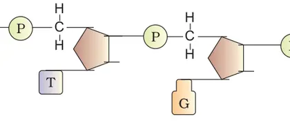
This chapter explains how genetic information is stored, replicated and expressed at the molecular level. Starting from classical experiments that established DNA as the hereditary material, it covers the chemical structure of nucleic acids, the double helix model, mechanisms of DNA replication, transcription and translation, and the regulation of gene expression (operon model). It also introduces molecular tools and techniques (restriction enzymes, vectors, PCR, recombinant DNA technology) and applications such as gene cloning and DNA fingerprinting. Importance: understanding the molecular basis of inheritance is essential for modern genetics, biotechnology, medicine and research. What the student will learn: major experiments and evidence for DNA as genetic material, structure–function relationships of DNA and RNA, steps and enzymes involved in replication/transcription/translation, features of the genetic code, control of gene expression in prokaryotes, and basic principles and applications of recombinant DNA technology.
Learning Objectives
- Define DNA as the genetic material and summarize the experimental evidence (Griffith, Avery, Hershey–Chase) that established it
- Describe the double helix structure of DNA, including sugar–phosphate backbone, antiparallel strands, complementary base pairing and significance of major/minor grooves
- Explain the semiconservative model of DNA replication and outline the roles of origin, helicase, primase, DNA polymerases, ligase and topoisomerase
- Compare replication in prokaryotes and eukaryotes with respect to origin(s), enzymes, and replication rate
- Apply complementary base‑pairing rules to predict the sequence of the complementary DNA strand, the mRNA transcript and the encoded polypeptide using the genetic code
- Describe transcription in prokaryotes and eukaryotes, noting promoter, RNA polymerase, initiation/elongation/termination and post‑transcriptional processing (capping, polyadenylation, splicing)
- Explain the process of translation—role of codons and anticodons, tRNA, ribosomes, and the stages of initiation, elongation and termination
- Interpret the properties of the genetic code (triplet, non‑overlapping, degenerate, universal, start/stop codons) and their biological implications
Topics in this chapter
9 topics · tap a topic title to jump straight to it.
Introduction and Historical Experiments
Introduction
The molecular basis of inheritance explains how genetic information is stored, replicated and transmitted from one generation to the next. The molecule that carries hereditary information is deoxyribonucleic acid (DNA). Understanding how DNA was identified as the genetic material and how its structure was discovered relies on several classical experiments. These experiments established (1) that a chemical carries genetic information, (2) that this chemical is DNA, and (3) how DNA is organized to encode information.
Key historical experiments (summary, methods and conclusions)
- Griffith (1928) — Transformation
- System: Streptococcus pneumoniae strains: virulent smooth (S) with capsule and non-virulent rough (R) without capsule.
- Experiment: Live R + heat-killed S injected into mice => mice died; live S bacteria recovered.
- Conclusion: Some “transforming principle” from dead S converted R into virulent S. Demonstrated genetic information could be transferred between bacteria (transformation).
- Avery–MacLeod–McCarty (1944) — Identifying the transforming principle
- Approach: Purified components (protein, RNA, DNA) from heat-killed S cells and tested which component could transform R cells.
- Key test: Destruction of each component by specific enzymes (protease, RNase, DNase).
- Conclusion: DNase abolished transforming activity while protease/RNase did not — DNA is the transforming principle (strong evidence that DNA carries genetic information).
- Hershey–Chase (1952) — Phage experiments confirm DNA is genetic material
- System: Bacteriophage T2 that infects E. coli.
- Labeling: Phage protein labeled with 35S (sulfur in proteins), phage DNA labeled with 32P (phosphorus in DNA).
- Result: After infection and blender-treatment, progeny phages contained 32P (DNA) but not 35S (protein).
- Conclusion: DNA, not protein, enters the bacterial cell and directs phage replication — DNA is genetic material.
- Chargaff (late 1940s–1950) — Base composition rules
- Observation: In DNA from any organism, amount of adenine (A) ≈ thymine (T) and guanine (G) ≈ cytosine (C).
- Chargaff's rules: A = T and G = C (in double-stranded DNA). Also overall base composition varies between species.
- Importance: Provided chemical constraints essential for correct DNA structure models.
- Rosalind Franklin & Maurice Wilkins (early 1950s) — X‑ray diffraction
- Method: X‑ray diffraction photographs of DNA fibers (Photo 51 by Franklin showed an X-shaped pattern).
- Conclusion: Pattern indicated a helical structure with regular repeating units; crucial evidence for a helical DNA model.
- Watson & Crick (1953) — Double helix model
- Model: DNA is a right-handed double helix of two antiparallel polynucleotide chains. Sugar-phosphate backbone on the outside; bases on the inside paired by hydrogen bonds: A with T (2 H‑bonds), G with C (3 H‑bonds).
- Implication: Complementary base pairing explains replication and storage of genetic information.
Conceptual consequences
- Complementary base pairing provides a mechanism for faithful DNA replication (each strand serves as a template).
- DNA is a stable molecule suitable for long-term information storage; its sequence encodes genes.
- These experiments laid the foundation for molecular genetics, biotechnology, forensic DNA analysis, and modern medicine.
Key terms to remember
- Transformation — uptake and expression of external DNA by a cell.
- Complementarity — specific A–T and G–C pairing.
- Antiparallel strands — the two DNA strands run in opposite directions (5'→3' and 3'→5').
- Hydrogen bonds — hold base pairs together (2 for A–T, 3 for G–C).
Tip: When studying experiments, focus on the question being asked, the experimental design (controls, labels, enzymes), the observations, and the logical conclusion — that sequence of reasoning is what proved DNA is the genetic material and how it could encode information.
- Natural bacterial transformation: Streptococcus pneumoniae and Streptococcus species can take up DNA from environment; this can spread antibiotic resistance genes between bacteria in clinical settings.
- Forensics and paternity testing: Identification methods (DNA fingerprinting) rely on the fact that DNA sequence is unique to individuals (except identical twins) and is stable across tissues.
- Genetic engineering / GMOs: Knowledge that DNA stores genetic information and that it can be transferred and expressed in other organisms underlies recombinant DNA technology (e.g., insulin production in E. coli).
- Phage therapy research: Understanding bacteriophage infection (Hershey–Chase) informs use of phages to target bacterial pathogens as an alternative to antibiotics.
- Chargaff's rules: %A = %T and %G = %C (for double-stranded DNA).
- GC content (%) = ((G + C) / (A + T + G + C)) × 100
- Wallace rule (approximate melting temperature for short oligos): Tm (°C) ≈ 2°C × (A + T) + 4°C × (G + C)
- Hydrogen bond counts per base pair: A–T = 2 H bonds, G–C = 3 H bonds
Structure of Nucleic Acids (DNA and RNA)
Overview
Nucleic acids (DNA and RNA) are biological polymers that store and transmit genetic information. Their monomers are nucleotides, each made of a nitrogenous base, a five‑carbon sugar and one or more phosphate groups.
Nucleotide components
- Sugar: Deoxyribose in DNA, ribose in RNA (RNA has an extra 2'‑OH group).
- Bases: Purines — adenine (A) and guanine (G); Pyrimidines — cytosine (C), thymine (T, only in DNA) and uracil (U, only in RNA).
- Phosphate: Links sugars by 3'→5' phosphodiester bonds to form a sugar‑phosphate backbone with polarity (5' end and 3' end).
DNA structure — Watson & Crick model
- DNA is a double helix of two antiparallel polynucleotide chains (one runs 5'→3', the other 3'→5').
- Complementary base pairing: A pairs with T (two H‑bonds); G pairs with C (three H‑bonds). This gives specificity and a uniform helix diameter (~2 nm).
- Helical parameters (B‑DNA, most common in cells): about 10 base pairs per turn, rise ≈ 0.34 nm per base pair, pitch ≈ 3.4 nm per turn, right‑handed helix.
- Major and minor grooves along the helix allow sequence‑specific protein binding (transcription factors, polymerases).
- Chargaff's rules: in double‑stranded DNA, %A ≈ %T and %G ≈ %C; total purines ≈ total pyrimidines.
- X‑ray diffraction (Rosalind Franklin) and model building (Watson & Crick) established the helical structure.
Alternate DNA forms
- B‑DNA: physiological form (right‑handed).
- A‑DNA: shorter, wider right‑handed form (observed in dehydrated samples and RNA‑DNA hybrids).
- Z‑DNA: left‑handed, zigzag backbone; occurs locally in GC‑rich stretches and can be involved in regulation.
DNA higher‑order packaging
- DNA in eukaryotes is wrapped around histone octamers to form nucleosomes (≈146 bp per nucleosome core). Nucleosomes fold into chromatin fibers and higher order loops to form chromosomes.
- DNA can be supercoiled (negative or positive) — important for compaction and for processes like replication and transcription.
RNA structure
- RNA is usually single‑stranded but folds into complex secondary and tertiary structures by intramolecular base pairing (hairpins, stem‑loops, bulges). Uracil (U) replaces thymine.
- Types and roles:
- mRNA (messenger RNA): carries coding information from DNA to ribosomes.
- tRNA (transfer RNA): cloverleaf secondary structure; carries amino acids and decodes codons during translation.
- rRNA (ribosomal RNA): structural and catalytic component of ribosomes (peptidyl transferase activity).
- Other RNAs: snRNA, miRNA, siRNA, lncRNA — regulatory and processing roles.
- RNA is more reactive (2'‑OH) and less stable than DNA, but the 2'‑OH enables catalytic RNAs (ribozymes) and complex folding.
Functional consequences of structure
- Complementarity allows accurate replication and transcription, and hybridization forms the basis of technologies (PCR primers, probes).
- Grooves and shape permit protein recognition — basis of gene regulation, DNA repair and recombination.
Historical/experimental support
- Chargaff's quantitative base composition rules.
- Franklin's X‑ray diffraction (X pattern indicating helix).
- Watson & Crick model explained complementary base pairing and replication mechanism.
Key numeric facts you should remember: 0.34 nm rise per bp, ~10 bp per turn (B‑DNA), helix diameter ≈2 nm, ~146 bp per nucleosome, A–T (2 H bonds), G–C (3 H bonds), human diploid DNA length (stretched) ≈ 2 meters per cell.
- Forensic DNA fingerprinting: uses sequence‑specific differences and PCR to match individuals using short tandem repeats (STRs) — relies on DNA double helix and complementarity for primer binding.
- mRNA vaccines (e.g., COVID‑19 vaccines): synthetic mRNA encodes a viral antigen; RNA structure, stability and 5' cap/3' poly(A) tail affect translation and immune response.
- Antibiotics targeting ribosomal RNA: many antibiotics bind rRNA in bacterial ribosomes, interfering with translation — exploits specific rRNA structure.
- DNA barcoding for species identification: short mitochondrial DNA regions (e.g., COI) are sequenced and matched — depends on conserved and variable regions arising from DNA structure/function.
- GC% = 100 × (G + C) / (A + T + G + C)
- Approximate melting temperature for short oligonucleotides (Wallace rule): Tm (°C) ≈ 2 × (A + T) + 4 × (G + C)
- Base pair axial rise: 1 bp ≈ 0.34 nm → length (nm) = 0.34 × number_of_bp
- Average molecular mass per base pair ≈ 660 g·mol⁻¹, so mass (g·mol⁻¹) ≈ 660 × number_of_bp
- Hydrogen bonds: A–T (2 H bonds), G–C (3 H bonds)
DNA Replication
Definition: DNA replication is the biological process by which a cell makes an identical copy of its DNA before cell division. It is semiconservative, bidirectional and highly accurate.
Key features & overview:
- Semiconservative: each daughter DNA contains one parental (old) strand and one newly synthesized strand (Meselson–Stahl experiment).
- Bidirectional: replication proceeds in both directions from the origin(s) of replication producing two replication forks.
- Directionality: DNA polymerases synthesize new DNA only in the 5'→3' direction; template is read 3'→5'.
Main steps (molecular mechanism):
- Initiation: Replication begins at specific DNA sites called origins (oriC in E. coli; multiple origins in eukaryotes). Initiator proteins (DnaA in bacteria; ORC in eukaryotes) bind origin and recruit helicase.
- Unwinding and stabilization: DNA helicase unwinds the double helix; single-strand binding proteins (SSB in prokaryotes; RPA in eukaryotes) stabilize single strands; topoisomerases (DNA gyrase in bacteria) relieve supercoiling.
- Primer synthesis: Primase (an RNA polymerase) makes short RNA primers complementary to the template because DNA polymerase cannot start de novo.
- Elongation: DNA polymerases add dNTPs to the 3' end of the primer, synthesizing the new strand. Leading strand is synthesized continuously; lagging strand is synthesized discontinuously as Okazaki fragments.
- Primer removal and joining: RNA primers are removed (DNA polymerase I and RNase H/FEN1 depending on organism), gaps filled and nicks sealed by DNA ligase to create a continuous backbone.
- Proofreading and repair: Many DNA polymerases have 3'→5' exonuclease proofreading reducing error rate; mismatch repair systems correct remaining errors.
- Termination: Replication stops when forks meet or at specific terminator sites; in eukaryotes, telomeres and telomerase solve the end-replication problem.
Key enzymes and proteins (summary):
- Helicase (unwinds DNA)
- Topoisomerase/gyrase (relieves torsional stress)
- Primase (synthesises RNA primer)
- DNA polymerase III (main bacterial elongase); DNA polymerase I (removes primers)
- DNA polymerases α, δ, ε (eukaryotic replication: α/primase initiates, δ mainly lagging, ε mainly leading)
- SSB / RPA (stabilizes ssDNA)
- Ligase (seals nicks)
- Telomerase (extends telomeres in germ/stem/cancer cells)
Proofreading and fidelity: DNA polymerases incorporate bases with high fidelity; 3'→5' exonuclease removes mismatched nucleotides. Residual mismatches are corrected by mismatch repair systems—this keeps mutation rate very low.
Special topics:
- Meselson–Stahl experiment: Showed semiconservative replication using 15N/14N density gradients: after one generation a hybrid band appears; after two generations hybrid + light bands appear.
- End-replication problem & telomeres: Linear chromosomes cannot be fully replicated at the 3' ends; telomeres (repeated sequences) and telomerase (a reverse transcriptase with RNA template) extend ends to prevent loss of coding DNA.
- Regulation: In eukaryotes replication is restricted to S phase and origins are ‘licensed’ once per cycle by controlled loading of MCM helicase and CDK regulation.
Biological significance: Accurate DNA replication is essential for genetic continuity; errors lead to mutations that can be neutral, harmful or occasionally beneficial (evolution). Telomerase dysfunction is linked to aging and uncontrolled telomerase activity is associated with cancer.
Class 12 level summary: DNA replication is semiconservative and bidirectional. It requires primers and many enzymes, produces Okazaki fragments on the lagging strand, has proofreading to ensure fidelity, and uses telomerase to maintain chromosome ends in certain cells. The Meselson–Stahl experiment provided classical evidence for the semiconservative model.
- Polymerase chain reaction (PCR): in vitro exponential amplification of a target DNA sequence using a heat-stable DNA polymerase (Taq); ideal copies after n cycles = 2^n.
- Antibiotics targeting replication: fluoroquinolones (e.g., ciprofloxacin) inhibit bacterial DNA gyrase/topoisomerase II, blocking replication and killing bacteria.
- Antiviral/antitumor nucleoside analogs: AZT (HIV) and certain chemotherapeutics act as chain terminators for viral or rapidly dividing cell DNA synthesis.
- Telomerase in cancer: many cancer cells reactivate telomerase to maintain telomeres and enable limitless replication.
- DNA damage & repair in everyday life: sunlight (UV) causes thymine dimers; nucleotide excision repair and DNA replication-related repair pathways preserve genomic integrity.
- Number of double-stranded DNA molecules after n rounds of perfect replication: 2^n
- Ideal PCR product copies after n cycles (perfect efficiency): copies = 2^n (practical efficiency E often < 1 → copies = (1+E)^n )
- Approximate replication time for a genome: T ≈ Genome size / (2 × fork speed × number_of_active_origins). Example: for a genome of size G bp, fork speed v bp/s and N origins active, T ≈ G / (2 v N).
- Expected number of replication errors per genome per replication: Errors ≈ genome_size × replication_error_rate_per_bp (after proofreading and initial corrections).
Transcription
Definition: Transcription is the process by which a single-stranded RNA molecule is synthesised from a DNA template by RNA polymerase. It is the first step of gene expression (DNA → RNA → Protein).
Transcription unit and strands: A transcription unit includes a promoter, the transcribed region (from start site to termination), and terminator. DNA has two strands: the template (antisense) strand used by RNA polymerase to build an RNA copy complementary to it, and the coding (sense) strand which has the same sequence as the RNA except T → U.
Base-pairing rules during transcription: A (DNA) → U (RNA), T (DNA) → A (RNA), C ↔ G.
Major steps:
- Initiation: RNA polymerase recognizes and binds the promoter (with sigma factor in prokaryotes or general transcription factors in eukaryotes). The start site (+1) is unwound to form the transcription bubble.
- Elongation: RNA polymerase moves 3'→5' on the template strand and synthesises RNA 5'→3' by adding ribonucleotides complementary to the template. The enzyme maintains a short RNA–DNA hybrid inside the bubble.
- Termination: In prokaryotes termination can be rho-dependent (requires rho protein) or rho-independent (intrinsic hairpin followed by U-run). In eukaryotes termination involves cleavage of pre-mRNA and subsequent polyadenylation; RNA Pol II often runs past the poly(A) site and is released later.
Prokaryotic vs Eukaryotic transcription (key differences):
- Prokaryotes: Single RNA polymerase; transcription and translation are coupled in cytoplasm. Promoters include -35 and -10 (Pribnow) regions; initiation assisted by sigma factor. Termination via rho-dependent/independent mechanisms.
- Eukaryotes: Three RNA polymerases (Pol I, II, III). RNA Pol II transcribes mRNA. Transcription occurs in nucleus; mRNA is processed before export and translation. Promoters include TATA box; basal/general transcription factors are needed. Complex regulation via enhancers/silencers and chromatin structure.
Post-transcriptional processing (eukaryotic mRNA):
- 5' capping: Addition of 7-methylguanosine cap to the 5' end soon after initiation—important for stability and translation initiation.
- Splicing: Removal of introns and joining of exons by the spliceosome (snRNPs). Alternative splicing produces multiple protein isoforms from one gene.
- 3' polyadenylation: Cleavage downstream of AAUAAA signal and addition of ~200 adenines (poly(A) tail) for stability and export.
Regulation of transcription: Transcription is controlled by transcription factors (activators/repressors), promoter strength, enhancers/silencers, epigenetic marks (DNA methylation, histone modifications), and in prokaryotes, operon systems (coordinated regulation of gene clusters).
Biological importance: Transcription determines which genes are expressed, when and how much. Errors or misregulation cause diseases (e.g., defective splicing can lead to β-thalassemia), while controlled transcriptional responses allow adaptation (e.g., bacterial lac operon induction).
Summary (compact): RNA polymerase recognises promoter → opens helix → synthesises RNA 5'→3' using the DNA template → RNA is released at terminator. In eukaryotes, the primary transcript is processed (cap, splice, polyA) before export and translation.
- Lac operon in E. coli: an inducible operon where presence of lactose (allolactose) relieves repressor binding allowing transcription of genes (lacZYA) for lactose metabolism.
- Trp operon in bacteria: a repressible operon that is switched off when tryptophan (co-repressor) is abundant.
- Beta-globin gene splicing: proper removal of introns and joining of exons is essential; defective splicing can cause β-thalassemia (reduced/absent β-globin).
- Alternative splicing in humans: a single gene produces multiple protein isoforms (e.g., tropomyosin, calcitonin/CGRP) — increases proteome diversity without increasing gene number.
- Viral transcription strategies: many RNA viruses use their own RNA-dependent RNA polymerases to transcribe genomes (e.g., influenza virus transcribes viral mRNAs in host nucleus/cytoplasm).
- Central dogma notation: DNA → RNA → Protein
- Complementarity during transcription: A (DNA) → U (RNA); T (DNA) → A (RNA); C ↔ G
- Transcription output (approximate): N = r × t, where N = number of nucleotides synthesised, r = transcription rate (nt/s), t = time (s). Typical rates: bacterial RNA polymerase ≈ 40–80 nt/s; eukaryotic RNA Pol II ≈ 20–40 nt/s (approximate values).
- Approximate relationship for mRNA length change by processing: Mature mRNA length = pre-mRNA length − (sum of intron lengths) + (length of 5' cap region + length of poly(A) tail)
Genetic Code
Definition: The genetic code is the set of rules by which the information encoded in the sequence of nucleotides in mRNA is translated into the sequence of amino acids in a polypeptide. A codon is a triplet of consecutive nucleotides in mRNA that specifies one amino acid or a translation stop signal.
Basic features:
- Triplet code: Each codon consists of three bases (nucleotides). Three bases give 64 possible codons (4^3), which is more than the 20 standard amino acids.
- Unambiguous: Each codon specifies only one amino acid (or a stop), i.e., a given codon has a single meaning.
- Degenerate (redundant): Most amino acids are encoded by more than one codon (synonymous codons).
- Non-overlapping: Codons are read one after another without overlap in a given reading frame.
- Commaless (no punctuation): The code is read continuously from a fixed start point without separators between codons.
- Start and stop signals: AUG is the usual start codon (codes for methionine in eukaryotes and for formylmethionine in bacteria). Three codons (UAA, UAG, UGA) are stop (termination) codons and do not code for amino acids.
- Nearly universal: The same code is used by most organisms, with minor variations (notable exceptions in mitochondrial genomes and some protozoa).
How translation uses the code: During translation, an mRNA is read in codons from the 5' to 3' end starting at the AUG start codon. Transfer RNAs (tRNAs) with anticodons complementary to mRNA codons bring specific amino acids. Aminoacyl-tRNA synthetases attach the correct amino acid to each tRNA; codon-anticodon pairing ensures the correct sequence of amino acids in the growing polypeptide.
Wobble hypothesis: Proposed by Francis Crick, the wobble hypothesis explains degeneracy via flexible base-pairing at the third (3') base of the codon (the first base of the tRNA anticodon). This allows one tRNA to recognize multiple synonymous codons, reducing the number of tRNAs required.
Reading frame and mutations: The correct reading frame is essential. A frameshift mutation (insertion or deletion of bases not in multiples of three) shifts the reading frame and usually produces a nonfunctional protein. Point mutations can be:
- Silent (synonymous): different codon, same amino acid
- Missense (nonsynonymous): different amino acid
- Nonsense: codon changed to a stop codon, truncating the protein
Exceptions and variations: Some organelles and certain organisms use slightly different genetic codes. For example, in human mitochondria, UGA codes for tryptophan instead of being a stop codon, and AUA can code for methionine.
Biological significance: The degeneracy of the code provides robustness against some point mutations. Codon usage bias (preference for certain synonymous codons) affects translation efficiency and is important in biotechnology (optimizing gene sequences for heterologous protein expression).
Summary table (short):
- Total codons: 64 (including 3 stop codons)
- Amino acids encoded: 20 standard amino acids
- Start codon: AUG (methionine)
- Stop codons: UAA, UAG, UGA
- Sickle cell anemia: A single base substitution in the beta-globin gene changes the codon GAG (glutamic acid) to GUG (valine) in DNA/mRNA, producing hemoglobin S with altered properties — an example of a missense mutation altering an amino acid.
- Nonsense mutation in Duchenne muscular dystrophy or some cases of beta-thalassemia: A point mutation converts a sense codon into a stop codon, producing a truncated nonfunctional protein.
- Mitochondrial genetic code variation: Human mitochondrial genome translates UGA as tryptophan instead of a stop, illustrating that the genetic code is nearly universal but not absolute.
- Codon optimization in biotechnology: When expressing a human gene in E. coli, synonymous codons are changed to match E. coli preferred codon usage to increase translation efficiency and protein yield (codon usage bias example).
- Number of possible codons = n^r where n = number of nucleotide bases (4) and r = length of codon (3); so 4^3 = 64 codons.
- Number of stop codons = 3 (UAA, UAG, UGA).
- Number of standard amino acids = 20, so average codon degeneracy ≈ 64/20 ≈ 3.2 (mean codons per amino acid).
- Number of possible polypeptides of length L = 20^L (combinatorial possibilities ignoring folding and function).
- Number of possible reading frames per single-stranded mRNA = 3; for double-stranded DNA (both strands translated) = 6.
Translation
Definition: Translation is the process by which the genetic information encoded in mRNA is decoded to build a polypeptide (protein). It occurs on ribosomes in the cytoplasm (prokaryotes and eukaryotes), often associated with the rough endoplasmic reticulum in eukaryotes for secreted/membrane proteins.
Major components:
- mRNA: contains codons (triplets) that specify amino acids.
- tRNA: adaptor RNA with anticodon and attached specific amino acid (aminoacyl-tRNA).
- Ribosomes: rRNA + proteins; sizes: prokaryotic 70S (50S + 30S), eukaryotic 80S (60S + 40S).
- Aminoacyl-tRNA synthetases: enzymes that charge tRNAs with their cognate amino acids.
- Translation factors: initiation, elongation and release (termination) factors; GTP provides energy for several steps.
Features of the genetic code (relevant to translation):
- Triplet codons (three bases per amino acid)
- Start codon: AUG (codes for methionine; in prokaryotes the initiator is formyl-Met)
- Stop codons: UAA, UAG, UGA (no tRNAs; recognized by release factors)
- Degenerate (redundant) but unambiguous
- Non-overlapping and nearly universal
Stepwise mechanism:
1. Charging of tRNA (Aminoacylation):
- Amino acid + tRNA + ATP —(aminoacyl-tRNA synthetase)—> aminoacyl-tRNA + AMP + PPi.
- This ‘‘charges’’ tRNA so it can deliver its amino acid to the ribosome.
2. Initiation:
- Small ribosomal subunit binds mRNA and locates start codon (often with initiation factors).
- Initiator tRNA (fMet-tRNA in prokaryotes; Met-tRNAi in eukaryotes) pairs with AUG at the P site.
- Large subunit joins to form a functional ribosome; initiation factors and GTP are involved.
3. Elongation:
- Charged tRNA corresponding to the A-site codon is delivered (by EF-Tu in bacteria) — GTP used.
- Peptide bond formation occurs via peptidyl transferase activity of the large subunit rRNA: the growing polypeptide on the P-site tRNA is transferred to amino acid on A-site tRNA.
- Translocation: ribosome moves one codon forward (EF-G in bacteria) shifting tRNAs from A to P to E sites — GTP used; empty tRNA leaves from E site.
- Cycle repeats, elongating the polypeptide from N-terminus to C-terminus.
4. Termination:
- When a stop codon enters the A site, release factors bind and catalyze hydrolysis of the bond between polypeptide and tRNA.
- Polypeptide is released; ribosomal subunits dissociate and can be reused.
Energy cost (summary): Charging each tRNA uses ATP -> AMP + PPi (equivalent of 2 ATP). During each elongation cycle at least 2 GTP are used (delivery and translocation). Overall, roughly 4 high-energy phosphate bonds are consumed per amino acid added (2 from charging + 2 from GTP use), plus additional GTPs for initiation and termination.
Post-translational events: Folding (often aided by chaperones), cleavage of signal peptides, covalent modifications (phosphorylation, glycosylation, acetylation), assembly into multimeric proteins and targeting to organelles/secretory pathway.
Biological importance and regulation: Translation controls protein amounts and is regulated at initiation, mRNA stability, availability of charged tRNAs, and via microRNAs in eukaryotes. Many antibiotics target bacterial translation (see examples).
- Insulin: synthesized as preproinsulin on ribosomes bound to rough ER, signal peptide cleaved to produce proinsulin, folded and cleaved to yield active insulin — illustrates translation, targeting and post-translational processing.
- Hemoglobin subunits: alpha and beta globin chains are translated in the cytosol, fold and assemble into tetramers — example of co- and post-translational folding and assembly.
- Antibiotics that inhibit translation: Chloramphenicol inhibits peptidyl transferase (prokaryotes), tetracycline blocks A-site tRNA binding, streptomycin causes misreading — clinical relevance of translation mechanism.
- Polysomes (polyribosomes): One mRNA molecule can be simultaneously translated by many ribosomes, increasing protein output — visible in electron micrographs as multiple ribosomes on a single mRNA.
- Aminoacylation: Amino acid + tRNA + ATP → aminoacyl-tRNA + AMP + PPi
- Approximate energy cost per amino acid added: 2 ATP equivalents (from charging) + ~2 GTP (elongation) ≈ 4 high-energy bonds
- Ribosome composition shorthand: Prokaryote 70S = 50S + 30S; Eukaryote 80S = 60S + 40S
Regulation of Gene Expression
What is regulation of gene expression?
Regulation of gene expression means controlling when, where, and how much a gene is transcribed and translated into a functional product (RNA or protein). It ensures correct cell specialization, development, response to environment and homeostasis.
Why is it needed?
All cells in a multicellular organism have (almost) the same DNA but different functions. Regulation allows cells to express only the genes needed for their role, conserve energy, respond to environmental cues (nutrients, heat, hormones) and maintain balance between synthesis and degradation of molecules.
Levels at which gene expression is regulated
- Epigenetic (chromatin level): DNA methylation and histone modifications change chromatin accessibility (euchromatin vs heterochromatin) and therefore transcription.
- Transcriptional: Control of initiation and rate of transcription by promoters, enhancers, silencers, transcription factors, and general transcription machinery.
- Post-transcriptional: RNA processing (capping, polyadenylation), alternative splicing, RNA editing and mRNA stability/decay.
- Translational: Control of initiation, ribosome binding, and regulatory RNAs that block translation.
- Post-translational: Protein folding, modification (phosphorylation, ubiquitination), activation/inactivation and degradation.
Key molecular mechanisms
- Promoters & operators: DNA sequences where RNA polymerase and regulators bind. Operators are typical in prokaryotic operons.
- Transcription factors (TFs): Proteins (activators or repressors) that bind specific DNA sequences and recruit or block the transcription machinery.
- Enhancers & silencers: Distant DNA elements that increase or decrease transcription via DNA looping to promoters.
- Chromatin remodellers and histone modifications: Acetylation (usually activates), methylation (can activate or repress depending on residue) and chromatin-remodelling complexes alter nucleosome positioning.
- Non-coding RNAs: miRNAs and siRNAs bind mRNAs to block translation or promote degradation; lncRNAs can scaffold chromatin modifiers or sequester factors.
- Feedback regulation: Negative and positive feedback loops maintain stable states or produce switches during development.
Prokaryotic example — Operon model (lac and trp)
In bacteria, contiguous structural genes with a single promoter form an operon. Regulatory proteins interact with operator/promoter to turn operons on/off in response to metabolites.
Key features of eukaryotic regulation
- Multiple TFs integrate signals at promoters and enhancers (combinatorial control).
- Chromatin state is central: heterochromatin = silent, euchromatin = active.
- RNA processing steps (especially alternative splicing) increase proteome diversity.
- Epigenetic changes (DNA methylation, histone marks) can be mitotically stable and sometimes heritable across generations.
Biological significance and consequences
- Development & cell differentiation (spatial and temporal gene expression patterns).
- Adaptation to environment (stress responses, nutrient sensing).
- Disease when misregulated: cancer (oncogene activation, tumour suppressor inactivation), imprinting disorders, developmental defects.
Summary (simplified)
Regulation of gene expression is a multi-layered process (epigenetic → transcriptional → post-transcriptional → translational → post-translational) that controls cellular phenotype, responses and organismal development by modulating production and activity of RNAs and proteins.
- Lac operon (E. coli): expression of genes needed to metabolize lactose is induced only when lactose is present and glucose is absent—controlled by the lac repressor and CAP–cAMP.
- Trp operon (E. coli): repressible operon where tryptophan binds repressor and turns transcription off when tryptophan is abundant.
- X-chromosome inactivation (mammals): one X chromosome in female cells is transcriptionally silenced (Barr body) by XIST lncRNA and chromatin modifications—example of epigenetic regulation leading to mosaicism (e.g., calico cats).
- Alternative splicing (eukaryotes): one gene can produce multiple protein isoforms; e.g., tropomyosin isoforms differ between muscle and non-muscle cells.
- RNA interference (RNAi): miRNAs/siRNAs regulate mRNA stability/translation. Used therapeutically (siRNA drugs) to downregulate disease genes.
- Genomic imprinting: parent-specific gene expression (e.g., Igf2 expressed only from paternal allele); defects cause Prader–Willi or Angelman syndromes.
- Central dogma (conceptual): DNA → RNA → Protein
- Steady-state protein level (simplified): [Protein] ≈ (k_transcription × k_translation) / (k_mRNA_decay × k_protein_decay)
- mRNA decay (first-order kinetics): N(t) = N0 × e^(−k t) ; half-life t1/2 = ln(2)/k
- Ligand–DNA binding equilibrium (for regulator binding): Kd = [Protein][DNA] / [Protein–DNA complex] (lower Kd = higher affinity)
- Hill equation (cooperativity, dose–response): Fraction bound = [L]^n / (Kd + [L]^n) — produces sigmoidal response when n>1
Mutations and DNA Repair
Definition: A mutation is a heritable change in the DNA sequence. Mutations can affect a single base (point mutation), a stretch of bases (insertions/deletions), whole chromosomes (structural changes) or chromosome number (aneuploidy). DNA repair comprises the cellular processes that detect and correct DNA damage to maintain genomic integrity.
Types of mutations
- Gene (point) mutations: substitution (transition, transversion) producing silent, missense or nonsense changes; frameshift mutations from small insertions/deletions that shift the reading frame.
- Chromosomal mutations: deletions, duplications, inversions, translocations.
- Aneuploidy / Polyploidy: loss or gain of whole chromosomes (e.g., trisomy 21 = Down syndrome).
Causes (Mutagens)
- Spontaneous: errors in DNA replication, tautomeric shifts, deamination, depurination.
- Induced: physical (UV light forms thymine dimers; ionizing radiation causes breaks), chemical (alkylating agents, base analogs, intercalating agents), biological (transposons, some viruses).
Consequences
- At molecular level: altered codons, truncated proteins, altered regulation.
- At organismal level: genetic diseases (e.g., sickle cell), cancers, sterility or developmental defects.
- At population level: source of genetic variation and raw material for evolution; can be beneficial, neutral or harmful.
DNA repair mechanisms
- Direct reversal: some damage is directly reversed (e.g., photoreactivation by photolyase breaks UV-induced thymine dimers in many organisms).
- Base excision repair (BER): glycosylase removes altered base → AP site → AP endonuclease cuts backbone → DNA polymerase fills gap → DNA ligase seals nick. Repairs small non-helix-distorting lesions (e.g., deaminated bases).
- Nucleotide excision repair (NER): recognises bulky helix-distorting lesions (e.g., thymine dimers) → removes an oligonucleotide segment → polymerase and ligase restore sequence. Essential in humans to prevent UV-induced skin cancers.
- Mismatch repair (MMR): corrects replication errors (mismatches, small indels) soon after replication using parental strand for reference (e.g., MutS/MutL in bacteria; MSH/MLH in eukaryotes).
- Double-strand break (DSB) repair: Homologous recombination (error-free, uses sister chromatid) and Non-homologous end-joining (NHEJ, faster but error-prone).
- SOS response and translesion synthesis (in bacteria): emergency systems allow replication across lesions using error-prone polymerases, increasing mutation rate but permitting survival.
Important molecular players: DNA glycosylases, AP endonuclease, DNA polymerases (repair types), DNA ligase, photolyase, RecA/Rad51 (strand exchange), ATM/ATR (damage sensing kinases in eukaryotes), MSH/MLH (MMR proteins).
Clinical relevance / Disorders from defective repair: Xeroderma pigmentosum (defective NER → extreme UV sensitivity, skin cancer), Hereditary nonpolyposis colorectal cancer (HNPCC; defective MMR), Ataxia-telangiectasia (defective ATM, problems sensing DSBs), Bloom and Fanconi anemia (DSB repair defects).
Summary: Mutations originate spontaneously or from mutagens and can be neutral, harmful, or occasionally beneficial. DNA repair systems constantly fix many lesions; failure of repair increases mutation load and disease risk. Understanding these processes explains genetic disorders, cancer etiology and evolutionary change.
- Sickle cell anaemia: a missense point mutation in the β‑globin gene (Glu → Val) alters haemoglobin structure and causes sickling of red blood cells.
- Cystic fibrosis: common ΔF508 mutation is a three-base deletion in the CFTR gene causing misfolded protein and chloride channel dysfunction.
- Down syndrome: trisomy 21 results from nondisjunction (an additional chromosome 21).
- UV-induced skin cancer: unrepaired thymine dimers (from UV radiation) can cause mutations that activate oncogenes or inactivate tumour suppressors.
- Antibiotic resistance in bacteria: point mutations or gene acquisition (plasmids) alter drug targets or increase drug efflux.
- Xeroderma pigmentosum: defective nucleotide excision repair causes extreme sensitivity to UV and high skin cancer incidence.
- Mutation frequency = (Number of mutants observed) / (Total number of individuals tested)
- Mutation rate per locus per generation ≈ (Number of new mutations) / (Number of gametes or individuals × number of generations)
- Luria–Delbrück / Poisson approximation (fluctuation test): P0 = e^(−m) , where P0 is fraction of cultures with zero mutants and m is the mean number of mutations per culture; m ≈ −ln(P0).
Applications and Modern Developments (overview)
Overview: Applications and modern developments in the molecular basis of inheritance concern how knowledge of DNA structure, replication and manipulation is used in medicine, agriculture, industry, forensics and research. Key molecular tools include restriction enzymes, ligases, vectors, PCR, DNA sequencing and genome editing systems (e.g., CRISPR-Cas). Advances in high-throughput sequencing, precise editing and bioinformatics have broadened applications from single-gene studies to whole genomes and populations.
Major techniques and how they are used:
- Recombinant DNA technology (cloning): insertion of a gene into a vector and host to produce proteins (eg. insulin) or to study gene function.
- PCR (Polymerase Chain Reaction): rapid amplification of specific DNA segments for diagnostics (infectious disease testing), cloning, and forensic identification.
- DNA sequencing: reading the nucleotide order. Sanger sequencing is used for small-scale work; Next-Generation Sequencing (NGS) enables whole genomes, exomes and transcriptomes.
- Genome editing (CRISPR-Cas9 and newer formats): targeted changes in genomes for research, potential therapies (gene correction) and crop improvement.
- DNA fingerprinting / forensic DNA typing: using VNTR/STR profiles for identity, paternity and criminal investigations.
- Transgenic organisms / GM crops & animals: adding genes for desirable traits (Bt cotton, herbicide tolerance, nutritional enhancement like Golden Rice).
Applications:
- Medicine: recombinant drugs (insulin, growth hormone), vaccines (recombinant and mRNA vaccines), molecular diagnostics (PCR tests for pathogens), gene therapy, CAR-T cell therapy, pharmacogenomics (drug response prediction), prenatal genetic testing (NIPT, PGD).
- Agriculture: genetically modified crops for pest resistance, herbicide tolerance, enhanced nutrition; marker-assisted selection; generation of transgenic animals for research and production traits.
- Forensics & legal: DNA profiling for criminal cases, paternity, disaster victim identification.
- Research & biotechnology industry: synthetic biology, industrial enzymes, bioremediation, microbial production of chemicals.
- Public health & epidemiology: pathogen sequencing for outbreak tracking, surveillance and vaccine design.
Recent and emerging developments:
- Next-Generation and Third-Generation sequencing: massively parallel sequencing (NGS) and long-read technologies (Nanopore, PacBio) make whole-genome, single-cell and metagenomic studies routine.
- CRISPR innovations: base editing and prime editing allow single-base changes without double-strand breaks; CRISPR diagnostics (e.g., SHERLOCK/DETECTR) enable rapid pathogen detection.
- Single-cell genomics and transcriptomics: resolve heterogeneity within tissues and tumors.
- Precision / personalized medicine: therapies and drug choices guided by an individual's genomic profile.
- Synthetic biology: design and construction of novel biological systems (biological parts, circuits and minimal genomes).
Ethical, legal and social considerations: benefits are paired with concerns — gene editing in germline cells, biosafety of GM organisms, privacy of genomic data, equitable access to genomic medicine and regulatory oversight are major topics.
Class 12 perspective — key takeaways: understand the principles of main techniques (PCR, cloning, sequencing, CRISPR), principal applications in everyday life (diagnostics, therapeutics, agriculture, forensics), and be aware of recent technological trends that are transforming healthcare and biology.
- Recombinant human insulin produced in E. coli for diabetes treatment.
- PCR-based RT-PCR tests for SARS-CoV-2 detection during the COVID-19 pandemic.
- Bt cotton — a transgenic crop expressing a bacterial toxin gene for insect resistance.
- DNA fingerprinting using STR profiles in forensic casework and paternity testing.
- CRISPR-Cas9 used in laboratory research to knock out genes to study function.
- Next-Generation Sequencing used in the Human Genome Project follow-ups and in cancer genomics to identify driver mutations.
- PCR amplification (ideal doubling): N = N0 × 2^n (N = number of copies after n cycles; N0 = initial copies)
- Approximate melting temperature for short oligonucleotides (14–20 nt): Tm (°C) ≈ 4 × (G + C) + 2 × (A + T)
- GC content (%) = [(G + C) / (A + T + G + C)] × 100
- DNA concentration from spectrophotometry (dsDNA): [DNA] (µg/mL) = A260 × 50 (where A260 is absorbance at 260 nm)
- Relationship in gel electrophoresis (qualitative): migration distance ∝ -log(molecular weight) (i.e., smaller fragments move farther)
Key Concepts
- DNA (Deoxyribonucleic Acid)
- Double-stranded polymer of deoxyribonucleotides that stores hereditary information.
- RNA (Ribonucleic Acid)
- Single-stranded polymer of ribonucleotides involved in transfer and expression of genetic information.
- Nucleotide
- Basic building block of nucleic acids composed of a nitrogenous base, a pentose sugar and one or more phosphate groups.
- Nucleoside
- Molecule consisting of a nitrogenous base attached to a sugar (no phosphate).
- Phosphodiester bond
- Covalent linkage joining the 3′-OH of one nucleotide sugar to the 5′-phosphate of the next, forming the nucleic acid backbone.
- Double helix
- Three-dimensional structure of DNA in which two antiparallel strands wind around each other forming a helical shape.
- Complementary base pairing
- Specific hydrogen-bonding between bases: A pairs with T (or U in RNA) and G pairs with C, ensuring accurate information transfer.
- Replication
- Process of copying DNA to produce two identical DNA molecules prior to cell division.
- Semi-conservative replication
- Mode of DNA replication where each daughter DNA molecule contains one parental strand and one newly synthesized strand.
- Replication fork
- The Y-shaped region where the DNA double helix is unwound and new strands are being synthesized.
- DNA polymerase
- Enzyme that synthesizes new DNA strands by adding nucleotides complementary to the template strand, usually in 5′→3′ direction.
- Primase
- RNA polymerase that synthesizes short RNA primers required to initiate DNA synthesis.
- DNA ligase
- Enzyme that joins DNA fragments by forming phosphodiester bonds, sealing nicks in the backbone.
- Transcription
- Synthesis of an RNA molecule using DNA as a template; first step of gene expression.
- mRNA (Messenger RNA)
- RNA that carries the genetic code from DNA in the nucleus to ribosomes for protein synthesis.
- tRNA (Transfer RNA)
- Small RNA that carries specific amino acids and contains an anticodon to recognize codons on mRNA during translation.
- Codon
- Triplet of nucleotides in mRNA that specifies a particular amino acid or a stop signal during translation.
- Anticodon
- Three-nucleotide sequence on tRNA complementary to an mRNA codon, enabling correct amino acid incorporation.
- Translation
- Process by which ribosomes synthesize polypeptides by decoding mRNA codons into a sequence of amino acids.
- Operon (e.g., lac operon)
- Cluster of functionally related genes under the control of a single promoter and regulatory sequences in prokaryotes.
End-of-Chapter Trial Paper & Test Questions
Topic-wise questions to test your understanding of every concept in this chapter.
-
How did the Hershey–Chase experiment confirm that DNA is the genetic material? / हर्षे–चेज प्रयोग ने कैसे सिद्ध किया कि DNA आनुवंशिक पदार्थ है?
Show answer
Using bacteriophage T2 with 32P-labeled DNA and 35S-labeled protein, they found only 32P (DNA) entered the bacteria and appeared in progeny phages, while 35S (protein) stayed outside, proving DNA carries genetic information. / बैक्टीरियोफेज T2 में 32P-चिह्नित DNA और 35S-चिह्नित प्रोटीन का प्रयोग कर उन्होंने पाया कि केवल 32P (DNA) जीवाणु में प्रवेश कर संतति फेज में आया, जबकि 35S (प्रोटीन) बाहर रहा, जो सिद्ध करता है कि DNA आनुवंशिक सूचना वहन करता है।
-
State Chargaff's rule and explain its structural significance. / चारगाफ का नियम बताइए और उसका संरचनात्मक महत्व समझाइए।
Show answer
Chargaff's rule states that in double-stranded DNA %A = %T and %G = %C; this reflects complementary base pairing (A–T, G–C) and supports the antiparallel double-helix model. / चारगाफ का नियम बताता है कि द्विरज्जुक DNA में %A = %T और %G = %C; यह पूरक क्षार युग्मन (A–T, G–C) को दर्शाता है और प्रतिसमानांतर द्विकुंडली मॉडल का समर्थन करता है।
-
Why is DNA replication described as semiconservative, and which experiment proved it? / DNA प्रतिकृति को अर्धसंरक्षी क्यों कहा जाता है, और किस प्रयोग ने इसे सिद्ध किया?
Show answer
It is semiconservative because each daughter DNA molecule contains one parental strand and one newly synthesized strand; the Meselson–Stahl experiment using 15N/14N density gradients proved this by showing a hybrid band after one generation. / यह अर्धसंरक्षी है क्योंकि प्रत्येक संतति DNA अणु में एक जनक रज्जु और एक नवसंश्लेषित रज्जु होती है; मेसेल्सन–स्टाल प्रयोग ने 15N/14N घनत्व प्रवणता का उपयोग कर एक पीढ़ी के बाद संकर बैंड दिखाकर इसे सिद्ध किया।
-
Why is the lagging strand synthesized discontinuously during replication? / प्रतिकृति के दौरान पश्चगामी (लैगिंग) रज्जु असतत रूप से क्यों संश्लेषित होती है?
Show answer
DNA polymerase can add nucleotides only in the 5'→3' direction, so on the lagging strand (whose template runs 5'→3' toward the fork) synthesis occurs in short Okazaki fragments that are later joined by DNA ligase. / DNA पॉलिमरेज केवल 5'→3' दिशा में न्यूक्लियोटाइड जोड़ सकता है, अतः पश्चगामी रज्जु पर (जिसका टेम्पलेट कांटे की ओर 5'→3' चलता है) संश्लेषण छोटे ओकाजाकी खंडों में होता है जिन्हें बाद में DNA लाइगेज जोड़ता है।
-
Differentiate between the template strand and the coding strand in transcription. / प्रतिलेखन में टेम्पलेट रज्जु और कूटलेखन (कोडिंग) रज्जु में अंतर बताइए।
Show answer
The template (antisense) strand is read 3'→5' by RNA polymerase to build the RNA, while the coding (sense) strand has the same sequence as the RNA except thymine is replaced by uracil. / टेम्पलेट (एंटीसेंस) रज्जु को RNA पॉलिमरेज 3'→5' पढ़कर RNA बनाता है, जबकि कूटलेखन (सेंस) रज्जु का अनुक्रम RNA जैसा ही होता है सिवाय इसके कि थायमीन की जगह यूरेसिल होता है।
-
List any four salient features of the genetic code. / आनुवंशिक कूट की कोई चार प्रमुख विशेषताएँ लिखिए।
Show answer
The genetic code is triplet (three bases per codon), degenerate (many amino acids have more than one codon), non-overlapping and commaless, and nearly universal with AUG as start and UAA/UAG/UGA as stop codons. / आनुवंशिक कूट त्रिक है (प्रति कोडॉन तीन क्षार), अपहासी/अपजनित है (कई अमीनो अम्लों के एक से अधिक कोडॉन), अनतिव्यापी व अल्पविरामहीन है, और लगभग सार्वभौमिक है जिसमें AUG प्रारंभ तथा UAA/UAG/UGA समाप्ति कोडॉन हैं।
-
Numerical: An mRNA contains the coding sequence AUG-GCU-UUU-UAA. How many amino acids will the polypeptide have? / संख्यात्मक: एक mRNA में कूटलेखन अनुक्रम AUG-GCU-UUU-UAA है। पॉलीपेप्टाइड में कितने अमीनो अम्ल होंगे?
Show answer
AUG (Met), GCU (Ala), UUU (Phe) code for 3 amino acids and UAA is a stop codon that codes for none, so the polypeptide has 3 amino acids. / AUG (मेट), GCU (एला), UUU (फेन) तीन अमीनो अम्ल कूटित करते हैं और UAA समाप्ति कोडॉन है जो किसी को कूटित नहीं करता, अतः पॉलीपेप्टाइड में 3 अमीनो अम्ल होंगे।
-
Explain how the lac operon is switched on in the presence of lactose. / लैक्टोज की उपस्थिति में लैक ऑपेरॉन कैसे चालू होता है, समझाइए।
Show answer
When lactose is present, allolactose (an inducer) binds the lac repressor and inactivates it, so the repressor leaves the operator and RNA polymerase can transcribe the structural genes (lacZYA) for lactose metabolism. / जब लैक्टोज उपस्थित होता है, एलोलैक्टोज (प्रेरक) लैक दमनकारी से जुड़कर उसे निष्क्रिय कर देता है, जिससे दमनकारी ऑपरेटर छोड़ देता है और RNA पॉलिमरेज लैक्टोज उपापचय के संरचनात्मक जीन (lacZYA) का प्रतिलेखन कर सकता है।
Related Laws & Principles
Explore allFoundational laws & principles behind this chapter. Each one opens a full page — what it says, why it matters, five practice questions and the mistakes to avoid.