Overview
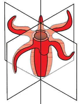
This chapter gives a structured account of the Animal Kingdom as presented in the NCERT Class 11 Biology textbook. It introduces why classification is necessary and the principles used to group animals, then examines the major animal phyla (non-chordates and chordates), their salient features and representative examples. Key organizing concepts include levels of organisation (cellular, tissue, organ), symmetry (radial, bilateral), germ layers (diploblastic, triploblastic), body cavity types (acoelomate, pseudocoelomate, coelomate), segmentation, embryonic development (protostome vs deuterostome), and notochord/nerve cord presence in chordates. The chapter develops skills in recognising diagnostic characters, constructing simple classification tables, and comparing phyla. Its importance lies in providing a framework to understand animal diversity, evolutionary relationships and how structure relates to function — foundational for later topics (physiology, ecology, evolution, and taxonomy). By the end of the chapter, students will be able to describe and distinguish major phyla, list representative animals, explain the basis of classification, and apply those principles to identify…
Learning Objectives
- Define the term 'Animal Kingdom' and related basic concepts such as phylum, class, and species for exam-style definitions.
- Classify animals up to phylum level using the major criteria (symmetry, body cavity, segmentation, germ layers) and list examples for each phylum.
- Differentiate between poriferan, coelenterate, platyhelminth, nematode, annelid, arthropod, molluscan, echinoderm and chordate plans based on salient features.
- Describe the general body organisation, symmetry, and level of organisation of major non-chordate phyla with one representative example each.
- Explain the diagnostic and distinguishing characters of chordates and non-chordates with emphasis on comparative features.
- Identify and state the type of body cavity (acoelomate, pseudocoelomate, coelomate) for selected phyla and explain its significance.
- Compare modes of nutrition, locomotion and respiration across representative phyla to answer comparative or reason-type questions.
- Outline the life cycles, parasitic adaptations and medical/veterinary significance of selected parasitic phyla (e.g., Platyhelminthes, Nematoda).
Topics in this chapter
30 topics · tap a topic title to jump straight to it.
Introduction and Need for Classification
What is classification? Classification is the process of arranging organisms into groups (taxa) on the basis of similarities and differences so that each group contains organisms that are more closely related to one another than to organisms in other groups. It provides an organized framework to name, identify and study the vast diversity of life.
Historical background (brief): Early attempts at grouping organisms were made by Aristotle and other ancient thinkers who grouped animals by habitat and morphology. Modern biological classification began with Carolus Linnaeus (18th century) who introduced hierarchical classification and binomial nomenclature (two-part scientific names).
Why do we need classification?
- Organization: It brings order to biodiversity so that millions of species can be studied systematically.
- Identification and communication: Standardized names (binomial nomenclature) let scientists worldwide unambiguously refer to the same species.
- Prediction: Knowing an organism’s group lets us infer likely characteristics (e.g., birds have feathers and lay eggs).
- Study of relationships and evolution: Classification reflects evolutionary relationships (phylogeny) and helps trace common ancestry.
- Conservation and biodiversity management: Identifying species and their relationships helps prioritize conservation (e.g., preserving keystone species, endemic taxa).
- Practical applications: Agriculture (crop breeding, pest control), medicine (drug discovery, pathogen identification), forensics, environmental monitoring and resource management all rely on classification.
- Avoids duplication: Standard taxonomic rules prevent multiple names for the same species and reduce confusion.
Types of classification approaches
- Artificial classification: Groups based on a few observable characters (e.g., plants with/without flowers). Simple but often misleading about relationships.
- Natural classification: Uses many characters (morphological, anatomical, embryological) to group organisms; aims to reflect "natural" affinities.
- Phylogenetic (cladistic) classification: Groups organisms based on evolutionary relationships inferred from shared derived characters and molecular data; results in cladograms/phylogenetic trees.
Basic principles and hierarchy
- Taxonomic hierarchy (major ranks): species < genus < family < order < class < phylum (division in plants) < kingdom < domain.
- Species is the basic unit: a group of organisms that can interbreed and produce fertile offspring (biological species concept).
- Binomial nomenclature: Each species is given a two-part Latin name: Genus name (capitalized) + specific epithet (lowercase), both italicized (for example, Homo sapiens).
Criteria and tools used in modern classification
- Morphology and anatomy (external and internal structures).
- Embryology (developmental stages and patterns).
- Cytology and karyology (chromosome number and structure).
- Biochemical traits (proteins, enzymes) and immunological methods.
- Molecular data: DNA/RNA sequences, genomic markers and molecular phylogenetics (now central to modern taxonomy).
- Tools: dichotomous keys for identification, cladograms and phylogenetic trees to depict relationships, databases and vouchers (type specimens) for reference.
Rules and standards (brief)
- International codes (e.g., ICZN for animals, ICN for plants) govern naming rules, priority, and types.
- Type specimens: a named species has a reference (type) specimen deposited in a museum/herbarium.
Summary: Classification is essential to make sense of biodiversity, communicate about organisms, infer relationships and evolution, and apply biological knowledge in agriculture, medicine, conservation and other fields. Modern classification increasingly relies on molecular data to reflect true evolutionary histories.
- Medicine: Correct identification of bacteria (e.g., Mycobacterium tuberculosis) guides appropriate antibiotic treatment and public health measures.
- Agriculture: Classifying pest insects helps select targeted biological control agents, reducing crop losses (e.g., identifying a specific stem borer species to choose an effective parasitoid).
- Conservation: Recognizing the tiger (Panthera tigris) subspecies and their distribution helps prioritize habitats for protection and anti-poaching efforts.
- Food safety: Distinguishing edible mushrooms from poisonous look-alikes prevents accidental poisoning (e.g., edible Agaricus spp. vs poisonous Amanita spp.).
- Drug discovery: Taxonomic identification of plants (e.g., Cinchona species for quinine) leads to discovery and sustainable use of medicinal compounds.
- Forensics and ecology: Species identification from hair, pollen, or DNA traces helps solve crimes or reconstruct past environments.
- Binomial format (nomenclature rule rather than algebraic formula): Genus species (e.g., Homo sapiens) — Genus capitalized, species lowercase, both italicized.
- Relative abundance of species i: p_i = n_i / N (where n_i is number of individuals of species i and N is total individuals sampled).
- Shannon diversity index (measures species diversity): H' = -Σ (p_i * ln p_i).
- Simpson's index (probability two random individuals are of different species): D = 1 - Σ (n_i(n_i - 1)) / (N(N - 1)).
- Percent sequence similarity (useful in molecular taxonomy): % similarity = (number of identical base matches / alignment length) × 100.
Introduction to Animal Kingdom
Definition: The Animal Kingdom (Kingdom Animalia) comprises multicellular, eukaryotic, heterotrophic organisms which generally show locomotion, complex tissue-level organisation and various body plans. This topic introduces the basis for classifying animals and the features used to distinguish major groups (phyla).
Major concepts
- Characteristics of animals: multicellularity; eukaryotic cells without cell walls; heterotrophy (ingestive nutrition); tissue/organ level organisation; ability to move at some stage; sexual reproduction is common.
- Levels of organisation:
- Cellular (e.g., Porifera - sponges)
- Tissue (e.g., Cnidaria - hydra)
- Organ (e.g., Platyhelminthes - planaria)
- Organ-system (e.g., Annelida, Arthropoda, Chordata)
- Body symmetry: asymmetry (sponges), radial symmetry (cnidarians, echinoderms at adult stage), bilateral symmetry (most higher animals). Symmetry relates to cephalisation and locomotion.
- Germ layers: diploblastic (two layers: ectoderm and endoderm — e.g., cnidarians) vs triploblastic (three layers: ectoderm, mesoderm, endoderm — most phyla). Germ layers determine organ complexity.
- Body cavity (coelom): acoelomate (no body cavity; e.g., Platyhelminthes), pseudocoelomate (false cavity; e.g., Nematoda), coelomate (true body cavity lined with mesoderm; e.g., Annelida, Mollusca, Arthropoda, Chordata).
- Segmentation: Metamerism or segmentation (e.g., Annelida, Arthropoda, Chordata) — repetition of body segments for specialization.
- Embryonic development: Patterns of cleavage and fate of blastopore — protostomes (blastopore → mouth; e.g., molluscs, annelids, arthropods) and deuterostomes (blastopore → anus; e.g., echinoderms, chordates).
- Taxonomic hierarchy & nomenclature: Domain > Kingdom > Phylum > Class > Order > Family > Genus > Species. Binomial nomenclature gives each species a two-part Latin name (Genus species).
Basis of classification (characters used)
- Organisation level (cellular → organ-system)
- Body symmetry and body plan
- Number of germ layers
- Presence/type of body cavity (coelom)
- Segmentation and appendages
- Developmental patterns (protostome vs deuterostome)
- Skeletal type (hydrostatic, exoskeleton, endoskeleton)
Overview of major animal phyla (brief)
- Porifera: sponges; cellular level; pores and canals; sessile filter-feeders.
- Cnidaria: hydra, jellyfish; diploblastic; radial symmetry; cnidae (stinging cells).
- Platyhelminthes: flatworms; triploblastic acoelomates; dorsoventrally flattened.
- Nematoda: roundworms; pseudocoelomates; tube-within-tube body plan.
- Annelida: segmented worms (earthworm); true coelom and metamerism.
- Mollusca: snails, octopus; muscular foot, mantle, visceral mass.
- Arthropoda: biggest phylum (insects, crustaceans, arachnids); segmented body, jointed appendages, exoskeleton of chitin.
- Echinodermata: starfish; pentaradial symmetry as adults, endoskeleton, deuterostomes.
- Chordata: animals with notochord at some stage (fishes, amphibians, reptiles, birds, mammals); dorsal hollow nerve cord, pharyngeal slits, post-anal tail.
Importance of classification: organizes biological diversity, helps predict characteristics, provides basis for study of evolution and relationships (phylogeny), and standardises names for communication.
Note for CBSE Class 11: Focus on the diagnostic features used to separate major phyla, examples of representative animals, and understanding the logical basis (levels of organisation, symmetry, coelom, segmentation, embryonic development) behind the classification.
- Porifera: Spongilla (freshwater sponge), Sycon
- Cnidaria: Hydra (freshwater polyp), Aurelia (jellyfish), Obelia
- Platyhelminthes: Planaria (free-living), Taenia (tapeworm, parasitic)
- Nematoda: Ascaris (roundworm), Caenorhabditis elegans (model organism)
- Annelida: Earthworm (Pheretima), Leech (Hirudinea)
- Mollusca: Pila (apple snail), Octopus, Loligo (squid)
- Taxonomic hierarchy (format): Domain > Kingdom > Phylum > Class > Order > Family > Genus > Species
- Binomial nomenclature (format): Genus species (italicized), e.g., Homo sapiens; genus name capitalized, species name lowercase
- Surface area to volume (important concept for size & organisation): SA = 4πr², V = (4/3)πr³ → SA/V = 3/r. As size (r) increases, SA/V decreases, affecting exchange with environment and influencing body plans and adaptations
Taxonomic Categories and Binomial Nomenclature
Overview: Taxonomy is the science of naming, describing and classifying organisms. Two central ideas are taxonomic categories (hierarchical ranks that group organisms by shared features) and binomial nomenclature (the two-part scientific naming system introduced by Carl Linnaeus).
Taxonomic categories (hierarchy): Organisms are placed into successive ranks from broadest to most specific. The usual sequence is:
- Domain (highest, not always covered in older texts)
- Kingdom — e.g. Animalia (animals)
- Phylum — groups of related classes; e.g. Chordata (animals with a notochord)
- Class — e.g. Mammalia
- Order — e.g. Primates
- Family — e.g. Hominidae
- Genus — a group of closely related species; e.g. Homo
- Species — the basic unit: organisms that can interbreed and produce fertile offspring (in the biological species concept); e.g. Homo sapiens
Mnemonic: 'Dear King Philip Came Over For Good Soup' (or DKPCOFGS including Domain).
Key points about hierarchical categories:
- Each higher category includes one or more lower categories. Categories are nested: species within a genus, genera within a family, etc.
- Categories group organisms by shared traits and evolutionary relationships. Modern classification seeks to reflect phylogeny (common ancestry).
- Additional ranks exist when needed (subphylum, subclass, infraorder, subspecies, variety, form).
Binomial nomenclature:
- Introduced by Carl Linnaeus (18th century). Each species is given a two-part Latinized name: the genus name and the specific epithet (species name).
- Format rules (conventions):
- The genus name begins with a capital letter; the specific epithet is written in lower case.
- Both words are italicized when typed (or underlined separately when handwritten): e.g. Homo sapiens or Homo sapiens (underlined if handwriting: Homo sapiens).
- After first use, the genus can be abbreviated to its initial with a period: e.g. H. sapiens.
- The name should be unique and universally accepted; an author name and year may follow (e.g. Panthera leo Linnaeus, 1758) but are not part of the binomial itself.
- Advantages: avoids ambiguous common names, provides a standard universal label, reflects relationships when genus placement is meaningful.
Species concept note: Several species concepts exist (biological, morphological, phylogenetic). In animals, the biological species concept (interbreeding producing fertile offspring) is commonly used but has limits for asexual organisms and fossils.
Practical rules and institutions: Naming in zoology follows the International Code of Zoological Nomenclature (ICZN). For plants and algae, the International Code of Nomenclature for algae, fungi, and plants applies.
Example of correct vs incorrect usage:
- Correct: Canis lupus (italicized, genus capitalized)
- Incorrect: canis lupus; CANIS LUPUS; Canis Lupus
How classification is determined: Morphology, anatomy, embryology, fossil records, biochemistry and molecular data (DNA sequences) are used to assign organisms to taxa and infer evolutionary relationships. Modern taxonomy increasingly emphasizes phylogenetic (cladistic) grouping.
Summary: Taxonomic categories provide a hierarchical framework to group organisms by relatedness; binomial nomenclature gives each species a two-part standardized name that is stable and internationally recognized.
- Homo sapiens — modern humans; Kingdom: Animalia, Phylum: Chordata, Class: Mammalia, Order: Primates, Family: Hominidae, Genus: Homo, Species: sapiens.
- Panthera tigris — tiger; shows genus Panthera (large cats) and species tigris.
- Felis catus — domestic cat; widely used example for genus and species format.
- Canis lupus — gray wolf; domestic dog is Canis lupus familiaris when treated as a subspecies.
- Mus musculus — common house mouse, used frequently in laboratory research.
- Anopheles stephensi — a mosquito species; demonstrates application to insects (genus Anopheles, species stephensi).
- Binomial (species name) = Genus name + specific epithet (e.g. Homo + sapiens = Homo sapiens).
- Hierarchy shorthand = Domain > Kingdom > Phylum > Class > Order > Family > Genus > Species (DKPCOFGS).
- Abbreviation rule = Genus can be shortened to initial after first use: Genus species -> G. species (e.g. H. sapiens).
Taxonomy, Systematics and Phylogeny
Taxonomy is the science of identifying, naming and classifying organisms into a hierarchical system. Its aims are to provide universal names (nomenclature), group organisms by shared characteristics, and produce stable, informative classifications. Key outputs are the taxonomic ranks (Domain, Kingdom, Phylum, Class, Order, Family, Genus, Species) and binomial names (Genus species).
Systematics is broader than taxonomy: it studies biological diversity and the relationships among organisms (both living and extinct). Systematics integrates taxonomy, phylogeny, biogeography and evolutionary history to reconstruct how lineages originated and diversified.
Phylogeny is the evolutionary history and relationships among species or groups. It is represented by phylogenetic trees (cladograms or phylograms) that show branching order (pattern of descent) and sometimes branch length (amount of change or time).
Important concepts
- Taxonomic hierarchy: Domain > Kingdom > Phylum > Class > Order > Family > Genus > Species. (Use mnemonics like: 'Dear King Philip Came Over For Good Soup'.)
- Binomial nomenclature: Two-part scientific name (Genus capitalized, species epithet lowercase, both italicized e.g. Homo sapiens), governed by codes (ICZN for animals, ICN for plants, etc.).
- Species concepts: biological (reproductive isolation), morphological (similar form), phylogenetic (smallest diagnosable monophyletic group). Different concepts apply in different contexts.
- Monophyly vs Paraphyly vs Polyphyly: Monophyletic group (clade) = ancestor + all descendants; Paraphyletic = ancestor + some descendants; Polyphyletic = grouping without common recent ancestor.
- Synapomorphy: shared derived character that defines a clade. Homology (shared ancestry) vs analogy/convergence (similarity by function, not ancestry).
Methods used
- Comparative morphology and anatomy (external and internal structures).
- Molecular systematics: DNA/RNA/protein sequences and genome data to infer relationships.
- Embryology, cytogenetics, biochemistry, behavioural traits, and fossil evidence.
- Cladistic analysis: use of character matrices to build most-parsimonious trees; or distance methods (neighbor-joining) from sequence distances.
Interpreting trees: A tree node represents a common ancestor. Branching order (topology) shows relationships; branch lengths may indicate genetic change or time. Rooted trees show direction (ancestral → derived); unrooted trees show relationships without implied ancestor.
Practical uses: classification for communication, identifying and conserving biodiversity, tracing origin and spread of diseases (molecular epidemiology), crop and livestock breeding, forensic identification, and understanding evolutionary patterns.
- Classification example — Homo sapiens: Domain Eukarya; Kingdom Animalia; Phylum Chordata; Class Mammalia; Order Primates; Family Hominidae; Genus Homo; Species sapiens.
- Canis lupus (wolf): shows how subspecies and common names relate to scientific names — e.g., Canis lupus familiaris (domestic dog) is a subspecies of C. lupus in some classifications.
- Drosophila melanogaster (fruit fly): model organism whose molecular phylogeny helps study gene function and evolution.
- Phylogeny example — humans and chimpanzees share a more recent common ancestor with each other than either does with gorillas; this is reflected in genetic sequence similarity and tree topology.
- Applied example — tracking viral outbreaks: sequencing viral genomes and building phylogenetic trees reveals transmission chains and mutation accumulation (used in influenza and SARS-CoV-2 surveillance).
- p-distance (proportion of nucleotide differences) = (number of differing nucleotide sites) / (total compared sites).
- Percent similarity ≈ (1 - p-distance) × 100.
- Jukes–Cantor correction (simple model to estimate substitutions per site): d = -3/4 × ln(1 - (4/3) × p), where p is the observed proportion of differences. (Use when multiple substitutions per site are likely.)
- Bootstrap support (conceptual): Bootstrap % = (number of times a clade appears among resampled trees ÷ total resamples) × 100. (Used to estimate confidence in tree branches.)
Basis of Classification
What it means: "Basis of classification" refers to the set of observable and measurable characters used to group animals into taxa so that closely related organisms are placed together. Classification may be artificial (based on few convenient characters), natural (based on many characters), or phylogenetic (based on evolutionary relationships).
Main criteria used:
- Level of organisation — cellular (Porifera) vs tissue-level (Cnidaria) vs organ-level (higher phyla).
- Body symmetry — asymmetry (many sponges), radial (Cnidaria, Echinodermata adults), bilateral (most phyla).
- Germ layers — diploblastic (ectoderm + endoderm) vs triploblastic (plus mesoderm).
- Body cavity (coelom) — acoelomate (Platyhelminthes), pseudocoelomate (Nematoda), coelomate (Annelida, Mollusca, Arthropoda, Chordata).
- Segmentation — metamerism present in Annelida, Arthropoda, Chordata.
- Appendages and locomotory structures — jointed appendages (Arthropoda), parapodia (Polychaeta), limbs (Tetrapoda).
- Exoskeleton vs endoskeleton — chitinous exoskeleton (arthropods) vs calcareous plates (echinoderms) vs bony/cartilaginous endoskeleton (vertebrates).
- Embryological development — protostome vs deuterostome development (mouth from blastopore vs anus from blastopore).
- Mode of nutrition and feeding structures — filter feeders (sponges), predators (many cnidarians), parasitic (tapeworms), herbivores, detritivores.
- Reproductive strategies — asexual vs sexual, direct vs indirect development (larval stages).
- Molecular and biochemical characters — DNA/RNA sequence similarity, immunological reactivity, protein sequences used to infer phylogeny.
How criteria are applied: Taxonomists compare characters (morphology, anatomy, embryology, physiology, molecular data) and use dichotomous keys, cladistics and phylogenetic trees to define groups that reflect common ancestry. More weight is now given to molecular (DNA/protein) evidence to resolve difficult cases.
Importance: Provides a universal framework for identification, communication, study of evolutionary relationships, biodiversity conservation and predicting characteristics of organisms.
Quick mapping to common phyla (CBSE-level):
- Porifera — cellular level, no true tissues, asymmetrical.
- Cnidaria (Coelenterata) — diploblastic, radial symmetry, cnidocytes (Hydra, jellyfish).
- Platyhelminthes — triploblastic, bilateral, acoelomate (flatworms).
- Nematoda — pseudocoelomate, unsegmented roundworms.
- Annelida — segmented coelomates (earthworms, leeches).
- Mollusca — unsegmented, soft-bodied, many with shells (snail, octopus).
- Arthropoda — jointed appendages, segmented, exoskeleton (insects, crustaceans).
- Echinodermata — pentaradial (adult), deuterostomes (starfish).
- Chordata — notochord, dorsal hollow nerve cord, pharyngeal slits (fish, amphibians, reptiles, birds, mammals).
- Sponges (Porifera) lack true tissues and organs — classified on cellular level of organisation.
- Hydra (Cnidaria) exhibits radial symmetry and has cnidocytes — basis for placing in Cnidaria.
- Planaria (Platyhelminthes) are acoelomates with bilateral symmetry — used to distinguish flatworms.
- Ascaris (Nematoda) has a pseudocoelom and a tubular digestive system — differentiates roundworms from flatworms.
- Earthworm (Annelida) shows metameric segmentation and coelom — key characters for annelids.
- Cockroach (Arthropoda) has jointed appendages and chitinous exoskeleton — basis for classifying arthropods.
- Surface area to volume ratio (important for physiology and constraints on size): SA/V ∝ 1/length. Example for a cube of side a: SA = 6a^2, V = a^3, so SA/V = 6/a.
- Percentage sequence similarity (used in molecular classification): % similarity = (number of matching bases or amino acids ÷ total length) × 100.
- Jaccard similarity coefficient (used for comparing presence/absence of characters): J = a / (a + b + c), where a = characters common to both taxa, b = characters unique to taxon 1, c = characters unique to taxon 2.
Five Kingdom Classification (Whittaker)
Whittaker's Five Kingdom Classification (1969) divides all living organisms into five kingdoms — Monera, Protista, Fungi, Plantae and Animalia — on the basis of fundamental differences in cellular structure, thallus organization, mode of nutrition and life cycle. It was developed to replace the two-kingdom (Plantae, Animalia) system and emphasises ecological and physiological differences as well as cell structure.
Criteria used by Whittaker
- Cell structure: prokaryotic or eukaryotic
- Cell organization: unicellular, colonial or multicellular
- Mode of nutrition: autotrophic (photosynthetic), heterotrophic (ingestion), saprophytic/absorptive
- Thallus organisation: level of tissue/organ differentiation
- Reproductive features: sexual/asexual/alternation of generations
- Ecological role: decomposers, producers, consumers
Summary of the five kingdoms
1. Kingdom Monera (Prokaryotae)
- Cell type: Prokaryotic (no true nucleus, no membrane-bound organelles)
- Organization: Unicellular (some form colonies)
- Cell wall: Peptidoglycan in bacteria (varies in blue-green algae/cyanobacteria)
- Nutrition: Autotrophic (photosynthetic cyanobacteria) or heterotrophic
- Reproduction: Asexual (binary fission), horizontal gene transfer common
- Examples: Escherichia coli, Nostoc (cyanobacteria)
2. Kingdom Protista
- Cell type: Eukaryotic, mostly unicellular (some colonial or simple multicellular)
- Organization: Single-celled or simple multicellular without tissue differentiation
- Cell wall: Present in some (algae), absent in many protozoans
- Nutrition: Autotrophs (algae) and heterotrophs (protozoans), or mixotrophic
- Reproduction: Asexual and sexual phases; complex life cycles in some
- Examples: Amoeba, Paramecium, Euglena, Chlamydomonas
3. Kingdom Fungi
- Cell type: Eukaryotic
- Organization: Mostly multicellular (except yeasts), filamentous body (hyphae, mycelium)
- Cell wall: Made of chitin
- Nutrition: Heterotrophic by absorption (saprophytic, parasitic, or mutualistic)
- Reserve food: Glycogen
- Reproduction: Both asexual (spores) and sexual
- Examples: Rhizopus, Penicillium, Saccharomyces (yeast)
4. Kingdom Plantae
- Cell type: Eukaryotic
- Organization: Multicellular with tissue and organ differentiation (roots, stems, leaves)
- Cell wall: Cellulose
- Nutrition: Autotrophic (photosynthesis) using chlorophyll
- Reserve food: Starch
- Reproduction: Sexual and asexual; alternation of generations common
- Examples: Spirogyra (alga), Marchantia, Pteris (fern), Pinus, flowering plants
5. Kingdom Animalia
- Cell type: Eukaryotic
- Organization: Multicellular with complex tissue and organ systems
- Cell wall: Absent (cells have membranes only)
- Nutrition: Heterotrophic (ingestion and internal digestion)
- Reserve food: Glycogen (in many animals)
- Reproduction: Mainly sexual, usually with embryonic development
- Examples: Hydra, Earthworm, Frog, Birds, Mammals
Significance and limitations
- Significance: Introduced ecological and nutritional criteria; highlighted prokaryote–eukaryote difference; useful teaching model.
- Limitations: Protista is a heterogeneous (paraphyletic) group; molecular phylogeny (Woese) separates Bacteria and Archaea; modern classifications use three domains (Bacteria, Archaea, Eukarya) and many more kingdoms or clades.
Quick classification key (simple)
If prokaryotic --> Kingdom Monera
Else (eukaryotic):
  If unicellular or simple multicellular --> Kingdom Protista
  Else (multicellular with absorptive nutrition, chitin wall) --> Kingdom Fungi
  Else (multicellular with photosynthesis, cellulose wall) --> Kingdom Plantae
  Else (multicellular, ingestive heterotrophs, no cell wall) --> Kingdom Animalia
Note for CBSE students: Whittaker’s five kingdom concept is part of the Class 11 syllabus; however, be aware that contemporary systematics builds on molecular data and may subdivide or reassign groups (e.g., Archaea as separate from Bacteria).
- Escherichia coli — Kingdom Monera (Bacteria)
- Nostoc (cyanobacterium) — Kingdom Monera
- Amoeba proteus — Kingdom Protista (protozoan)
- Paramecium caudatum — Kingdom Protista (protozoan)
- Euglena — Kingdom Protista (mixotrophic protist)
- Chlamydomonas / Spirogyra — Kingdom Protista (algae)
- Photosynthesis (key equation for Plantae and many Protista): 6 CO2 + 6 H2O + light energy -> C6H12O6 + 6 O2
- Cellular respiration (common to Fungi and Animalia): C6H12O6 + 6 O2 -> 6 CO2 + 6 H2O + energy (ATP)
- Simple decision-rule (classification flow): Prokaryote -> Monera Eukaryote + unicellular/simple -> Protista Eukaryote + multicellular + absorptive nutrition + chitin wall -> Fungi Eukaryote + multicellular + photosynthetic + cellulose wall -> Plantae Eukaryote + multicellular + ingestive heterotrophy -> Animalia
- Cell wall composition (useful mnemonic/formula-like note): Monera (bacteria) -> Peptidoglycan Fungi -> Chitin Plantae -> Cellulose Animalia -> None
Hierarchical Taxonomic Categories and Binomial Nomenclature
Overview
Taxonomy is the science of naming, describing and classifying organisms. Hierarchical taxonomic categories arrange organisms from broad to specific groups based on shared characteristics. Binomial nomenclature is the two-part scientific naming system introduced by Carl Linnaeus to give every species a unique, universal name.
Hierarchical Taxonomic Categories (from broadest to most specific)
- Domain – Highest rank; separates life into Archaea, Bacteria and Eukarya (animals belong to Eukarya).
- Kingdom – Large groups such as Animalia (animals), Plantae, Fungi, Protista, Monera (older systems).
- Phylum – Major body plans or organization (e.g., Chordata: animals with notochord).
- Class – Grouping within a phylum (e.g., Mammalia: animals with mammary glands and hair).
- Order – Further subdivision (e.g., Primates: monkeys, apes, humans).
- Family – Related genera (e.g., Hominidae: great apes and humans).
- Genus – One or more closely related species (e.g., Homo).
- Species – Basic unit of classification; individuals that can interbreed to produce fertile offspring (e.g., sapiens).
Mnemonic: "Dear King Philip Came Over For Good Soup" (Domain, Kingdom, Phylum, Class, Order, Family, Genus, Species).
How ranks are assigned
Each rank groups organisms by shared morphological, developmental, ecological and increasingly molecular (DNA) traits. Higher ranks are broader and include more diversity; lower ranks are more specific and show very close relationships.
Binomial Nomenclature (scientific naming)
- Each species receives a two-part Latinized name: Genus name (capitalized) + specific epithet (lowercase). Example: Homo sapiens.
- Format rules: write in italics when typed (or underline when handwritten); genus name may be abbreviated to its initial after first use (e.g., H. sapiens).
- A scientific name is unique and internationally accepted, avoiding local common-name confusion.
- Trinomial names are used for subspecies: Genus species subspecies (e.g., Canis lupus familiaris for domestic dog as a subspecies of wolf).
- Authority and year (optional) follow the name to indicate who described the species first, e.g., Homo sapiens Linnaeus, 1758.
Advantages of the system
- Universality: same name used worldwide.
- Stability: governed by international codes (ICZN for animals) to reduce arbitrary changes.
- Precision: avoids ambiguity of common names.
Limitations and modern updates
Classical Linnaean ranks are sometimes adjusted when molecular phylogenetics reveals different evolutionary relationships. Concepts like clades and phylogenetic trees complement hierarchical categories to reflect evolutionary history.
Practical connections (real life)
Scientific names are used in research papers, conservation laws (endangered species lists), medical literature (pathogen identification), agriculture (pest control), and biodiversity inventories.
- Human: Domain Eukarya; Kingdom Animalia; Phylum Chordata; Class Mammalia; Order Primates; Family Hominidae; Genus Homo; Species sapiens → Homo sapiens
- Domestic dog: Canis lupus familiaris (often treated as domestic form or subspecies of the wolf Canis lupus)
- Tiger: Panthera tigris (Genus Panthera, Family Felidae)
- Housefly: Musca domestica (important in disease transmission; scientific name avoids confusion with other 'flies')
- Common crow (Indian house crow): Corvus splendens (example of a globally recognized name used in ecology and management)
- Hierarchy notation: Domain > Kingdom > Phylum > Class > Order > Family > Genus > Species
- Binomial format: Genus (capitalized) + specific epithet (lowercase) → e.g., Genus species (italicized)
- Trinomial (subspecies): Genus species subspecies → e.g., Canis lupus familiaris
- Abbreviation rule: After first full mention, Genus can be abbreviated: Homo sapiens → H. sapiens
Parazoa and Eumetazoa
Overview
In the CBSE classification of the Animal Kingdom, animals are broadly divided into Parazoa and Eumetazoa. This division is based on the level of body organisation and presence/absence of true tissues.
Parazoa
- Definition: Parazoa (literally 'near animals') include organisms that lack true tissues and organs. Their cells are relatively independent and often totipotent.
- Main features:
- Level of organisation: cellular (no true tissues).
- Symmetry: absent.
- Germ layers: none (no gastrulation to form distinct germ layers).
- Body plan: simple with canal system for water flow (ostia, spongocoel, osculum).
- Cell types: choanocytes (collar cells), pinacocytes, porocytes, archaeocytes/amoebocytes.
- Support: spicules (calcareous or siliceous) and/or spongin fibres.
- Digestion: mostly intracellular.
- Nervous/muscle cells: absent.
- Reproduction: both asexual (budding, fragmentation) and sexual; larvae free-swimming in many species.
- Typical phylum: Porifera (sponges).
Eumetazoa
- Definition: Eumetazoa ('true animals') possess true tissues organised into organs and organ systems.
- Main features:
- Level of organisation: tissue level and above (tissues, organs, organ systems).
- Symmetry: radial (e.g., cnidarians) or bilateral (most other phyla).
- Germ layers: diploblastic (2 layers: ectoderm and endoderm) in Radiata; triploblastic (3 layers: ectoderm, mesoderm, endoderm) in Bilateria.
- Digestive system: usually an extracellular digestive cavity; may be incomplete (one opening) or complete (mouth and anus).
- Nervous and muscle tissues: present (nerve nets in radially symmetrical forms; centralized nervous systems in bilaterians).
- Body cavity (in triploblasts): acoelomate, pseudocoelomate or coelomate.
- Reproduction and development: sexual reproduction common; gastrulation leads to formation of germ layers and embryonic axes.
- Major groups include Cnidaria (Coelenterata), Ctenophora, Platyhelminthes, Nematoda, Annelida, Mollusca, Arthropoda, Echinodermata, Chordata, etc.
Key differences (summary)
| Feature | Parazoa | Eumetazoa |
|---|---|---|
| Level of organisation | Cellular | Tissue/Organ |
| Germ layers | None | Diploblastic or Triploblastic |
| Symmetry | Absent | Radial or Bilateral |
| Digestive type | Intracellular | Extracellular (usually) |
| Nervous system | Absent | Present (varies) |
| Example phyla | Porifera | Cnidaria, Platyhelminthes, Mollusca, Arthropoda, Chordata, etc. |
Developmental notes
Eumetazoan embryos undergo gastrulation to form germ layers; the number of germ layers determines tissue complexity. Triploblasts (most eumetazoans) develop a mesoderm that gives rise to muscles and internal organs, allowing complex body plans and organ systems.
Practical/CBSE focus
Remember: sponges (Parazoa) are the odd group that lack true tissues and organs; all other animals studied in Class 11 (from Hydra to chordates) are Eumetazoa with tissue-level organisation.
Note on modern phylogeny: Molecular studies have refined relationships (e.g., placement of ctenophores); for CBSE-level classification, use the Parazoa vs Eumetazoa distinction and the diploblastic vs triploblastic subdivision within Eumetazoa.
- Parazoa: Phylum Porifera — species examples: Sycon, Spongilla, Euspongia (bath sponge), Euplectella
- Eumetazoa (diploblastic/radiata): Phylum Cnidaria — examples: Hydra, Aurelia (jellyfish), Obelia
- Eumetazoa (triploblastic): Phylum Platyhelminthes — Planaria; Phylum Annelida — Earthworm (Pheretima); Phylum Mollusca — Pila (apple snail), Octopus; Phylum Arthropoda — Prawn, Insects; Phylum Echinodermata — Starfish; Phylum Chordata — Fish, Amphibians, Birds, Mammals (human)
- Tissue layers formula: Parazoa = 0 ; Eumetazoa = 2 (diploblastic) or 3 (triploblastic)
- Symmetry rule: Parazoa → no symmetry ; Eumetazoa → radial OR bilateral
- Digestive mode shorthand: Parazoa → intracellular digestion ; Eumetazoa → extracellular digestion (usually)
- Body-plan shorthand for triploblasts: Triploblastic → mesoderm → muscles + organs → higher complexity
Basis of Classification in Animals
Purpose of classification: Organising animal diversity into groups makes identification, study and communication easier. Modern classification aims to reflect evolutionary (phylogenetic) relationships rather than only superficial similarity.
Types of classification: Artificial (based on a single or few characters, e.g., habitat), natural (based on many characters), and phylogenetic (reflecting common ancestry).
Main bases of classification (with concise explanation):
1. Morphology and external characters: Body symmetry, segmentation, presence/absence of limbs, type of appendages, body covering (scales, fur, feathers). Example: fish (scales, fins) vs amphibians (moist skin, no scales).
2. Anatomy (internal structure): Organization of organ systems (digestive, circulatory, nervous), presence of coelom, type of coelom (acoelomate, pseudocoelomate, coelomate), body cavity, skeleton (endoskeleton vs exoskeleton). Example: vertebrates have an internal bony or cartilaginous skeleton; arthropods have an exoskeleton.
3. Embryology and development: Early developmental stages and patterns (cleavage, gastrulation, fate of blastopore). Example: Deuterostomes (echinoderms, chordates) show blastopore → anus; protostomes (arthropods, molluscs) show blastopore → mouth.
4. Fossil record and palaeontology: Geological age and transitional forms help place groups on evolutionary timelines. Example: Archaeopteryx links reptiles and birds.
5. Biochemical and molecular characters: DNA/RNA sequences, protein similarities, molecular markers. These provide precise measures of relatedness and often revise traditional groups. Example: DNA comparisons show humans and chimpanzees have ~98–99% similarity.
6. Chromosomal characters and karyotypes: Number, size and morphology of chromosomes can distinguish species and indicate relationships.
7. Physiology and metabolism: Thermoregulation (endothermy vs ectothermy), respiratory mechanisms, reproductive physiology. Example: birds and mammals are endothermic; reptiles are ectothermic.
8. Behaviour and ecology: Nesting habits, social structure, feeding behavior, habitat preference. Example: eusocial behavior groups certain insects (ants, bees) together.
9. Reproductive characters: Modes of reproduction (oviparous, viviparous), type of fertilization, larval forms (tadpole vs direct development). Example: amphibians typically have aquatic larvae (tadpoles) while many reptiles hatch as miniature adults.
Key concept — Homology vs Analogy: Homologous structures arise from common ancestry (e.g., forelimb bones of human, whale, bat). Analogous structures arise by convergent evolution, not common ancestry (e.g., wings of insects vs wings of birds). Classification should prefer homologies for grouping.
Cladistics and phylogenetic trees: Modern classification often uses shared derived characters (synapomorphies) to build cladograms that show branching evolutionary relationships.
Practical tools used in classification: Dichotomous keys for identification, molecular phylogenetic methods (sequence alignment, tree-building algorithms), and statistical measures of similarity/distance.
Summary: A robust classification integrates morphological, anatomical, embryological, ecological and molecular evidence to reflect evolutionary relationships. Taxonomic ranks (species → genus → family → order → class → phylum → kingdom) organise organisms hierarchically.
- Homologous structures: forelimb bones of human, bat, whale — similar bone plan due to common ancestry.
- Analogous structures: wings of insects (exoskeletal outgrowths) and wings of birds (forelimb modification) — similar function, different origin.
- Embryology: pharyngeal pouches present in vertebrate embryos show relationships among chordates.
- Molecular: Humans and chimpanzees share ~98–99% DNA sequence similarity, supporting close evolutionary relationship.
- Ecology/behavior: Eusocial behavior groups ants, bees and termites as social insects, but molecular data clarify deeper relationships.
- Binomial nomenclature format: Genus species (Genus capitalized, species lowercase; both italicized) — e.g., Homo sapiens.
- Percentage sequence similarity = (Number of matching positions / Total aligned positions) × 100
- Jaccard similarity index (for presence/absence characters) = a / (a + b + c), where a = characters present in both, b = present in A only, c = present in B only.
- Genetic distance (simple approximation) = -ln(similarity) — used to transform similarity into a distance for some phylogenetic methods.
Levels of Organisation
Definition: Levels of organisation describe increasing structural and functional complexity in living organisms: protoplasmic, cellular, tissue, organ and organ-system levels. Each higher level shows greater division of labour, specialisation and integration.
Levels and key features
- Protoplasmic (unicellular) — All life functions occur within a single cell (protoplasm). Example: amoeba, Paramecium. No true tissues or organs.
- Cellular level — Cells are aggregated and may show division of labour but tissues are absent. Example: Porifera (sponges) — choanocytes, pinacocytes, archaeocytes perform specialized functions.
- Tissue level — Cells organised into tissues (a group of similar cells performing a common function). Typically diploblastic animals (two germ layers). Example: Cnidaria (Hydra, jellyfish) with epidermis and gastrodermis and a simple nerve net.
- Organ level — Different tissues form organs that perform specific functions. Usually triploblastic (three germ layers). Example: Platyhelminthes (flatworms) have a simple excretory and digestive organs.
- Organ-system level — Organs work together in systems (circulatory, digestive, nervous, etc.). Found in advanced animals (Annelida, Arthropoda, Mollusca, Echinodermata, Chordata). Example: human respiratory and circulatory systems.
Why levels evolve? Increased body size and complexity require specialization to maintain efficiency (e.g., transport of nutrients and gases). Multicellularity and organisation reduce metabolic constraints (surface area to volume limits) and allow specialised functions.
Associated concepts
- Germ layers: Diploblastic (2 layers: ectoderm, endoderm) vs triploblastic (3 layers: ectoderm, mesoderm, endoderm) — mesoderm allows formation of organs and organ systems.
- Tissue types in animals: Epithelial, connective, muscular, nervous — combination of these forms organs.
- Division of labour: Specialisation of cells/tissues increases overall fitness and efficiency.
- Protoplasmic: Amoeba — single cell performs ingestion, digestion, excretion, respiration and reproduction.
- Cellular: Sponges (Porifera) — different cell types (choanocytes, archaeocytes) but no true tissues or organs.
- Tissue: Hydra (Cnidaria) — epidermis and gastrodermis with a simple nerve net; tissues but no organs.
- Organ: Planaria (Platyhelminthes) — distinct organ systems like excretory tubules and a ladder-like nervous system.
- Organ-system: Earthworm (Annelida) and Humans (Chordata) — complete organ systems (digestive, circulatory, nervous, reproductive).
- Hierarchical relation (conceptual): Cell < Tissue < Organ < Organ system < Organism
- Surface area and volume for a sphere (illustrates SA:V constraint driving multicellularity): SA = 4πr^2, V = (4/3)πr^3, so SA/V = 3/r. As radius r increases, SA/V decreases, limiting diffusion across surface.
Levels of Organization
Definition: Levels of organization describe the hierarchical arrangement of biological systems from simple molecules to the entire biosphere. Each higher level exhibits emergent properties that are not present at lower levels.
Main levels (with animal emphasis):
- Molecular level: Biomolecules (DNA, proteins, lipids) — building blocks for cell structure and function.
- Cellular level: The cell is the basic unit of life. Single-celled animals (protozoa) and cellular-level organization in some metazoans (e.g., Porifera where cells perform independent functions).
- Tissue level: Groups of similar cells performing a specific function (in animals: epithelial, connective, muscular, nervous). Example: Hydra (Cnidaria) shows tissue-level organization.
- Organ level: Different tissues combine to form organs with specific functions (e.g., heart, liver). Many flatworms (Platyhelminthes) show organ-level organization.
- Organ-system level: Organs working together form systems (digestive, circulatory, nervous). Complex animals (Annelida, Arthropoda, Chordata) display this level.
- Organism: An individual living entity composed of integrated organ systems (e.g., a human, an earthworm).
- Population: A group of organisms of the same species in a given area (e.g., a school of fish).
- Community: Different populations interacting in an area (e.g., pond community with fish, frogs, algae).
- Ecosystem: Community plus its abiotic environment (energy flow and nutrient cycles; e.g., coral reef ecosystem).
- Biosphere: All ecosystems on Earth — the global sum of life and their environments.
Key concepts:
- Emergent properties: New functions/behaviours arise at higher levels that cannot be predicted from lower-level components (e.g., consciousness from neural networks).
- Specialization and integration: Cells differentiate (e.g., nerve vs muscle) and integrate into tissues and organs for efficient functioning; organ systems coordinate via nervous and endocrine control to maintain homeostasis.
- Constraints and scaling: Physical limits such as surface-area-to-volume ratio and diffusion distances influence cell size and shape and thus the necessity for multicellularity and specialized transport systems.
- Examples from Animal Kingdom taxonomy: Porifera — cellular; Cnidaria — tissue; Platyhelminthes — organ; Annelida/Arthropoda/Chordata — organ-system.
Why it matters: Understanding levels of organization helps explain structural complexity, physiological integration, evolutionary transitions (unicellular → multicellular), and how organisms interact with each other and their environment.
- Porifera (sponges) — cellular level: individual cell types (choanocytes, amoebocytes) perform distinct functions without true tissues.
- Hydra (a cnidarian) — tissue level: two germ layers form tissues (epidermis, gastrodermis) and a nerve net but no true organs.
- Planaria (flatworms) — organ level: distinct organs such as excretory flame cells and primitive digestive structures.
- Earthworm (Annelida) — organ-system level: complete digestive tract, circulatory and nervous systems enabling higher integration.
- Human — organism composed of organ systems (digestive, respiratory, circulatory, nervous, endocrine) maintaining homeostasis.
- Pond ecosystem — multiple species (algae, insects, fish, microbes) interacting with abiotic factors such as light, temperature, and nutrients.
- Surface area of a sphere: SA = 4πr^2 (useful when modelling spherical cells)
- Volume of a sphere: V = (4/3)πr^3
- Surface area to volume ratio (sphere): SA/V = 3/r — as radius r increases, SA/V decreases; explains limits on cell size and need for transport systems
- Diffusion time (approximate): t ∝ L^2 / D (diffusion time increases with the square of distance L; D = diffusion coefficient) — explains why cells must be small or have specialized transport
- Metabolic scaling (empirical): Metabolic rate ∝ mass^(3/4) (Kleiber's law) — shows how physiology changes with organism size and relates to level-of-organization constraints
Symmetry
Definition: Symmetry in animals means the presence of one or more planes or axes that divide the body into similar or mirror-image parts. It is a basic morphological character used to classify animal body plans.
Major types of symmetry:
- Asymmetry: No definite symmetry plane or axis. Example: most Porifera (sponges) — body irregular or porous.
- Radial symmetry: Body parts arranged around a central longitudinal axis so that many planes through the axis produce similar halves. Common in sessile/slow-moving animals. Example phyla: Cnidaria (jellyfish, sea anemones), many adult Echinodermata show derived radial symmetry (pentaradial).
- Bilateral symmetry: Single plane (sagittal plane) divides the body into right and left mirror-image halves. Associated with directional movement and cephalization (concentration of sense organs at the head). Common in Platyhelminthes, Annelida, Mollusca, Arthropoda, Chordata.
- Biradial symmetry: Combination of radial and bilateral — two planes of symmetry only. Seen in some ctenophores and a few cnidarians.
- Spherical symmetry: Body is a sphere and any plane through center produces similar halves. Rare in multicellular animals; seen in some protozoa (e.g., some radiolarians) and theoretical descriptions.
Body axes and planes:
- Axes: Anterior–posterior (head–tail), dorsal–ventral (back–belly), left–right (lateral).
- Planes: Sagittal (midline, single for bilateral), frontal/coronal (divides dorsal and ventral), transverse (cross-section). For radial animals, planes of symmetry are those that contain the longitudinal axis.
Biological significance:
- Bilateral symmetry favors streamlined movement and cephalization — important for active locomotion, predation, and sensory integration.
- Radial symmetry suits sessile or drifting lifestyles where environment is encountered from all sides.
- Symmetry affects internal organ arrangement, development, and evolutionary pathways (e.g., echinoderms are secondary radial from bilateral ancestors).
Special/derived cases: Adult echinoderms (starfish) show pentaradial symmetry derived from bilateral larvae. Some sponges may appear symmetrical but are often irregular. Irregular echinoids (sea urchins, sand dollars) show secondary bilateral adaptations.
How to recognise symmetry in a specimen: Observe the external outline and try to place planes through the body: if exactly one mirror plane exists, it is bilateral; if many planes through a central axis give similar halves, it is radial; if none, asymmetrical.
- Asymmetry: Most sponges (Porifera) — irregular form with no definite plane.
- Radial symmetry: Jellyfish (Scyphozoa), sea anemone (Actiniaria); adult echinoderms show pentaradial symmetry — starfish (Asteroidea).
- Bilateral symmetry: Earthworm (Annelida), cockroach (Arthropoda), frog (Chordata).
- Biradial symmetry: Comb jellies (Ctenophora) — two planes of symmetry.
- Spherical symmetry (micro/rare): Some radiolarians (protists) — body approximates a sphere.
- Rotational (n-fold) symmetry: smallest rotation angle that maps the body onto itself = 360° / n (where n = number of repeated sectors). Example: pentaradial n = 5 → rotation angle = 72°.
- Plane-count summary (conceptual): radial → infinite planes through longitudinal axis (or n-fold if discrete), bilateral → 1 median plane, biradial → 2 planes, spherical → infinite planes in all orientations.
- Symmetry test (practical): If f(x,y) describes a 2-D body outline in coordinate plane, bilateral symmetry about the y-axis satisfies f(x,y) = f(−x,y) for all points; radial symmetry about origin satisfies invariance under rotation: f(r,θ) = f(r,θ+Δθ) for many Δθ.
Germ Layers: Diploblastic vs Triploblastic
Overview
Germ layers are primary tissue layers formed during gastrulation in the embryo. They are the basis for tissues, organs and body plans. Animals may be diploblastic (two germ layers) or triploblastic (three germ layers).
Developmental sequence
Zygote → Morula → Blastula → Gastrula → Germ layers
Diploblastic (2 layers)
- Germ layers: ectoderm and endoderm.
- Formed by animals in which gastrulation produces two basic layers; there is no true mesoderm.
- Ectoderm derivatives: epidermis, nerve cells (simple nerve net in cnidarians).
- Endoderm derivatives: gastrodermis (gut lining), digestive cells.
- Organization level: tissue level; organs generally absent or simple; body symmetry usually radial (e.g., cnidarians).
- Examples: Hydra, jellyfish, sea anemones (Phylum Cnidaria) and commonly ctenophores.
Triploblastic (3 layers)
- Germ layers: ectoderm, mesoderm, and endoderm.
- Mesoderm appears during gastrulation and gives rise to muscles, circulatory system, skeleton, excretory and reproductive organs — enabling organ-level organization.
- Organisation level: organ and organ-system levels; usually bilateral symmetry (exceptions: adult echinoderms are radial but larvae are bilateral).
- Allows evolution of body cavities (coelom) and complex organ systems.
- Examples: Platyhelminthes (flatworms), Annelida (earthworms), Arthropoda (insects, crustaceans), Mollusca, Echinodermata (starfish), Chordata (including humans).
Functional significance — what each layer gives rise to (typical derivatives)
- Ectoderm: epidermis, hair, nails (in vertebrates), nervous system (brain, spinal cord), sensory organs.
- Mesoderm: muscles, skeleton, blood and blood vessels, kidneys, gonads, connective tissues.
- Endoderm: lining of gut and associated glands (liver, pancreas), respiratory epithelium (lungs in vertebrates), urinary bladder lining.
Relation to body cavity (coelom) and complexity
Triploblastic animals may be acoelomate (no body cavity), pseudocoelomate (body cavity not fully lined by mesoderm) or coelomate (true coelom completely lined by mesoderm). Presence of a mesoderm enables body cavities that permit organ development and movement.
Key comparative points (quick)
- Number of germ layers: diploblastic = 2; triploblastic = 3.
- Complexity: diploblastic < triploblastic.
- Symmetry: diploblastic often radial; triploblastic usually bilateral.
- Levels of organisation: tissue (diploblastic) vs organ/organ system (triploblastic).
Class 11 tips
Memorize typical derivatives of each germ layer and the representative examples of diploblastic vs triploblastic phyla. Understand the developmental sequence and how mesoderm presence correlates with more complex organ systems.
- Diploblastic: Hydra (freshwater cnidarian) — ectoderm (epidermis) and endoderm (gastrodermis); simple nerve net and gastrovascular cavity.
- Diploblastic: Jellyfish (Scyphozoa) — radial symmetry, two tissue layers, specialised stinging cells (cnidocytes).
- Triploblastic: Earthworm (Annelida) — ectoderm, mesoderm (muscles, coelomic lining), endoderm; segmented body and closed circulatory system.
- Triploblastic: Cockroach (Arthropoda) — well-developed mesoderm-derived muscles and organ systems; bilateral symmetry.
- Triploblastic: Human (Chordata, Mammalia) — ectoderm (nervous system, skin), mesoderm (muscles, heart, kidneys), endoderm (gut, lungs' lining).
- Developmental sequence: Zygote → Morula → Blastula → Gastrula → Germ layers
- Diploblastic = Ectoderm + Endoderm
- Triploblastic = Ectoderm + Mesoderm + Endoderm
- Symmetry correlation (rule of thumb): Diploblastic → radial symmetry (mostly); Triploblastic → bilateral symmetry (mostly)
- Coelom types (in triploblasts): Acoelomate (no coelom) / Pseudocoelomate (partial lining) / Coelomate (true coelom fully lined by mesoderm)
Symmetry and Body Plan
Overview
Symmetry and body plan describe the overall shape, internal organisation and arrangement of tissues and organs in animals. These features are fundamental to classification, mode of life (sessile vs motile), and evolutionary complexity.
Symmetry — definition and types
- Asymmetry: No plane divides the body into mirror-image halves. Example: many sponges (Phylum Porifera).
- Radial symmetry: Body parts arranged around a central axis; multiple planes through the axis give mirror halves. Typical of sessile or drifting animals. Example phyla: Cnidaria (jellyfish, sea anemones) and adult Echinodermata (starfish show secondary radial symmetry).
- Bilateral symmetry: A single sagittal plane divides the body into right and left mirror-image halves. Associated with active movement and cephalization (concentration of sense organs and nerve ganglia at the anterior end). Examples: Platyhelminthes, Annelida, Arthropoda, Mollusca, Chordata.
Planes and axes
In bilateral animals, common anatomical planes are sagittal (left-right), frontal/coronal (dorsal-ventral), and transverse (anterior-posterior). The main body axis is anterior–posterior (head to tail) in bilaterians; radial animals have an oral–aboral axis.
Body plan — key components
- Levels of organisation: Cellular (Porifera), tissue (Cnidaria), organ and organ-system levels (most higher animals).
- Germ layers: Diploblastic animals have two layers—ectoderm and endoderm (e.g., Cnidaria). Triploblastic animals have three—ectoderm, mesoderm and endoderm (most bilaterians).
- Body cavity (coelom): The fluid-filled space between the body wall and gut. Major types:
- Acoelomate — no body cavity (e.g., Platyhelminthes).
- Pseudocoelomate — cavity partially lined by mesoderm (e.g., Nematoda).
- Coelomate (eucoelomate) — cavity fully lined by mesoderm (e.g., Annelida, Mollusca, Arthropoda, Chordata; note arthropods have a reduced true coelom and a haemocoel).
- Coelom formation: Two embryological modes—schizocoely (mesoderm splits to form coelom; typical of protostomes like annelids) and enterocoely (coelom forms from outpocketing of the archenteron; typical of deuterostomes like echinoderms and chordates).
- Segmentation (metamerism): Repetition of body units (segments) seen in Annelida, Arthropoda and Chordata; aids in locomotion and specialization.
Functional significance
- Symmetry influences mobility, feeding, sensory organ placement and behaviour: bilateral symmetry favours directional movement and cephalization; radial symmetry suits sessile or drifting lifestyles.
- Body cavities allow organ development and movement of internal fluids (hydrostatic skeleton), provide space for organ growth, and permit independent movement of body wall and gut.
- Germ layers determine tissue and organ complexity: triploblasty (presence of mesoderm) allows formation of muscles, circulatory systems and more complex organs.
Summary
Symmetry (asymmetry, radial, bilateral) and body plan features (level of organisation, germ layers, coelom type, segmentation) together define the architecture of animals, their functional capabilities and are key characters used in their classification and evolutionary interpretation.
- Porifera (sponges) — asymmetrical, cellular-level organisation.
- Hydra, jellyfish (Cnidaria) — radial symmetry, diploblastic (ectoderm + endoderm).
- Starfish (Echinodermata) — adult pentaradial symmetry (secondary); larvae bilateral; triploblastic, coelomate (deuterostome).
- Planaria (Platyhelminthes) — bilateral symmetry, acoelomate, triploblastic.
- Ascaris (Nematoda) — bilateral symmetry, pseudocoelomate (body cavity not fully lined by mesoderm).
- Earthworm (Annelida) — bilateral symmetry, true coelomate (schizocoely), segmented (metameric) body plan.
- Surface area and volume (useful for understanding constraints on size): - Cube with side a: Surface area (SA) = 6a^2 ; Volume (V) = a^3 ; SA:V = 6/a.
- Sphere with radius r: SA = 4πr^2 ; V = (4/3)πr^3 ; SA:V = 3/r (shows SA:V decreases as size increases).
- General scaling relationship: SA ∝ L^2 and V ∝ L^3 , so as linear size (L) increases, volume grows faster than surface area (limits diffusion-based exchange and affects body plan evolution).
Body Cavity (Coelom)
Definition
The coelom (body cavity) is a fluid-filled space between the digestive tract and the body wall, present in many animals. In true coelomates the cavity is completely lined by mesoderm on both sides. The body cavity provides space for organs, a medium for internal transport, and allows independent movement of organs relative to the body wall.
Types of body cavities
- Acoelomate: No body cavity between gut and body wall. (Example: Platyhelminthes — planaria.)
- Pseudocoelomate: Body cavity present but only partly lined by mesoderm (mesoderm on body wall but not around gut). Called pseudocoel or false coelom. (Example: Nematoda — Ascaris.)
- Coelomate (Eucoelomate): Body cavity completely lined by mesoderm on both sides. True coelom usually contains peritoneum and mesenteries. (Examples: Annelida, Mollusca, Echinodermata, Chordata.)
Embryonic origin and formation
Two main ways a coelom is formed during embryogenesis:
- Schizocoely — coelom forms by splitting of mesodermal masses (typical of many protostomes such as annelids and molluscs).
- Enterocoely — coelom forms as outpocketings of the archenteron (typical of deuterostomes such as echinoderms and chordates).
Structure & components
In eucoelomates the cavity is lined by mesothelium (peritoneum). Mesenteries (folds of peritoneum) suspend organs, carry blood vessels and nerves, and compartmentalize the cavity. In segmented animals (e.g., annelids), septa can partition the coelom into repeated compartments.
Functions
- Cushioning and protection of internal organs against mechanical shocks.
- Space for growth, development and movement of internal organs (e.g., gut elongation).
- Hydrostatic skeleton: coelomic fluid under pressure provides support and assists locomotion in soft-bodied animals (e.g., annelids).
- Transport medium: coelomic fluid distributes nutrients, wastes and cells (immune cells, gametes) in some groups.
- Facilitates independent movement of body wall and organs (improved mobility and specialization).
Comparative and evolutionary significance
Evolution of a true coelom is associated with increased organ complexity, larger body size and improved locomotion. Pseudocoelomates show intermediate complexity; acoelomates are relatively simple and often flattened (to facilitate diffusion).
Special cases
- Hemocoel: In arthropods and some molluscs the primary body cavity is a haemocoel (part of open circulatory system) that is not a true coelom; true coelomic spaces are reduced.
- Coelomic cells: In many animals coelomic fluid contains cells involved in immunity, storage and gamete transport.
Key points for students
- Remember lining: acoelomate = no cavity; pseudocoelomate = partial mesoderm lining; coelomate = full mesoderm lining.
- Schizocoely versus enterocoely distinguishes major developmental pathways (protostome vs deuterostome tendencies).
- Functions include hydrostatic support, organ protection, space for organ systems and transport.
- Planaria (Phylum Platyhelminthes) — acoelomate: no body cavity
- Ascaris (Phylum Nematoda) — pseudocoelomate: false coelom between gut and body wall
- Earthworm (Pheretima, Phylum Annelida) — coelomate: true coelom used as hydrostatic skeleton
- Octopus (Phylum Mollusca, Class Cephalopoda) — coelomate with complex organs
- Starfish (Phylum Echinodermata) and Human (Phylum Chordata) — coelomates formed by enterocoely
- Surface area to volume ratio: SA ∝ L^2, V ∝ L^3, so SA:V ∝ 1/L — explains why internal cavities and transport systems become important as size increases
- Hydrostatic pressure relation: P = F / A (pressure = force divided by area) — used to explain how coelomic fluid under pressure can act as a hydrostatic skeleton
- Relative compartment volume (conceptual): Coelom volume (%) = (Volume of coelomic cavity / Total body volume) × 100 — useful for comparing cavity prominence across species
Germ Layers and Embryonic Development
Overview
Germ layers are primary cell layers formed during early embryonic development. They are the source of all tissues and organs in an animal. Key processes: fertilisation → cleavage → blastula → gastrulation → formation of germ layers → organogenesis.
Stages of Early Embryonic Development
- Zygote – single-celled fertilised egg.
- Cleavage – rapid mitotic divisions without growth; produces a multicellular morula.
- Morula – compact ball of cells.
- Blastula (blastocyst in mammals) – hollow ball with a blastocoel; in mammals inner cell mass and trophoblast form.
- Gastrulation – cell movements (invagination, ingression, involution, delamination) produce germ layers and form the archenteron (primitive gut) and blastopore.
- Organogenesis – germ layers differentiate into tissues and organs; neurulation (neural tube formation) occurs in chordates.
Germ Layers and Their Main Derivatives
- Ectoderm: epidermis, nervous system (brain, spinal cord), sense organs, hair, nails, enamel of teeth.
- Endoderm: lining of digestive tract and respiratory tract, liver, pancreas, thyroid, urinary bladder lining.
- Mesoderm: muscles, skeletal system, circulatory system (heart, blood vessels), kidneys, gonads, connective tissues, dermis.
Diploblastic vs Triploblastic
Diploblastic animals (e.g., Cnidaria: hydra, jellyfish) form two germ layers — ectoderm and endoderm. Triploblastic animals (most advanced phyla including flatworms, annelids, molluscs, arthropods, echinoderms, chordates) form three germ layers including a mesoderm.
Body Cavity (Coelom) Types
- Acoelomate – no body cavity (e.g., Platyhelminthes).
- Pseudocoelomate – body cavity not fully lined by mesoderm (e.g., Aschelminthes/Nematoda).
- Coelomate (true coelom) – cavity completely lined by mesoderm (e.g., Annelida, Mollusca, Arthropoda, Chordata).
Formation of Coelom
- Schizocoely (protostomes): coelom forms by splitting of mesodermal masses.
- Enterocoely (deuterostomes): coelom forms from outpocketing of the archenteron.
Protostome vs Deuterostome (key differences)
- Cleavage: protostome = spiral and determinate; deuterostome = radial and indeterminate.
- Blastopore fate: protostome → mouth; deuterostome → anus.
- Coelom formation: protostome = schizocoely; deuterostome = enterocoely.
Clinical/Relevance Notes
Many congenital defects arise from early embryonic errors. Example: failure of neural tube closure (ectodermal derivative) leads to neural tube defects such as spina bifida; folic acid supplementation in pregnancy reduces risk. Implantation of the blastocyst (mammals) into the uterine wall is a critical step in pregnancy (~day 6–7 in humans).
Mnemonic / Sequence Reminder
Zygote → Cleavage → Morula → Blastula → Gastrula → Organogenesis (Neurulation in chordates).
- Hydra (Cnidaria) — diploblastic (ectoderm + endoderm), lacks mesoderm.
- Jellyfish — diploblastic; gastrovascular cavity derived from endoderm.
- Planaria (Platyhelminthes) — triploblastic acoelomate; mesoderm present but no coelom.
- Ascaris (Nematoda) — triploblastic pseudocoelomate; body cavity not completely lined by mesoderm.
- Earthworm (Annelida) — triploblastic coelomate; coelom formed by schizocoely (protostome).
- Starfish (Echinodermata) and Human (Chordata) — triploblastic coelomates; deuterostomes with enterocoely; in humans neurulation produces the central nervous system.
- Number of cells after n rounds of equal cleavage: Cells = 2^n
- Developmental sequence (compact form): Zygote → Cleavage → Morula → Blastula → Gastrula → Organogenesis
- Blastopore fate rule: Protostome → mouth; Deuterostome → anus
- Coelom formation summary: Protostome (schizocoely) vs Deuterostome (enterocoely)
Protostomes and Deuterostomes
Overview: In animal embryology, bilaterally symmetrical animals are divided into two major developmental groups — protostomes and deuterostomes — based on early embryonic events: type of cleavage, fate of the blastopore, and mode of coelom formation. These differences reflect fundamental developmental pathways and evolutionary relationships.
Early embryonic stages (brief): zygote → cleavage (series of cell divisions) → morula → blastula (hollow ball) → gastrula (formation of germ layers and blastopore).
Key distinguishing characters:
- Blastopore fate: Protostomes: blastopore becomes the mouth (proto = first, stoma = mouth). Deuterostomes: blastopore becomes the anus (deutero = second), mouth forms later.
- Type of cleavage: Protostomes: spiral and determinate (cells divide at oblique angles and early cell fates are fixed). Deuterostomes: radial and indeterminate (cells divide parallel/perpendicular producing tiered arrangement; early cells retain potential to form a complete embryo).
- Coelom formation: Protostomes: schizocoely — mesoderm splits to form the coelomic cavity. Deuterostomes: enterocoely — coelom forms by outpocketing of the archenteron (primitive gut).
- Examples of phyla: Protostomes include Arthropoda, Mollusca, Annelida, Nematoda (and two major clades: Lophotrochozoa and Ecdysozoa). Deuterostomes include Echinodermata, Hemichordata and Chordata (including vertebrates).
Developmental and biological implications:
- Indeterminate cleavage in deuterostomes allows identical twins and embryonic stem cell experiments because early blastomeres can form complete embryos.
- Determinate cleavage in many protostomes means removal of a particular early blastomere can cause missing structures in the adult.
- These embryological traits correlate with major evolutionary divergences and are used in animal classification and phylogeny.
Important notes and exceptions: While the protostome/deuterostome division is widely useful, some taxa show variations in cleavage or coelomogenesis. Modern molecular phylogenetics refines relationships (e.g., grouping protostomes into Lophotrochozoa and Ecdysozoa), so embryological patterns are one of several classification criteria.
Summary:
- Protostomes: blastopore → mouth; spiral, determinate cleavage; schizocoelous coelom formation (examples: insects, molluscs, annelids).
- Deuterostomes: blastopore → anus; radial, indeterminate cleavage; enterocoelous coelom formation (examples: echinoderms, chordates including humans).
- Earthworm (Annelida) — protostome
- Octopus (Mollusca) — protostome
- Butterfly (Arthropoda) — protostome
- Roundworm (Nematoda) — protostome
- Starfish (Echinodermata) — deuterostome
- Sea urchin (Echinodermata) — deuterostome
- Blastopore fate: Protostome => blastopore becomes mouth; Deuterostome => blastopore becomes anus
- Cleavage pattern: Protostome = Spiral + Determinate; Deuterostome = Radial + Indeterminate
- Coelom formation: Protostome = Schizocoely (mesoderm splits) ; Deuterostome = Enterocoely (archenteron pouches)
Protostome and Deuterostome Development
Definition: Animal embryos develop along two fundamental pathways — protostome and deuterostome — that differ in cleavage pattern, fate of the blastopore, coelom formation and embryonic cell determination. These differences are central to grouping major animal phyla.
General developmental sequence (common): zygote → cleavage (mitotic divisions) → morula → blastula → gastrula (formation of germ layers) → organogenesis / larva → adult.
- Key differences (quick summary):
- Blastopore fate: Protostome: blastopore becomes mouth; Deuterostome: blastopore becomes anus (mouth forms secondarily).
- Cleavage pattern: Protostomes: typically spiral cleavage (cells divide at oblique angles); Deuterostomes: radial cleavage (cells divide parallel or perpendicular to polar axis).
- Cleavage determinacy: Protostomes: often determinate (mosaic) — early cells have restricted fates; Deuterostomes: often indeterminate (regulative) — early cells can adjust and form complete embryos.
- Coelom formation: Protostomes: schizocoely (mesoderm splits to form coelom); Deuterostomes: enterocoely (coelom forms from pouches of archenteron).
Detailed points:
- Cleavage:
- Cleavage is the early cell division without growth between divisions. The number of cells after n synchronous cleavage divisions is N = 2^n (e.g., after 3 divisions → 8 cells).
- Protostomes: stereotyped spiral, often unequal cleavages (tiered 4-cell, 8-cell patterns). Examples: many annelids, molluscs, arthropods (though some arthropods, like insects with yolky eggs, have modified cleavage).
- Deuterostomes: radial, generally equal cleavages producing tiers of cells aligned above one another (e.g., echinoderms, chordates).
- Gastrulation and blastopore:
- Gastrulation creates the primary gut (archenteron) and the blastopore. Fate of blastopore is a major distinguishing feature: Protostome → mouth; Deuterostome → anus.
- Gastrulation movements may include invagination, ingression, delamination or epiboly; patterns differ by taxa.
- Coelom formation:
- Schizocoely (protostomes): mesoderm arises as a solid mass that splits to form the coelomic cavity.
- Enterocoely (deuterostomes): mesoderm forms from outpocketings of the archenteron that pinch off to become coelomic pouches.
- Embryonic cell fate:
- Mosaic (determinate) development means removal of an early blastomere leads to missing structures — common in many protostomes.
- Regulative (indeterminate) development means isolated early blastomeres can regulate and form complete embryos (characteristic of many deuterostomes, enabling monozygotic twinning).
Exceptions and modifications: Biology is not absolute: some protostomes have modified or variable cleavage, and some deuterostome features appear in parts of protostome lineages. Insects (a protostome group) often show superficial/meroblastic cleavage because of large yolky eggs.
Evolutionary significance: These developmental modes reflect deep evolutionary divergences and are used, alongside molecular data, to infer phylogenetic relationships among animal phyla.
Important terms to remember: blastopore, archenteron, gastrula, coelom, schizocoely, enterocoely, spiral cleavage, radial cleavage, determinate (mosaic), indeterminate (regulative).
- Protostomes: Arthropoda — Drosophila melanogaster (fruit fly); Mollusca — Pila or snail; Annelida — Earthworm (Pheretima).
- Deuterostomes: Echinodermata — Starfish (Asterias) showing radial cleavage and enterocoely; Chordata — Frog (Rana) and human (Homo sapiens) exhibiting deuterostome features.
- Cell number after n synchronous cleavage divisions: N = 2^n (e.g., 3 divisions → 2^3 = 8 cells).
- Blastopore fate (mnemonic/formula-like): Protostome: blastopore → mouth; Deuterostome: blastopore → anus.
- Coelom formation shorthand: Protostome: mesoderm splits → coelom (schizocoely); Deuterostome: archenteron pouches → coelom (enterocoely).
Segmentation (Metamerism)
Definition: Metamerism (segmentation) is the serial repetition of similar body units (metameres or segments) along the antero–posterior axis of an animal. Each metamere may contain repeated parts of organ systems (muscles, excretory organs, nervous ganglia and coelomic compartments).
Types / Patterns:
- Homonomous segmentation — segments are similar in form and function (typical of annelids like earthworms).
- Heteronomous segmentation (tagmatization) — segments are grouped and specialized into functional units (tagmata) such as head, thorax, abdomen in arthropods (insects, crustaceans).
- Internal/embryonic metamerism — in chordates segmentation is seen in somites during embryonic development; external segmentation may not be obvious in adults.
Structural features associated with metamerism:
- Repeated coelomic compartments (in coelomates like annelids)
- Segmental nervous ganglia and ventral nerve cord
- Segmental excretory organs (nephridia in annelids)
- Muscle blocks (myotomes) enabling coordinated movement
- Septa dividing segments internally (e.g., annelids)
Advantages:
- Improved locomotion by local control of muscle contractions (peristalsis in annelids).
- Redundancy — damage to one segment does not incapacitate entire organism.
- Allows specialization — segments can be modified for feeding, reproduction, locomotion.
- Facilitates increase in body size by adding serial units.
Examples in major animal groups:
- Annelida: True metamerism — earthworms (Pheretima), Nereis; each segment has setae, nephridia and ganglia.
- Arthropoda: Metamerism present but often modified into tagmata (head, thorax, abdomen) — insects, crustaceans. Segments bear paired appendages (legs, antennae) which may be specialized.
- Chordata: Embryonic segmentation as somites (vertebrates). Adult segmentation is internal (vertebrae, muscles) rather than externally obvious.
- Hirudinea (leeches): Segmentation reduced or modified; internal septa may be lost.
Key distinctions to remember (CBSE focus):
- Metamerism is different from simple body division — it implies serial repetition of organ systems.
- True metamerism (homonomous) = repeated similar segments (annelids). Heteronomous = specialization and fusion of segments (arthropods).
- Coelomic segmentation (division of body cavity) is typically associated with true metamerism.
- Earthworm (Pheretima) — clear homonomous metamerism: repeating segments with setae, nephridia, and ganglia; septa divide coelomic compartments.
- Nereis (marine annelid) — each segment has parapodia (limb-like appendages) and repeated organs.
- Leech (Hirudinaria) — segmentation is modified/reduced; externally appears segmented but internal septa are reduced.
- Grasshopper (insect) — segments fused into tagmata: head, thorax (bearing legs and wings), abdomen; demonstrates heteronomous segmentation.
- Prawn/crayfish (crustacean) — segmented body with specialized appendages on different segments.
- Vertebrate embryo (human) — somites are paired segmental blocks of mesoderm; give rise to vertebrae, ribs and segmental muscles.
- Average segment length = Total body length / Number of segments
- Percentage of specialized segments = (Number of specialized segments / Total number of segments) × 100
- Segment density (segments per cm) = Number of segments / Body length in cm
Organization of Body Wall and Locomotion
Overview
Organization of the body wall and locomotion describes how animal body coverings, supporting structures (skeletons) and muscles are arranged and coordinated to produce movement. Different phyla show characteristic combinations of body wall layers, types of skeletons (hydrostatic, exo- and endoskeleton) and locomotory mechanisms (ciliary, muscular, hydraulic, jet propulsion, appendage-driven).
1. Body wall: basic layers and components
- Epidermis/epithelium: outer cellular layer derived from ectoderm in many animals — may be ciliated (cnidarians, some molluscs, larvae) or glandular (mucus in gastropods).
- Basement membrane: thin extracellular layer separating epidermis from deeper tissues.
- Connective tissue / mesoglea / mesenchyme: non-cellular (mesoglea in cnidarians) or cellular connective tissue (e.g., loose connective tissue in higher animals) that houses fibres, cells and extracellular matrix.
- Muscle layer(s): sheets or bands of contractile cells (myocytes) arranged as circular, longitudinal, oblique and/or dorsoventral muscles depending on the group.
- Inner lining: in coelomates the body cavity is lined by mesothelium (peritoneum); in cnidarians an inner gastrodermis lines the gut.
2. Types of skeletal support
- Hydrostatic skeleton: fluid-filled cavity (coelom or pseudocoelom) provides incompressible medium against which muscles act (e.g., annelids, nematodes, many soft-bodied invertebrates).
- Exoskeleton: external hard covering that provides attachment for muscles (e.g., arthropods — chitinous cuticle; molluscs — calcareous shell). Requires moulting (ecdysis) in growing arthropods.
- Endoskeleton: internal support (calcareous ossicles in echinoderms; bones/cartilage in vertebrates) with muscles attached internally.
3. Muscle types and arrangements
- Smooth (invertebrate) and striated (vertebrate/in some invertebrates): muscle cells shorten to produce movement.
- Arrangement patterns: longitudinal muscles shorten body; circular muscles constrict and lengthen body; oblique and dorsoventral muscles provide bending and flattening.
- Antagonistic pairs: muscles work in opposition (flexors vs extensors) to control limbs and segments; essential in arthropod and vertebrate locomotion.
4. Mechanisms of locomotion
- Ciliary locomotion: coordinated beating of cilia propels small animals or larvae (e.g., ctenophores, many molluscan larvae, planula larvae of cnidarians).
- Amoeboid movement: pseudopodia extend and flow of cytoplasm produces movement (protozoa, some white blood cells).
- Peristalsis: sequential circular and longitudinal muscle contraction produce waves that move the body forward (earthworms).
- Undulatory swimming: lateral waves along the body or tail propel fish and many elongated invertebrates (e.g., leeches, nematodes).
- Parapodial or limb-driven locomotion: parapodia in polychaetes or jointed legs in arthropods produce crawling/walking; muscles acting across joints produce controlled movement.
- Hydraulic locomotion: water vascular system in echinoderms powers tube feet by changing internal pressure (starfish locomotion).
- Jet propulsion: contraction of mantle cavity forces water out (cephalopods like squid, the medusae to some degree).
- Flight: specialized appendages (wings) powered by direct or indirect flight muscles (insects) or pectoral muscles (birds, bats). In insects, indirect flight muscles deform thorax causing wing beats.
5. Examples by phylum (brief)
- Porifera: mostly sessile; choanocytes move water but no true locomotion.
- Coelenterata (Cnidaria): diploblastic body wall (epidermis + mesoglea + gastrodermis); locomotion by ciliary action (larvae) or muscular contraction of bell (jellyfish).
- Platyhelminthes: ciliated epidermis + longitudinal and circular muscles; glide on surfaces using cilia and mucus, or undulate.
- Nematoda: pseudocoelom as hydrostatic skeleton; longitudinal muscles produce whip-like movements.
- Annelida: segmented body with coelom as hydrostatic skeleton; setae and peristaltic waves produce crawling.
- Mollusca: muscular foot for crawling (gastropods) or jet propulsion (cephalopods); shell acts as exoskeleton in many.
- Arthropoda: chitinous exoskeleton with jointed appendages; complex limb musculature for walking, swimming, flying.
- Echinodermata: endoskeleton of calcareous ossicles; water vascular system and tube feet for locomotion.
6. Functional principles and coordination
Movement requires coordinated neural control of muscle contractions and sensory feedback. Antagonistic muscle actions, synergies (groups acting together), and segmental coordination (metamerism) are common themes. Energy efficiency, size and environment (aquatic vs terrestrial) shape the evolution of locomotory systems.
Key takeaways
- Body wall organization = epidermis + supporting/connective tissue + muscle layers (+ internal lining in coelomates).
- Skeleton types (hydrostatic/exo/endo) determine how muscles produce movement.
- Different phyla use distinct locomotion mechanisms adapted to their body plans and habitats.
- Earthworm (Annelida): Peristalsis using alternating contraction of circular and longitudinal muscles and anchoring setae; coelomic fluid acts as hydrostatic skeleton.
- Snail (Gastropod mollusc): Pedal waves across muscular foot plus mucus secretion produce creeping locomotion.
- Squid (Cephalopod): Jet propulsion by rapid contraction of mantle cavity forcing water out of siphon.
- Starfish (Echinodermata): Water vascular system operates tube feet hydraulically to adhere and pull the body along surfaces.
- Cockroach (Arthropoda): Jointed legs with antagonistic muscle pairs produce rapid walking and running.
- F = P × A — Force generated by a hydraulic system (e.g., tube foot): force equals pressure times area of the podium.
- Mechanical advantage (MA) = length of effort arm / length of load arm — useful to analyse lever action in limbs; MA > 1 amplifies force, MA < 1 amplifies speed.
- Wave speed relation for undulatory motion: v = f × λ — wave speed (v) equals frequency (f) times wavelength (λ); applies to body waves in snakes, fish and worms.
Digestive and Circulatory Systems
Overview
Digestive and circulatory systems are essential for nutrient acquisition and internal distribution in animals. The digestive system breaks down food into absorbable molecules; the circulatory system transports those molecules, gases and wastes between cells and organs.
Digestive Systems — types and organization across animal groups
- Intracellular digestion: Food is taken up by phagocytosis and digested in food vacuoles (some unicellular protists and sponges).
- Gastrovascular cavity (incomplete gut): Single opening serves ingestion and egestion; digestion partly extracellular and partly intracellular (Cnidaria like Hydra, Platyhelminthes like Planaria).
- Complete digestive tract / alimentary canal: Two openings (mouth and anus), regional specialization (esophagus, stomach, intestine); typical of coelomates (Annelida, Mollusca, Arthropoda, Echinodermata, Chordata). Advantages: unidirectional flow, compartmentalized digestion and absorption.
- Special adaptations: Ruminant fore-stomachs for cellulose fermentation (cow), crop and gizzard in birds and some insects for storage and grinding, parasitic tapeworms lack digestive tract and absorb nutrients across body surface.
Key organs and processes (vertebrates, esp. human example)
- Ingestion (mouth): mechanical breakdown (teeth), salivary amylase starts starch digestion.
- Transport: pharynx → esophagus (peristalsis).
- Stomach: protein digestion (pepsin), acidic medium (HCl) for denaturation and antimicrobial action.
- Small intestine: duodenum (pancreatic enzymes, bile emulsification of fats), jejunum & ileum (absorption of nutrients via villi and microvilli).
- Large intestine: water and salt absorption, formation of feces; gut microbiota assist in fermentation and vitamin synthesis.
- Accessory glands: salivary glands, liver (bile, metabolism), pancreas (digestive enzymes and bicarbonate).
Digestive enzymes (examples): salivary amylase (starch → maltose), pepsin (proteins → peptides), pancreatic lipase (triglycerides → fatty acids + monoglycerides), trypsin/chymotrypsin (proteins → peptides), disaccharidases (maltase, sucrase) at intestinal brush border.
Circulatory Systems — types and organization across animal groups
- No specialised circulatory system: Small or thin animals rely on diffusion (Porifera, some cnidarians).
- Gastrovascular cavity: Also functions in internal distribution (Cnidaria, Platyhelminthes).
- Open circulatory system: Hemolymph bathes organs in hemocoel; pumped by heart(s) into sinuses (Arthropoda, most Mollusca). Lower pressure, less efficient for high metabolic rates.
- Closed circulatory system: Blood confined to vessels; more efficient exchange and higher pressures (Annelida, Cephalopod Molluscs, Vertebrates).
Heart structure and circulation patterns (vertebrates)
- Fish: Single circulation, 2-chambered heart (1 atrium + 1 ventricle): heart → gills (oxygenation) → body → heart.
- Amphibians: Double circulation but incomplete separation (3-chambered heart: 2 atria, 1 ventricle); mixing of oxygenated & deoxygenated blood.
- Reptiles: Mostly 3-chambered with partial septum (improved separation); crocodilians have 4-chambered heart.
- Birds & Mammals: Complete double circulation with 4-chambered heart (2 atria + 2 ventricles) — no mixing, allows high metabolic rates.
Blood composition & functions
- Plasma (water, ions, proteins), erythrocytes (oxygen transport via hemoglobin), leukocytes (immune defense), platelets (clotting).
- Functions: transport (O2, CO2, nutrients, hormones, waste), homeostasis (temperature, pH, osmotic balance), immunity, clotting.
Physiological principles
- Pressure-driven flow: circulation depends on pressure gradients generated by the heart and resisted by vessel resistance.
- Gas exchange coupling: respiratory surfaces (gills, lungs) cooperate with circulation for O2 uptake and CO2 removal.
Comparative importance
Across the Animal Kingdom, increasing complexity of gait, activity level and body size correlates with increasingly efficient digestive processing (specialization, microbial fermentation) and circulatory systems (closed circulation, chambered hearts) to meet oxygen and nutrient demands.
- Hydra (Cnidaria): gastrovascular cavity with single opening; extracellular digestion in the cavity and intracellular finishing in food vacuoles.
- Planaria (Platyhelminthes): branched gastrovascular cavity distributes digested nutrients to the body.
- Earthworm (Annelida): complete alimentary canal + closed circulatory system with dorsal and ventral blood vessels; hemoglobin dissolved in plasma.
- Grasshopper (Arthropoda): complete gut (crop, midgut, hindgut) and open circulatory system; malpighian tubules for excretion.
- Starfish (Echinodermata): complete digestive tract and water-vascular system for locomotion; stomach eversion during feeding on bivalves.
- Cow (mammal, ruminant): multi-chambered stomach (rumen, reticulum, omasum, abomasum) for microbial cellulose digestion; rumination (cud chewing).
- Cardiac Output (CO) = Heart Rate (HR) × Stroke Volume (SV)
- Mean Arterial Pressure (MAP) ≈ Cardiac Output (CO) × Total Peripheral Resistance (TPR)
- Flow (Q) = ΔP / R (blood flow is pressure difference divided by resistance)
- Fick principle for Cardiac Output: CO = VO2 / (Ca - Cv) where VO2 = rate of O2 consumption, Ca and Cv = arterial and venous O2 content
- Oxygen delivery ≈ CO × CaO2 (CaO2 = arterial O2 content, depends on hemoglobin concentration and saturation)
Major Phyla — General Approach
Introduction: The 'general approach' to studying major animal phyla is a systematic way to identify and compare animals using a set of morphological, developmental and functional characters. This approach helps place an organism into the correct phylum and understand evolutionary relationships.
Stepwise approach (key characters to examine)
- Level of organization: cellular (Porifera), tissue (Cnidaria), organ/organ-system (most other phyla).
- Symmetry: asymmetry (e.g., many sponges), radial (Cnidaria, Echinodermata adult pentaradial), bilateral (most phyla).
- Germ layers: diploblastic (ectoderm + endoderm — Cnidaria) vs triploblastic (adds mesoderm — most other phyla).
- Body cavity (coelom): acoelomate (Platyhelminthes), pseudocoelomate (Nematoda), coelomate (Annelida, Mollusca, Arthropoda, Echinodermata, Chordata).
- Digestive tract: incomplete (one opening — mouth only; e.g., Cnidaria, Platyhelminthes) vs complete (mouth and anus — most triploblastic phyla).
- Segmentation: present (Annelida, Arthropoda, Chordata somites) vs absent (Mollusca, Nematoda).
- Appendages and locomotion: parapodia, setae, jointed legs, tube feet, cilia, tentacles—helpful for identification.
- Skeletal type: hydrostatic (many worms), exoskeleton (arthropods), endoskeleton (echinoderms, chordates), calcareous shells (molluscs).
- Nervous and circulatory systems: nerve nets (Cnidaria), ladder-like cords (Platyhelminthes), closed circulation (Annelida, vertebrates) vs open circulation (many arthropods, molluscs).
- Reproduction and development: asexual vs sexual, direct development vs larval stages (trochophore in annelids/molluscs, nauplius in crustaceans), deuterostome vs protostome pattern (important for grouping).
- Habitat and feeding: aquatic vs terrestrial, parasitic vs free-living, filter feeders, predators, herbivores—gives ecological context.
How to apply the approach (short decision flow)
- Check for multicellularity and tissues → if only cellular level, likely Porifera.
- If tissues present, check symmetry → radial (Cnidaria) or bilateral (proceed).
- Count germ layers → diploblastic vs triploblastic.
- If triploblastic, check body cavity → acoelomate/pseudocoelomate/coelomate.
- Use segmentation, appendages, skeleton and development to narrow to phylum.
Common pitfalls: Convergent features can mislead (e.g., similar appendages evolved independently). Some groups show secondary modifications (e.g., echinoderm larvae bilateral but adults pentaradial). Use multiple characters together rather than a single trait.
Summary tip: Memorize a small set of diagnostic characters for each major phylum (level of organization, symmetry, germ layers, body cavity, segmentation, appendages/development). Use a simple flowchart when identifying specimens.
- Porifera — Sponges (e.g., Sycon): cellular organization, pores and canals, no true tissues.
- Cnidaria (Coelenterata) — Hydra, jellyfish, corals: diploblastic, radial symmetry, cnidocytes (stinging cells), gastrovascular cavity.
- Platyhelminthes — Planaria, tapeworms: triploblastic acoelomates, dorsoventrally flattened, incomplete gut (or absent in parasites).
- Nematoda — Ascaris, C. elegans: pseudocoelomates, complete gut, unsegmented cylindrical body, longitudinal muscles only.
- Annelida — Earthworm, leech: coelomate, segmented body (metamerism), closed circulatory system, setae (in many).
- Mollusca — Snail, clam, octopus: coelomate, muscular foot, mantle and shell (many), trochophore/veliger larvae in some.
- Surface area to volume ratio (sphere): SA:V = (4πr^2) : (4/3πr^3) = 3/r (so SA:V ∝ 1/size). Useful for understanding limits of diffusion and necessity of specialised transport systems as animals get larger.
- Diffusion time (approximate): t ∝ x^2 / D (diffusion time increases with the square of distance x). Explains why thick bodies need circulatory systems.
- Fick's first law (flux for diffusion): J = -D (dC/dx). Indicates flux J depends on concentration gradient dC/dx and diffusion coefficient D; relevant when comparing gas/nutrient exchange mechanisms.
Nervous System and Sense Organs
Overview: The nervous system detects internal and external stimuli, integrates information and coordinates responses. It comprises the central nervous system (CNS: brain and spinal cord) and the peripheral nervous system (PNS: cranial and spinal nerves, autonomic nerves).
Neuron structure: Neurons are the functional units. A typical neuron has a cell body (soma), dendrites (receive signals), an axon (conducts impulses) and axon terminals (transmit to next cell). Many axons are myelinated by Schwann cells (PNS) or oligodendrocytes (CNS); nodes of Ranvier allow saltatory conduction.
Types of neurons: Sensory (afferent) neurons bring information to CNS, motor (efferent) neurons carry commands to effectors, and interneurons (association neurons) process information within CNS.
Electrical basis of signaling:
- Resting membrane potential: the inside of a neuron is negative (~-70 mV) relative to outside, maintained by ion gradients (Na+, K+) and Na+/K+ ATPase.
- Action potential (AP): a rapid, all-or-none change in membrane potential with characteristic phases: depolarization (voltage-gated Na+ channels open, Na+ inflow), repolarization (voltage-gated K+ channels open, K+ outflow), and often hyperpolarization before returning to rest.
- Conduction: APs travel along axons. In unmyelinated fibers conduction is continuous; in myelinated fibers conduction is saltatory (APs jump from node to node), increasing speed and energy efficiency.
Synapses and neurotransmission:
- C hemical synapses: arrival of an AP causes Ca2+ influx into axon terminal, triggering vesicle fusion and neurotransmitter release. Neurotransmitter binds receptors on postsynaptic cell, producing excitatory or inhibitory postsynaptic potentials (EPSPs or IPSPs).
- Electrical synapses: direct ionic current flow through gap junctions (faster, bidirectional).
- Summation: temporal and spatial summation of EPSPs/IPSPs determines whether postsynaptic neuron reaches threshold to fire an AP.
Reflex arc: A rapid, involuntary response pathway: receptor → sensory neuron → integrating center (may include interneuron) → motor neuron → effector (muscle or gland). Example: knee-jerk (stretch) reflex, withdrawal reflex from hot object.
Sense organs and receptors:
- Receptor types by stimulus: mechanoreceptors (touch, pressure, hearing), photoreceptors (vision: rods and cones), chemoreceptors (taste, smell), thermoreceptors (temperature), nociceptors (pain), proprioceptors (muscle/ joint position).
- General senses: distributed receptors in skin, muscles and viscera. Special senses: concentrated organs — eye (vision), ear (hearing and balance), nose (olfaction), tongue (taste).
Eye (brief): The eye focuses light on the retina. Key parts: cornea and lens (refractive media), iris (controls pupil size), retina (photoreceptors). Rods: dim-light vision, high sensitivity; cones: colour and high-acuity vision (red, green, blue types). Accommodation is achieved by ciliary muscle changing lens curvature. Common defects: myopia (near-sighted), hypermetropia (far-sighted), astigmatism, presbyopia; corrected by lenses or surgery.
Ear (brief): Outer ear (pinna, auditory canal) funnels sound; middle ear contains tympanum and ossicles (malleus, incus, stapes) that amplify vibrations; inner ear (cochlea) converts mechanical vibrations into nerve impulses via hair cells in organ of Corti. Semicircular canals and vestibule (utricle/saccule) detect balance and acceleration.
Taste and smell: Taste buds (papillae on tongue) detect sweet, sour, salty, bitter and umami via chemoreceptors. Olfactory epithelium in nasal cavity contains receptor neurons detecting many odorant molecules; signals are sent to olfactory bulb and higher centers.
Integration and higher functions: Brain regions (cerebral cortex, limbic system, cerebellum, brainstem) process sensory information, produce perception, memory, emotion, voluntary movement and autonomic control.
Clinical and real-life relevance: Reflexes protect from injury (withdrawal reflex), vision and hearing guide behavior (crossing roads, hearing alarms), loss/dysfunction of receptors or nerves causes sensory deficits (e.g., deafness, blindness, neuropathy).
Summary: The nervous system is a fast, highly organized network using electrical and chemical signals to sense the environment, integrate information and coordinate rapid responses. Sense organs transduce specific physical or chemical stimuli into neural signals that the CNS interprets.
- Knee-jerk reflex: tapping the patellar tendon stretches muscle spindles → sensory neuron → spinal cord → motor neuron → quadriceps contraction.
- Withdrawal from hot object: nociceptors detect heat → sensory neuron → spinal interneuron → motor neuron → muscle withdrawal (protective reflex).
- Seeing a red traffic light: photoreceptors (cones) in retina transduce light → optic nerve → visual cortex → decision to stop.
- Hearing a fire alarm: sound waves → tympanic membrane → ossicles → cochlea hair cells → auditory nerve → brainstem and cortex → alarm recognition and response.
- Smelling gas leak: olfactory receptors detect molecules at low concentrations → olfactory bulb → cortex → alert and ventilate area.
- Nernst equation (equilibrium potential for ion X): E_X = (RT / zF) * ln([X]_outside / [X]_inside). At 37°C (approx): E_X (mV) ≈ (61.5 / z) * log10([X]_out / [X]_in).
- Goldman–Hodgkin–Katz (GHK) equation (resting membrane potential considering multiple ions): V_m = (RT / F) * ln((P_K[K+]_out + P_Na[Na+]_out + P_Cl[Cl-]_in) / (P_K[K+]_in + P_Na[Na+]_in + P_Cl[Cl-]_out)).
- Length constant (axon passive spread): λ = sqrt(r_m / r_i) where r_m is membrane resistance per unit length and r_i is internal (axial) resistance per unit length — larger λ → farther spread of graded potentials.
- Time constant: τ = r_m * c_m (membrane resistance × membrane capacitance) — determines how fast membrane potential changes in response to current.
- Approximate relation (qualitative) for conduction velocity: CV increases with axon diameter and myelination; unmyelinated CV ∝ sqrt(diameter) (qualitative) while myelinated fibers conduct much faster via saltatory conduction.
Phylum Porifera
Introduction: Porifera (sponges) are primitive, mostly aquatic animals belonging to the Animal Kingdom (Class XI CBSE). They show cellular level of organisation, are mainly marine (few freshwater), sessile as adults and are filter feeders.
General characteristics:
- Level of organisation: Cellular (no true tissues or organs).
- Symmetry: Usually asymmetrical or radially symmetrical.
- Habitat: Mostly marine; freshwater (e.g., Spongilla) occurs rarely.
- Nutrition: Heterotrophic filter feeders — water current brings in food particles and oxygen.
- Body wall and cavities: Body perforated by many pores (ostia), an internal cavity (spongocoel), and an excurrent opening (osculum).
- Support: Skeleton of spicules (calcareous or siliceous) and/or spongin fibers.
- Reproduction: Asexual (budding, fragmentation, gemmules) and sexual (usually monoecious; internal fertilisation with free-swimming larvae).
Cell types and functions:
- Choanocytes (collar cells): create water current and trap food by phagocytosis.
- Pinacocytes: flat cells forming outer layer (pinacoderm).
- Porocytes: tubular cells forming ostia (in simpler body plans).
- Archaeocytes (amoebocytes): totipotent cells — digestion, transport, secretion of spicules and gemmules.
Canal systems / body plans (important CBSE topic):
- Asconoid — simplest; spongocoel lined by choanocytes (e.g., Leucosolenia). Small size only.
- Syconoid — choanocytes line radial canals; increased surface area (e.g., Sycon).
- Leuconoid — most complex; choanocytes in chambers, highly folded, found in large sponges (e.g., Spongia). Most efficient for filter feeding.
Skeleton and classification (major classes):
- Calcarea: spicules calcareous (CaCO3), small, all three canal types present.
- Hexactinellida (glass sponges): siliceous spicules, typically six-rayed (hexactinal), syncytial tissues, mainly deep-sea.
- Demospongiae: most species (~90%), siliceous spicules (not six-rayed) and/or spongin; all freshwater sponges are in this group.
Reproduction and life cycle (brief):
- Asexual: budding, fragmentation; gemmules (internal buds of archaeocytes) in freshwater sponges survive adverse conditions.
- Sexual: Usually monoecious — choanocytes or archaeocytes differentiate into gametes; sperm released into water and taken in by another sponge; fertilisation often internal; development produces a free-swimming larva (type varies: amphiblastula, parenchymella, etc.) that settles and metamorphoses into sessile adult.
Ecological and economic importance:
- Filter water — help maintain water clarity and nutrient cycles.
- Provide habitat for many small marine organisms.
- Commercial bath sponges (Demospongiae), source of bioactive compounds with pharmaceutical potential.
- Indicator organisms for water quality in freshwater systems.
Distinctive CBSE points to remember:
- Porifera exhibit the cellular level of organisation — no true tissues.
- Three canal types: asconoid, syconoid and leuconoid — efficiency increases from asconoid to leuconoid.
- Skeleton composition distinguishes major classes (calcareous vs siliceous spicules vs spongin).
- Gemmules are important asexual structures in freshwater sponges for survival in harsh conditions.
Suggested diagram labels for study: ostium, osculum, spongocoel, choanocyte chamber, choanocyte, flagellum, collar, pinacoderm, archaeocyte, spicule, spongin fibres, gemmule.
- Leucosolenia (Calcarea) — asconoid type sponge
- Sycon (Calcarea) — syconoid canal system
- Spongilla (Demospongiae) — freshwater sponge (forms gemmules)
- Spongia or Euspongia (Demospongiae) — commercial bath sponges
- Euplectella (Hexactinellida) — glass sponge with siliceous six-rayed spicules
- Surface area to volume ratio (SA:V) = Surface area / Volume — (explains why increased folding/chambers (syconoid & leuconoid) helps filter feeding and diffusion).
- Volumetric flow rate (approx.) Q = A × v, where A = cross-sectional area of canal/opening, v = average velocity of water (useful to relate ostia/osculum size to flow).
- Fick's law (diffusion) — J = -D × (ΔC/Δx), where J is diffusion flux, D is diffusion coefficient, ΔC is concentration difference and Δx is diffusion distance (shows why thin choanocyte collars and high surface area aid exchange).
Phylum Coelenterata / Cnidaria
Definition and scope: Phylum Coelenterata (modern name: Cnidaria) comprises mostly aquatic, mostly marine, diploblastic animals characterised by radial symmetry, a single opening leading to a gastrovascular cavity, and specialised stinging cells called cnidocytes.
Body organisation and tissues: Body is diploblastic with two cellular layers — outer epidermis (ectoderm) and inner gastrodermis (endoderm) — separated by an acellular gelatinous mesoglea. The body cavity is a single cavity (coelenteron or gastrovascular cavity) that serves for digestion, circulation and sometimes respiration. There is only one mouth opening that may be surrounded by tentacles.
Symmetry and body forms: Adults show radial symmetry. Two basic body forms occur: polyp (cylindrical, usually sessile, mouth/trunk up; e.g., Hydra, corals) and medusa (umbrella-shaped, free-swimming, mouth down; e.g., jellyfish). Many cnidarians show alternation of generations between polyp (asexual) and medusa (sexual) phases.
Cnidocytes and nematocysts: Unique to Cnidaria are cnidocytes (stinging cells). Each cnidocyte contains a capsule (nematocyst) with coiled tubule and toxin. Mechanical/chemical stimulation triggers rapid nematocyst discharge to capture prey or for defence.
Feeding and digestion: Predatory or symbiotic (many corals house zooxanthellae). Prey is immobilised by nematocysts and captured by tentacles, then extracellular digestion occurs in the gastrovascular cavity; intracellular digestion finishes inside gastrodermal cells.
Nervous and muscular systems: A simple diffuse nerve net (no central brain) coordinates movements. Muscular activity is provided by epithelial-muscle cells enabling contraction of the body and tentacles; medusae swim by rhythmic contractions of the bell.
Reproduction and life cycle: Asexual reproduction: budding, fragmentation, and strobilation. Sexual reproduction: gametes (often produced by medusae or polyps), external fertilisation giving rise to a free-swimming ciliated planula larva which settles to form a polyp. Examples of life cycles: Hydra (polyp-dominant; budding), Obelia (colonial polyp stage alternates with medusa stage), Aurelia (typical scyphozoan with planula → scyphistoma → strobila → ephyra → medusa).
Classification (major classes): Hydrozoa (Hydra, Obelia, Physalia), Scyphozoa (true jellyfish, e.g., Aurelia), Cubozoa (box jellies), Anthozoa (sea anemones, corals — polyp-only, no medusa stage). Many textbooks include these four; older texts sometimes use different subdivisions.
Respiration, excretion and circulation: Lack specialised organs; gas exchange and excretion occur by diffusion across body surface and through the gastrovascular cavity.
Skeletal support: Hydrostatic skeleton (body fluid in gastrovascular cavity and mesoglea). Anthozoans (corals) secrete calcareous exoskeletons (CaCO3) forming coral reefs.
Ecological and economic importance: Coral reefs support biodiversity and fisheries but are threatened by bleaching and acidification. Jellyfish blooms can disrupt fisheries and tourism; some species are medically important (stings) or a source of toxins used in research. Aquaria and tourism also rely on cnidarians.
Key distinguishing features (quick summary): radial symmetry; diploblastic (epidermis + gastrodermis); mesoglea present; single gastrovascular cavity with mouth but no anus; cnidocytes with nematocysts; polyp and medusa body forms; nerve net; mostly marine.
- Hydra (freshwater, class Hydrozoa) — solitary polyp, reproduces by budding
- Obelia (marine, class Hydrozoa) — colonial polyp stage and free-swimming medusae
- Aurelia (moon jelly, class Scyphozoa) — well-known medusa with strobilation life cycle
- Physalia (Portuguese man o' war, class Hydrozoa) — colonial float with specialised zooids
- Sea anemone (e.g., Metridium, class Anthozoa) — solitary polyp, symbiotic relationships
- Hard corals (class Anthozoa) — colonial polyps secreting CaCO3 skeletons forming reefs
- Surface area of sphere: SA = 4πr^2 ; Volume of sphere: V = (4/3)πr^3 ; SA/V ratio = 3/r (explains why small size favors diffusion-based exchange)
- Surface area and volume for a cube (illustrative): SA = 6a^2 ; V = a^3 ; SA/V = 6/a (shows how surface-to-volume ratio decreases with size)
- Diffusion time approximation: t ≈ x^2 / (2D) where x is diffusion distance and D is diffusion coefficient (explains constraints on thickness of tissues and reliance on diffusion)
- Exponential population growth (useful to model jellyfish blooms): N(t) = N0 · e^(rt), where N0 = initial population, r = intrinsic growth rate, t = time
Respiratory and Excretory Systems
Overview
The respiratory and excretory systems are essential for gas exchange, removal of metabolic wastes, and maintenance of internal homeostasis. Both systems vary widely across the animal kingdom according to body size, level of activity, habitat (aquatic or terrestrial) and evolutionary adaptations.
Respiratory Systems (Across Animal Phyla)
Principle: uptake of O2 and removal of CO2 using diffusion, specialized surfaces and transport pigments. Gas exchange efficiency depends on surface area, thickness, partial pressure gradients and transport mechanisms.
- Unicellular and simple metazoans: Gas exchange by diffusion across the body surface (protozoa, poriferans, cnidarians, flatworms).
- Annelids and some molluscs: Cutaneous respiration (earthworms) or gills (many aquatic molluscs).
- Arthropods: Aquatic crustaceans use gills; insects use a tracheal system (air-filled tubes delivering O2 directly to tissues); spiders have book lungs.
- Fishes: Gills with filament-lamellae and countercurrent exchange maximize O2 uptake from water.
- Amphibians: Combination of cutaneous respiration and lungs (buccal pumping).
- Reptiles: More efficient lungs with increased surface area and rib ventilation.
- Birds: Highly efficient flow-through lungs with air sacs and parabronchi producing unidirectional airflow and continuous gas exchange.
- Mammals: Lungs with branching bronchioles ending in alveoli; tidal ventilation driven by diaphragm and intercostal muscles.
Respiratory pigments: Hemoglobin (in vertebrates and many invertebrates), hemocyanin (arthropods, molluscs) and hemerythrin (some marine invertebrates) increase O2-carrying capacity. Hemoglobin shows a sigmoid oxygen dissociation curve; shifts to the right (Bohr effect) indicate decreased affinity (easier O2 release) with increased CO2/H+ or temperature.
Key Mechanisms
- Diffusion: Gas exchange across thin, moist surfaces; follows Fick's law.
- Countercurrent exchange (fishes): Blood and water flow in opposite directions across gill lamellae to maintain a favorable gradient along the entire exchange surface.
- Tidal vs unidirectional ventilation: Mammalian tidal flow reverses direction in alveoli each breath; avian unidirectional flow provides continuous fresh air.
Excretory Systems (Across Animal Phyla)
Principles: removal of nitrogenous wastes (products of protein and nucleic acid metabolism), osmoregulation and ionic balance. Major nitrogenous wastes: ammonia (NH3/NH4+), urea, uric acid.
- Ammonotelic: Excrete ammonia directly (most aquatic animals, many fishes, many invertebrates) — low energy cost but high water requirement.
- Ureotelic: Convert ammonia to urea (mammals, amphibians, some fishes) — less toxic, more water-soluble, moderate energy cost.
- Uricotelic: Excrete uric acid (birds, many reptiles, insects) — low water loss, high energy cost; excreted as paste/crystals.
Types of excretory organs:
- Protonephridia: Flame cells or solenocytes (flatworms) — filtration and excretion by beating cilia and tubule networks.
- Metanephridia (annelids): Open at both ends; coelomic fluid filtered into tubule and processed.
- Malpighian tubules (insects): Secrete wastes into gut lumen where water and ions are reabsorbed; conserve water.
- Vertebrate kidneys: Functional unit = nephron. Processes: glomerular filtration, tubular reabsorption, tubular secretion and excretion. Types of nephrons: cortical and juxtamedullary (the latter with long loops of Henle for concentrated urine).
Kidney Functional Details
Countercurrent multiplication in the loop of Henle and the countercurrent exchange in the vasa recta create and preserve a hyperosmotic medullary interstitium. This gradient allows collecting ducts, under control of antidiuretic hormone (ADH), to reabsorb water and produce concentrated urine. Aldosterone increases Na+ reabsorption (and K+ secretion) in distal tubules and collecting ducts.
Physiological Integration and Adaptations
- Freshwater fish: tend to hyperhydrate and excrete large volumes of dilute urine; they actively uptake ions across gills.
- Marine fish: tend to lose water osmotically and drink seawater; marine bony fishes excrete salts at gills and produce small volumes of concentrated urine (elasmobranchs retain urea).
- Desert animals (camel, kangaroo rat): kidneys and behavior reduce water loss; produce very concentrated urine and dry feces; insects use uric acid.
Clinical/Practical Notes
Human respiratory control involves central chemoreceptors (sensitive to arterial CO2 via CSF pH) and peripheral chemoreceptors (carotid/aortic bodies sensing low O2, high CO2, low pH). Kidney function is assessed by urine volume, specific gravity, blood urea nitrogen (BUN), creatinine and glomerular filtration rate (GFR).
- Fish gills: countercurrent exchange allows efficient O2 uptake from water even at low oxygen levels.
- Insect tracheal system: air-filled tubes deliver oxygen directly to tissues without circulatory transport.
- Human lungs: alveolar sacs provide large surface area and thin barrier for gas exchange; diaphragm-driven tidal ventilation.
- Earthworm nephridia: metanephridia filter coelomic fluid and excrete nitrogenous wastes while conserving ions.
- Bird respiratory system: air sacs and parabronchi enable unidirectional airflow and high metabolic support for flight.
- Kangaroo rat kidneys: produce highly concentrated urine to survive with minimal water intake.
- Fick's law (diffusion rate): Rate = (D × A × (P1 - P2)) / T where D = diffusion coefficient, A = surface area, P1-P2 = partial pressure difference, T = thickness of barrier.
- Alveolar gas equation (approximate): PAO2 = PIO2 - (PaCO2 / R) where PAO2 = alveolar O2 partial pressure, PIO2 = inspired O2 partial pressure, PaCO2 = arterial CO2 partial pressure, R = respiratory exchange ratio (~0.8).
- Respiratory minute volume: V_E = TV × RR where V_E = minute ventilation, TV = tidal volume, RR = respiratory rate.
- Renal clearance: C = (U × V) / P where C = clearance of substance, U = concentration in urine, V = urine flow rate, P = plasma concentration. (Creatinine clearance approximates GFR.)
- Filtration fraction: FF = GFR / RPF where GFR = glomerular filtration rate, RPF = renal plasma flow.
Phylum Platyhelminthes
Definition and overview: Phylum Platyhelminthes (flatworms) are dorsoventrally flattened, bilaterally symmetrical, unsegmented invertebrates that are acoelomate and triploblastic. They show organ-level organization and range from free‑living forms (mainly aquatic) to important parasitic forms infecting humans, livestock and other animals.
General characteristics:
- Body: dorso‑ventrally flattened, unsegmented, soft-bodied.
- Symmetry and germ layers: bilateral symmetry; three germ layers (ectoderm, mesoderm, endoderm).
- Coelom: acoelomate (no body cavity; mesenchyme/ parenchyma fills interior).
- Body cavity & gut: incomplete digestive system (mouth only) in many; complete loss of gut in Cestoda (tapeworms).
- Respiration & circulation: no specialized respiratory or circulatory systems—gas exchange and distribution occur by diffusion across body surface (advantage: flattened shape increases surface area).
- Excretory/osmoregulatory system: protonephridia with flame cells (ciliated structures) for removal of wastes and osmoregulation.
- Nervous system: ladder‑like with cerebral ganglia (simple brain) and longitudinal nerve cords with transverse commissures; degree of cephalization present.
- Reproduction: many are hermaphroditic (monoecious) with internal fertilization; parasitic forms often have complex life cycles with sexual and asexual stages and one or more intermediate hosts.
Classification (main classes) and key features:
- Turbellaria – mostly free‑living (e.g., Planaria/Dugesia). Have ciliated epidermis, well‑developed gastrovascular cavity with one opening.
- Monogenea – mostly ectoparasitic on fish; simple direct life cycle, opisthaptor (attachment organ).
- Trematoda (flukes) – endoparasites with complex life cycles involving one or more intermediate hosts (e.g., Fasciola hepatica, Schistosoma species). Leaf‑shaped body, oral and ventral suckers.
- Cestoda (tapeworms) – endoparasitic in vertebrate intestines (e.g., Taenia solium, Taenia saginata). Body composed of scolex, neck and many proglottids; no digestive tract; absorb nutrients through tegument.
Physiological adaptations: flattened body increases surface area to volume ratio facilitating diffusion of gases and nutrients; tegument in parasitic forms is specialized for protection and absorption; reproductive adaptations (high fecundity, resistant eggs, multiple hosts) aid transmission.
Life cycle examples (typical patterns):
- Fasciola hepatica (liver fluke, Trematoda): egg → miracidium (larva) infects snail (first intermediate host) → sporocyst/redia → cercaria → encysts on vegetation as metacercaria → ingested by herbivore/human → adult in bile ducts.
- Taenia solium (pork tapeworm, Cestoda): eggs/proglottids in feces → ingested by pig (intermediate host) → cysticercus in muscle → human eats undercooked pork → adult in human intestine. (Humans can also become intermediate hosts by ingesting eggs → cysticercosis.)
- Dugesia/Planaria (Turbellaria): free‑living, regenerate body parts, reproduce sexually and by fission (asexual).
Medical and economic importance: Several platyhelminths are medically important parasites: Schistosoma spp. cause schistosomiasis; Fasciola causes fascioliasis in livestock and humans; Taenia spp. cause taeniasis and cysticercosis. Economic losses occur in livestock; public‑health measures and proper cooking/sanitation are key to control.
Diagnostic and control measures: diagnosis often by detection of eggs or proglottids in feces, serology or imaging (for tissue cysts). Control: sanitation, intermediate host control (snail control), meat inspection and thorough cooking, antihelminthic drugs (praziquantel, albendazole), health education.
Evolutionary notes: Flatworms represent an early branch of bilaterian animals showing beginnings of organ complexity, cephalization and bilateral symmetry. Parasitic lineages show morphological reduction (loss of digestive tract in cestodes) and remarkable life‑cycle specialization.
Summary (concise): Platyhelminthes are acoelomate, flattened bilaterians with organ systems, including flame cells for excretion, a ladder nervous system, and varied reproductive and life‑history strategies. They include free‑living turbellarians and medically important parasites (trematodes and cestodes).
- Dugesia (Planaria) – free‑living freshwater turbellarian, shows regeneration
- Fasciola hepatica – liver fluke (trematode) causing fascioliasis; snail intermediate host
- Schistosoma spp. – blood flukes (trematodes) causing schistosomiasis; complex life cycle with snail host
- Taenia solium – pork tapeworm (cestode); causes taeniasis and can cause cysticercosis in humans
- Surface area to volume ratio (important for diffusion): SA:V = surface area / volume
- Fick's law (diffusion rate ≈ proportional to surface area / diffusion distance): Rate ∝ (A * ΔC) / Δx (A = area, ΔC = concentration difference, Δx = thickness)
- Basic exponential population growth (useful for parasite population modeling): N(t) = N0 * e^(r t) (N0 = initial population, r = growth rate, t = time)
- Magnification for microscopy (helpful when observing specimens): M = image size / object size
Reproduction and Life Cycles
Overview: Reproduction is the biological process by which organisms produce new individuals of the same species. In animals, reproduction and life cycles determine how a species persists, adapts and evolves.
Types of reproduction
- Asexual reproduction: A single parent produces offspring without fusion of gametes. Offspring are genetically similar to the parent. Common modes: binary fission, budding, fragmentation, regeneration, spore formation, parthenogenesis. Typical in many protists, some invertebrates and some vertebrate exceptions (e.g., some reptiles and fishes).
- Sexual reproduction: Involves formation and fusion of gametes (sperms and eggs) resulting in a zygote. It increases genetic variation and often involves specialised reproductive organs and behaviour. Common in most animals (invertebrates and vertebrates).
Key processes in sexual reproduction
- Gametogenesis: Formation of male and female gametes—spermatogenesis (sperm) and oogenesis (eggs).
- Fertilization: Fusion of gametes. Can be external (e.g., many aquatic animals like frogs, many fishes) or internal (e.g., mammals, birds, reptiles, many insects).
- Development: After fertilization, the zygote undergoes cleavage, gastrulation and organogenesis leading to an embryo and then juvenile/adult.
Developmental types and life cycles
- Direct development: Juvenile resembles the adult (no distinct larval stage). Example: many reptiles, birds, mammals.
- Indirect development: Includes distinct larval stages that differ morphologically and ecologically from the adult. Larvae often undergo metamorphosis (radical transformation) into the adult form.
- Metamorphosis: Can be partial (incomplete) or complete. Incomplete metamorphosis (hemimetabolous): egg -> nymph -> adult (grasshopper). Complete metamorphosis (holometabolous): egg -> larva (caterpillar) -> pupa -> adult (butterfly).
Complex life cycles: Some animals, especially parasites, have multi-host or alternation-type cycles combining sexual and asexual phases. Example: Plasmodium (malaria parasite) alternates between sexual reproduction in the mosquito (definitive host) and asexual reproduction in the vertebrate host (human). Many helminths (tapeworms, flukes) have intermediate and definitive hosts.
Ecological and evolutionary significance
- Asexual reproduction is rapid and efficient in stable environments; sexual reproduction generates genetic diversity useful in changing environments.
- Larval stages often exploit different habitats or food sources than adults, reducing intraspecific competition (e.g., tadpoles are aquatic herbivores while adult frogs are terrestrial insectivores).
- Complex life cycles can increase transmission success for parasites or improve dispersal (e.g., many marine invertebrates use planktonic larvae for dispersal).
Practical examples to study: Compare frog life cycle (egg -> tadpole -> metamorphosis -> adult) with insect life cycles (butterfly vs grasshopper); study Plasmodium and tapeworm as examples of parasitic multi-host cycles; observe budding in Hydra or binary fission in Amoeba as simple asexual reproduction models.
- Binary fission in Amoeba and Paramecium (asexual reproduction).
- Budding in Hydra and yeast (asexual reproduction producing a bud that detaches).
- Fragmentation and regeneration in Planaria and starfish (part of the body regenerates a whole organism).
- Parthenogenesis in aphids, some lizards and rotifers (development of egg without fertilization).
- External fertilization and indirect development in frogs (eggs -> aquatic tadpoles -> metamorphosis -> adult frog).
- Internal fertilization and direct development in mammals (humans: embryo develops inside uterus; live birth in most mammals).
- \[Exponential (continuous) population growth: N(t) = N0 * e^{r t}\]\[where N0 = initial population\]\[r = intrinsic rate of increase\]\[t = time.\]
- Doubling time (continuous growth): t_d = ln(2) / r.
- \[Geometric (discrete generations) growth: N_t = N0 * R0^{t}\]\[where R0 = net reproductive rate per generation.\]
- Relation between net reproductive rate and intrinsic rate: r ≈ ln(R0) / T, where T = generation time (average age of reproduction).
- Logistic (limited) growth differential equation: dN/dt = r N (1 - N/K), where K = carrying capacity; growth slows as N approaches K.
Phylum Nematoda
Overview: Phylum Nematoda (roundworms) comprises unsegmented, bilaterally symmetrical, triploblastic animals with a pseudocoelom. They occur as free‑living decomposers in soil and water and as parasites of plants, animals and humans.
General characters
- Body: elongated, cylindrical, tapering at both ends; covered by a multilayered non‑cellular cuticle secreted by the hypodermis.
- Symmetry and germ layers: bilateral symmetry; triploblastic (ectoderm, mesoderm, endoderm).
- Body cavity: pseudocoelomate—body cavity incompletely lined by mesoderm.
- Musculature: only longitudinal muscle fibres (no circular muscles) producing characteristic thrashing movement.
- Digestive system: complete alimentary canal with mouth, muscular pharynx, intestine and anus.
- Nervous system: circumpharyngeal nerve ring, dorsal and ventral nerve cords, sensory organs (amphids/anterior chemoreceptors; phasmids in some groups).
- Excretory system: renette cells or lateral excretory canals/glands depending on group.
- Growth: ecdysis (cuticle moults) — typically four molts from larva to adult.
- Reproduction: mostly dioecious with sexual dimorphism (males usually smaller, possess spicules); some species hermaphroditic (e.g., Caenorhabditis elegans).
- Development: direct in many species (no intermediate host) or involving intermediate hosts in parasitic species; some show eutely (fixed cell number).
Morphology (internal structure)
- Cuticle – tough, flexible, periodically shed.
- Hypodermis – syncytial in many nematodes, forms lateral cords.
- Pseudocoelomic cavity – fluid‑filled hydrostatic skeleton distributing nutrients and waste.
- Digestive tract – tubular, often with specialized pharynx for feeding.
- Reproductive organs – well developed gonads; males have accessory copulatory structures; females often have paired ovaries and uteri.
Classification (major groups used in introductory texts)
- Secernentea (Phasmidia): many common parasitic forms (e.g., Ascaris, Ancylostoma, Enterobius).
- Adenophorea (Aphasmidia): includes some free‑living and some parasitic taxa (e.g., Trichinella, Trichuris).
Economic and medical importance
- Human and animal parasites: cause ascariasis, hookworm disease, enterobiasis, filariasis, trichinosis, etc.; lead to malnutrition, anemia, lymphatic damage and socioeconomic burden.
- Agricultural pests: plant‑parasitic nematodes (root‑knot Meloidogyne, cyst nematodes) reduce crop yields.
- Ecological role: abundant in soils; contribute to nutrient cycling and decomposition; serve as food for other organisms.
- Model organisms: Caenorhabditis elegans is a key genetic and developmental biology model (compact genome, invariant cell lineage).
Control and prevention
- Sanitation, clean water, hygiene education, deworming programs for humans.
- Crop rotation, resistant cultivars and nematicides for plant nematodes.
- Integrated vector and intermediate host control for filarial and other vector‑borne nematodes.
Notable biological facts: many nematodes show eutely (fixed number of somatic cells in the adult); C. elegans hermaphrodite has about 959 somatic cells. Movement depends on alternating contraction of longitudinal muscles against the pseudocoelomic hydrostatic skeleton.
- Ascaris lumbricoides — human intestinal roundworm; causes ascariasis (oral ingestion of eggs).
- Ancylostoma duodenale / Necator americanus — hookworms; cause anemia by blood‑feeding in the intestine.
- Enterobius vermicularis — pinworm; common childhood perianal itching due to egg laying.
- Wuchereria bancrofti — filarial nematode; causes lymphatic filariasis (elephantiasis); transmitted by mosquitoes.
- Trichinella spiralis — causes trichinosis; acquired by eating undercooked pork containing cysts.
- Dracunculus medinensis — guinea worm; transmitted via drinking water with copepods; emerging disease nearing eradication.
- Prevalence (%) = (Number of infected individuals / Total population) × 100
- Incidence rate = (New cases during time period / Population at risk during period) × 1000 (or appropriate factor)
- Volume of a cylindrical nematode (approx.) = π × r^2 × h (r = radius, h = length)
- Surface area (lateral) ≈ 2 × π × r × h ; Surface area to volume ratio (lateral) = (2 × π × r × h) / (π × r^2 × h) = 2 / r (shows SA/V increases as radius decreases)
- Basic reproductive number (SIR model) R0 ≈ β / γ (where β is transmission rate and γ is recovery rate) — used to estimate spread potential of parasitic infections
Porifera (Sponges)
Overview: Porifera (sponges) are simple, mostly marine, sessile, filter‑feeding animals. They show cellular level of organisation (no true tissues or organs), asymmetrical or radially symmetrical body, and a body perforated by pores for water circulation.
Body plan and organisation:
- Layers: External pinacoderm (pinacocytes), internal choanoderm (choanocytes) and an intermediate gelatinous matrix called mesohyl (contains archaeocytes, spicules, spongin).
- Cells: Choanocytes (collar cells) create water current and capture food, pinacocytes form the outer covering, porocytes form ostia in asconoids, archaeocytes are totipotent and involved in digestion, reproduction and spicule formation.
- Canal systems: Three types — asconoid (simplest, straight spongocoel lined by choanocytes), syconoid (canal walls folded increasing choanocyte area), leuconoid (most complex, flagellated chambers, efficient filtration). Complexity increases surface area for filtering.
- Skeleton: Made of spicules (calcareous or siliceous) and/or spongin (a collagenous protein). Spicule types differ between classes: calcareous spicules (Calcarea), six‑rayed siliceous spicules (Hexactinellida), and varied siliceous spicules with spongin (Demospongiae).
Classes (major, CBSE level):
- Calcarea — calcareous spicules, small, e.g., Sycon, Leucosolenia.
- Hexactinellida (glass sponges) — siliceous 6‑rayed spicules, syncytial tissues, e.g., Euplectella (Venus' flower basket).
- Demospongiae — mostly siliceous spicules and/or spongin, includes most bath and freshwater sponges, e.g., Spongia, Spongilla.
Feeding and water flow: Water enters through numerous ostia (pores), flows via canals into choanocyte chambers where food particles are trapped, and exits through a large opening, the osculum. Choanocytes generate current by beating flagella and phagocytose food which is then transferred to archaeocytes for intracellular digestion.
Reproduction:
- Asexual: Budding, regeneration; freshwater and some marine sponges form gemmules — internal resistant aggregates of archaeocytes encased in a tough coat for surviving adverse conditions.
- Sexual: Mostly monoecious. Choanocytes or archaeocytes produce gametes. Sperm are released, taken in with incoming water and fertilize eggs internally. Development produces a free‑swimming larva (e.g., amphiblastula or parenchymula) that settles and metamorphoses into a sessile adult.
Ecological and economic importance:
- Important suspension feeders — maintain water quality and nutrient cycling.
- Provide habitat and shelter for many marine organisms; many have symbiotic algae or bacteria.
- Commercial uses: natural bath sponges (Demospongiae) and biomedical compounds (antibiotics, enzymes) isolated from sponge metabolites.
Limitations and adaptive significance: The absence of true tissues limits complexity but the canal systems and cell‑level specialisation allow efficient filtration. Increase in body size requires evolution from asconoid to leuconoid type to maintain adequate surface area for feeding.
- Leucosolenia (class Calcarea) – simple asconoid/colonial sponge
- Sycon (class Calcarea) – syconoid body plan; common in textbooks
- Euplectella (class Hexactinellida) – 'Venus' flower basket', silica 6‑rayed spicules
- Spongia (class Demospongiae) – commercial bath sponge (historical source)
- Spongilla (freshwater sponge) – forms gemmules to survive winter
- Surface area of a sphere: SA = 4·π·r²
- Volume of a sphere: V = (4/3)·π·r³
- Surface area to volume ratio: SA:V = (4·π·r²) / ((4/3)·π·r³) = 3/r — shows that SA:V decreases as size (r) increases, explaining why simple asconoid forms are small
- Continuity equation for flow: A1·v1 = A2·v2 (area×velocity constant) — used to explain how narrowing canals/chambers increase water velocity through choanocyte regions
- Simple flow/throughput estimate: Q = A·v (volume flow rate = cross‑sectional area × velocity); total filtration ≈ number of choanocytes × flow per choanocyte
Key Concepts
- Kingdom Animalia
- Multicellular, eukaryotic, heterotrophic organisms lacking cell walls; usually capable of movement at some life stage and show complex organization.
- Porifera
- Aquatic, mostly marine organisms (sponges) with porous bodies, choanocytes and no true tissues or organs.
- Cnidaria
- Radially symmetrical, diploblastic animals with a gastrovascular cavity and specialised stinging cells (cnidocytes).
- Platyhelminthes
- Dorsoventrally flattened, triploblastic, acoelomate and bilaterally symmetrical worms often with incomplete digestive system.
- Nematoda
- Unsegmented, cylindrical worms with a pseudocoelom, bilateral symmetry and a complete digestive tract.
- Annelida
- Segmented (metameric) worms with a true coelom (eucoelom), closed circulatory system and often chaetae.
- Arthropoda
- Largest animal phylum characterised by jointed appendages, segmented body and an exoskeleton of chitin.
- Mollusca
- Soft-bodied animals typically with a muscular foot, mantle and often a calcareous shell; many possess a radula.
- Echinodermata
- Marine deuterostomes with pentaradial symmetry in adults, endoskeleton of calcareous ossicles and a water vascular system.
- Chordata
- Animals that at some stage have a notochord, dorsal hollow nerve cord, pharyngeal slits and a post-anal tail.
- Symmetry
- The pattern of arrangement of body parts around an axis; major types are radial and bilateral symmetry.
- Asymmetry
- Absence of any definite symmetry or plane dividing the body into similar halves.
- Radial symmetry
- Body plan in which parts are arranged around a central axis so any longitudinal plane through the axis divides the body into similar halves.
- Bilateral symmetry
- Body plan with a single plane that divides the organism into right and left mirror-image halves, often associated with cephalization.
- Germ layers
- Primary tissue layers (ectoderm, mesoderm, endoderm) formed during embryogenesis that give rise to all organs and tissues.
- Diploblastic
- Organisms whose embryos develop two primary germ layers: ectoderm and endoderm.
- Triploblastic
- Organisms whose embryos develop three germ layers: ectoderm, mesoderm and endoderm, allowing greater organ complexity.
- Coelom
- A true body cavity entirely lined by mesoderm, providing space for organ development and suspension.
- Acoelomate
- Organisms that lack a body cavity between gut and body wall; space is filled with mesenchyme or parenchyma.
- Pseudocoelom
- A body cavity that is not fully lined by mesoderm (partially lined or lined only by endoderm on one side).
End-of-Chapter Trial Paper & Test Questions
Topic-wise questions to test your understanding of every concept in this chapter.
-
Explain why classification of animals is necessary, giving two reasons. / पशुओं के वर्गीकरण की आवश्यकता को दो कारण देते हुए समझाएं।
Show answer
Classification brings order to vast biodiversity so that millions of species can be studied systematically, and through standardized binomial names it allows scientists worldwide to refer unambiguously to the same organism. / वर्गीकरण विशाल जैव विविधता में व्यवस्था लाता है ताकि लाखों प्रजातियों का क्रमबद्ध अध्ययन हो सके, और मानकीकृत द्विपद नामों के माध्यम से यह विश्व भर के वैज्ञानिकों को एक ही जीव का अस्पष्टता रहित उल्लेख करने देता है।
-
Differentiate between diploblastic and triploblastic animals with one example each. / द्विकोरकी तथा त्रिकोरकी जंतुओं में एक-एक उदाहरण सहित अंतर बताएं।
Show answer
Diploblastic animals have two germ layers, ectoderm and endoderm (e.g., Cnidaria such as Hydra), whereas triploblastic animals have three germ layers—ectoderm, mesoderm and endoderm (e.g., Annelida such as earthworm). / द्विकोरकी जंतुओं में दो जनन स्तर—बाह्यत्वचा तथा अंतःत्वचा होते हैं (जैसे सीलेन्ट्रेट Hydra), जबकि त्रिकोरकी जंतुओं में तीन जनन स्तर—बाह्यत्वचा, मध्यत्वचा तथा अंतःत्वचा होते हैं (जैसे एनेलिडा केंचुआ)।
-
Compare protostomes and deuterostomes on the basis of the fate of the blastopore, with examples. / ब्लास्टोपोर के भविष्य के आधार पर प्रोटोस्टोम तथा ड्यूटेरोस्टोम की तुलना उदाहरण सहित करें।
Show answer
In protostomes the blastopore develops into the mouth (e.g., molluscs, annelids, arthropods), while in deuterostomes the blastopore develops into the anus and the mouth forms later (e.g., echinoderms, chordates). / प्रोटोस्टोम में ब्लास्टोपोर मुख में विकसित होता है (जैसे मोलस्का, एनेलिडा, आर्थ्रोपोडा), जबकि ड्यूटेरोस्टोम में ब्लास्टोपोर गुदा में विकसित होता है तथा मुख बाद में बनता है (जैसे एकाइनोडर्मेटा, कॉर्डेटा)।
-
Why are sponges placed under Parazoa and not Eumetazoa? / स्पंजों को यूमेटाज़ोआ के बजाय पैराज़ोआ के अंतर्गत क्यों रखा जाता है?
Show answer
Sponges are placed under Parazoa because they lack true tissues and organs; they show only cellular-level organisation with no germ layers, symmetry or nervous tissue, unlike Eumetazoa which have tissue-level or higher organisation. / स्पंजों को पैराज़ोआ में रखा जाता है क्योंकि इनमें सच्चे ऊतक तथा अंग नहीं होते; इनमें केवल कोशिकीय स्तर का संगठन होता है, कोई जनन स्तर, सममिति या तंत्रिका ऊतक नहीं होता, जबकि यूमेटाज़ोआ में ऊतक स्तर या उससे उच्च संगठन होता है।
-
State the three diagnostic chordate features found in chordates at some stage of life. / जीवन की किसी अवस्था में कॉर्डेट्स में पाए जाने वाले तीन नैदानिक कॉर्डेट लक्षण बताएं।
Show answer
Chordates possess a notochord, a dorsal hollow nerve cord and pharyngeal slits at some stage of their life; these distinguish them from non-chordates. / कॉर्डेट्स में जीवन की किसी अवस्था में पृष्ठरज्जु, पृष्ठीय खोखली तंत्रिका रज्जु तथा ग्रसनी क्लोम-दरारें पाई जाती हैं; ये उन्हें अकॉर्डेट्स से अलग करते हैं।
-
Classify the following on the basis of body cavity: Planaria, Ascaris and earthworm. / शरीर गुहा के आधार पर वर्गीकृत करें: Planaria, Ascaris तथा केंचुआ।
Show answer
Planaria (Platyhelminthes) is acoelomate with no body cavity, Ascaris (Nematoda) is pseudocoelomate with a false cavity not lined by mesoderm, and earthworm (Annelida) is coelomate with a true body cavity lined by mesoderm. / Planaria (प्लेटीहेल्मिन्थीज) अगुहिक है जिसमें शरीर गुहा नहीं होती, Ascaris (नेमेटोडा) कूटगुहिक है जिसमें मध्यत्वचा से अस्तरित न होने वाली कूट गुहा होती है, तथा केंचुआ (एनेलिडा) प्रगुहिक है जिसमें मध्यत्वचा से अस्तरित सच्ची शरीर गुहा होती है।
-
How does the surface-area-to-volume ratio change as an animal increases in size, and why is this biologically significant? / जैसे-जैसे किसी जंतु का आकार बढ़ता है, सतह क्षेत्रफल-आयतन अनुपात कैसे बदलता है और यह जैविक रूप से क्यों महत्वपूर्ण है?
Show answer
As size increases, the surface-area-to-volume ratio decreases (SA/V is proportional to 1/r), so larger animals cannot rely on simple diffusion across the body surface and must evolve specialised transport, respiratory and circulatory systems. / आकार बढ़ने पर सतह क्षेत्रफल-आयतन अनुपात घटता है (SA/V, 1/r के समानुपाती होता है), अतः बड़े जंतु शरीर सतह पर सरल विसरण पर निर्भर नहीं रह सकते और उन्हें विशेषीकृत परिवहन, श्वसन तथा परिसंचरण तंत्र विकसित करने पड़ते हैं।
-
Why is the biological species concept difficult to apply to asexually reproducing organisms? / अलैंगिक रूप से जनन करने वाले जीवों पर जैविक प्रजाति संकल्पना लागू करना कठिन क्यों है?
Show answer
The biological species concept defines a species as a group that can interbreed to produce fertile offspring, but asexual organisms do not interbreed, so this criterion cannot be tested and other concepts (such as morphological or phylogenetic) must be used. / जैविक प्रजाति संकल्पना प्रजाति को ऐसे समूह के रूप में परिभाषित करती है जो परस्पर प्रजनन कर उपजाऊ संतति उत्पन्न कर सके, परंतु अलैंगिक जीव परस्पर प्रजनन नहीं करते, अतः यह कसौटी जाँची नहीं जा सकती और अन्य संकल्पनाओं (जैसे आकारिकीय या जातिवृत्तीय) का उपयोग करना पड़ता है।
Related Laws & Principles
Explore allFoundational laws & principles behind this chapter. Each one opens a full page — what it says, why it matters, five practice questions and the mistakes to avoid.