Overview
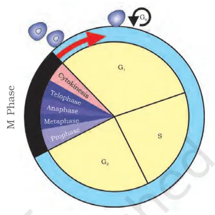
This chapter introduces the cell cycle and the two major types of cell division — mitosis and meiosis — as presented in NCERT Class 11 Biology. Introduction: cells reproduce by an ordered series of events called the cell cycle, which ensures growth, maintenance, and reproduction. Importance: understanding the cell cycle is fundamental to topics such as growth and development, tissue repair, asexual and sexual reproduction, genetic variation, and disorders such as cancer. Key themes: phases of the cell cycle (G1, S, G2, M), detailed events and stages of mitosis and meiosis, regulation of the cell cycle (checkpoints, cyclins/CDKs), cytokinesis (plant vs animal), and the significance of meiotic processes (synapsis, crossing over, independent assortment) in generating genetic variation. What the student will learn: recognise and describe each stage of interphase, mitosis and meiosis; explain mechanisms that ensure accurate chromosome segregation; compare mitosis and meiosis; understand the biological significance of cell division; identify examples and practical observations (e.g., onion root tip mitosis, pollen mother cell meiosis); and appreciate causes and consequences of errors…
Learning Objectives
- Define cell cycle, interphase, mitosis and meiosis with appropriate technical terms
- Describe the events and significance of G1, S and G2 phases of interphase
- Explain sequential stages of mitosis and the role of each stage in nuclear division
- Draw and label clear diagrams of mitosis and meiosis indicating chromosome behavior at each stage
- Differentiate mitosis and meiosis with respect to purpose, number of divisions, chromosome number and genetic outcome
- Explain the processes and key events of meiosis I and meiosis II, including synapsis and crossing over
- Illustrate formation of chiasmata and explain its role in genetic recombination and variation
- Explain the role of cell-cycle regulators (cyclins, CDKs) and checkpoints in control of cell division
Topics in this chapter
27 topics · tap a topic title to jump straight to it.
Introduction to Cell Division
What is Cell Division?
Cell division is the biological process by which a parent cell divides into two or more daughter cells. It is fundamental to growth, development, tissue repair, asexual reproduction and gamete formation. There are two main types of eukaryotic cell division: mitosis (nuclear division producing genetically identical somatic cells) and meiosis (two successive divisions producing genetically varied haploid gametes).
Why cells divide
- Growth of multicellular organisms (more cells, larger body).
- Tissue repair and replacement (e.g., skin, blood).
- Asexual reproduction in some organisms (budding, binary fission in single-celled organisms).
- Genetic continuity and variation: mitosis maintains chromosome number; meiosis reduces chromosome number and introduces variation.
Overview of the cell cycle
The cell cycle is a controlled sequence of stages leading to cell division. It has two major parts: interphase (G1, S, G2) when the cell grows and replicates DNA, and M phase (mitosis or meiosis plus cytokinesis) when the cell divides. Checkpoints (G1/S, G2/M, spindle checkpoint) ensure correct progression.
Phases at a glance
- G1 (Gap 1): cell growth and preparation for DNA synthesis.
- S (Synthesis): DNA replication; each chromosome becomes two sister chromatids.
- G2 (Gap 2): further growth and preparation for mitosis; repair of DNA errors.
- M phase (mitosis): prophase, metaphase, anaphase, telophase — segregation of chromosomes followed by cytokinesis (division of cytoplasm).
Control and errors
Progress through the cell cycle is regulated by cyclins and cyclin-dependent kinases (CDKs). Failures in control (DNA damage not repaired, checkpoint failure) can cause uncontrolled division — cancer — or cell death. Meiosis introduces variation by recombination and independent assortment; errors can cause aneuploidy (e.g., Down syndrome).
Key points to remember
- Mitosis: produces two genetically identical diploid somatic cells (in diploid organisms).
- Meiosis: produces four genetically diverse haploid gametes; involves recombination and two divisions (Meiosis I and II).
- Cell division balances growth, replacement and heredity while being tightly regulated.
- Human skin repair: epidermal cells divide rapidly to close a wound (mitosis).
- Embryonic development: a single fertilized egg divides repeatedly to form a multicellular embryo.
- Bacterial binary fission: one bacterium divides into two genetically similar bacteria — simple cell division in prokaryotes.
- Yeast budding: a new daughter cell grows from the parent (asexual reproduction involving mitotic-like division).
- Meiosis in animals: formation of sperm and eggs, producing genetic variation in offspring.
- N = N0 × 2^n — Number of cells after n rounds of division (for synchronous binary divisions).
- N = N0 × 2^(t/τ) — Cell population after time t, where τ is the doubling (generation) time.
- n = log2(N / N0) — Number of divisions that have occurred given initial (N0) and final (N).
- τ = t / n — Average generation (doubling) time if n divisions occurred in time t.
- Mitotic index (%) = (Number of cells in mitosis / Total number of cells observed) × 100 — measure of proliferative activity.
Overview of Cell Cycle
What is the Cell Cycle? The cell cycle is an ordered series of events by which a cell grows, duplicates its DNA and divides to produce two daughter cells. It ensures accurate transmission of genetic material and coordinates growth with division.
Main phases
- Interphase (preparation for division) — consists of three subphases:
- G1 (Gap 1): cell grows, makes RNA and proteins; prepares for DNA replication. Typical length varies (several hours).
- S (Synthesis): DNA replication; each chromosome is duplicated (DNA content increases from 2C to 4C).
- G2 (Gap 2): further growth, protein synthesis, and preparation for mitosis; checks DNA replication completeness.
- M phase (Mitosis): nuclear division (prophase, metaphase, anaphase, telophase) followed by cytokinesis (cytoplasm divides) to give two daughter cells.
- G0: a quiescent/non-dividing state entered by some cells (e.g., many neurons, some liver cells). Cells in G0 are metabolically active but not cycling.
Regulation
- The cell cycle is controlled by cyclins and cyclin-dependent kinases (CDKs). Different cyclin–CDK complexes become active at specific phases to drive the cell forward.
- Checkpoints ensure fidelity: the G1/S checkpoint (is environment favorable? is DNA intact?), the G2/M checkpoint (is all DNA replicated and undamaged?), and the spindle (metaphase) checkpoint (are chromosomes properly attached to spindle?).
- DNA damage triggers repair pathways or, if damage is severe, apoptosis to prevent propagation of errors.
Key measurable concepts
- DNA content (C-value): G1 cells contain 2C DNA, S-phase cells range from 2C to 4C, and G2/M cells contain 4C.
- Mitotic index: percentage of cells in mitosis; a quick measure of proliferative activity.
- Cell cycle time (Tc): total time required for one complete cycle (varies by cell type — e.g., typical mammalian cell ~24 h; rapidly dividing embryo cells much shorter).
Biological importance
- Growth and development (embryogenesis, organ growth).
- Tissue repair and regeneration (wound healing, blood cell formation).
- Maintenance of tissue homeostasis by balancing proliferation and cell death.
- When regulation fails, uncontrolled division leads to cancer.
Methods to study the cell cycle: time-lapse microscopy, labeling of S-phase (e.g., BrdU or EdU), flow cytometry (DNA histogram), mitotic index counts, molecular assays for cyclins/CDKs.
Typical durations (approximate for many animal cells): G1 ~6–12 h, S ~6–8 h, G2 ~4–6 h, M ~0.5–1 h; total ≈ 24 h (varies widely).
Summary: The cell cycle is a tightly regulated sequence of growth, DNA replication and division. Checkpoints and regulatory proteins (cyclins/CDKs) maintain fidelity. Understanding the cell cycle explains growth, repair and diseases like cancer.
- Wound healing: skin cells near a cut are stimulated to leave G0, re-enter the cell cycle and proliferate to replace lost tissue.
- Embryonic development: very rapid cell cycles (short or absent G1/G2) allow fast increase in cell number during early cleavage stages.
- Bone marrow hematopoiesis: stem and progenitor cells actively cycle to produce large numbers of blood cells.
- Plant root meristem: root tip cells divide continuously for root growth, showing active cycles in apical meristem tissues.
- Cancer: loss of checkpoint control (e.g., mutated p53) leads to uncontrolled cell cycles and tumor growth.
- Exponential growth of cell number: N(t) = N0 × 2^(t / Tc) (N0 = initial cell number, Tc = cell cycle time, t = time)
- Doubling time relation: k = ln(2) / Tc, where k is the growth rate constant in an exponential model
- Fraction of cells in a phase = (duration of phase) / (total cell cycle time) => Duration of phase = (number of cells in that phase / total cells) × Tc
- DNA content rules: G1 = 2C, S = between 2C and 4C, G2/M = 4C
- Mitotic index (%) = (number of cells in mitosis / total number of cells observed) × 100
Cell Cycle
Definition: The cell cycle is an ordered series of events by which a cell grows, duplicates its DNA and organelles, and divides to produce two daughter cells. It ensures faithful transmission of genetic material and appropriate cell number in tissues.
Phases of the Cell Cycle
The cell cycle is broadly divided into two major periods:
- Interphase (preparatory phase) — the longest phase, when the cell grows and duplicates its DNA. It has three subphases:
- G1 (Gap 1): Cell grows, synthesises proteins and organelles; cell monitors environment to decide whether to divide.
- S (Synthesis): DNA replication occurs; each chromosome becomes two sister chromatids attached at the centromere.
- G2 (Gap 2): Further growth and synthesis of proteins (including those needed for mitosis); cell checks DNA replication for errors.
- M phase (Mitotic phase) — nuclear division (mitosis) followed by cytokinesis (division of the cytoplasm):
- Prophase: Chromosomes condense and become visible; mitotic spindle begins to form; nuclear envelope starts to break down.
- Prometaphase: Nuclear envelope fragments; spindle microtubules attach to kinetochores.
- Metaphase: Chromosomes align at the cell equator (metaphase plate).
- Anaphase: Sister chromatids separate and move toward opposite poles.
- Telophase: Chromatids arrive at poles; nuclear envelopes re-form; chromosomes decondense.
- Cytokinesis: Division of cytoplasm producing two genetically identical daughter cells (in animal cells via a contractile ring; in plant cells via cell plate formation).
Control of the Cell Cycle
- Checkpoints: G1/S (restriction point), G2/M, and spindle (metaphase) checkpoint ensure integrity of DNA, completion of replication and correct chromosome attachment.
- Molecular regulators: Cyclins and cyclin-dependent kinases (CDKs) drive progression; tumour suppressors (e.g., p53, Rb) can halt the cycle or trigger apoptosis if damage is irreparable.
Biological Significance
- Tissue growth and development, wound healing, and asexual reproduction.
- Maintenance of correct chromosome number and genomic stability.
- Misregulation leads to diseases such as cancer (uncontrolled cell division).
Typical Duration
Duration varies by cell type: early embryonic cells may divide very rapidly (minutes to hours), many mammalian cells have a ~24-hour cycle, while some differentiated cells (neurons, muscle) are permanently in G0 (quiescent) or divide rarely.
Measurement and Experimental Notes
- Mitotic index measures the proportion of cells in mitosis and is used to estimate proliferative activity of a tissue.
- Flow cytometry (DNA content histograms) can estimate percentage of cells in G1, S and G2/M phases.
- Wound healing: skin cells near a cut re-enter the cell cycle from G0, proliferate to replace lost cells and close the wound.
- Embryonic development: rapid, synchronous cell cycles without G1 and G2 in early embryos (cleavage divisions) to increase cell number quickly.
- Hair and intestinal epithelium renewal: high mitotic index tissues where constant cell division replaces lost cells.
- Cancer: mutations in checkpoint genes (e.g., p53) remove control, causing uncontrolled cell division and tumour formation.
- Plant meristems: apical meristem cells divide actively to enable stem and root growth.
- Mitotic index (%) = (Number of cells in mitosis / Total number of observed cells) × 100
- Exponential growth of cell population: Nt = N0 × 2^(t/T) (Nt = cell number at time t, N0 = initial cell number, T = generation time/doubling time)
- Doubling time (T) from observations: T = t × log(2) / log(Nt/N0) (or using natural logs: T = t × ln2 / ln(Nt/N0))
Interphase
Interphase is the longest and metabolically most active stage of the cell cycle that occurs between two successive cell divisions. It is the period when the cell grows, carries out normal functions and prepares for mitosis (or meiosis) by duplicating its DNA and organelles. Interphase is conventionally divided into three subphases: G1 (Gap 1), S (Synthesis), and G2 (Gap 2). Some cells may exit the cycle into a non-dividing state called G0.
Subphases and major events
- G1 phase: Cell grows in size, synthesizes RNAs and proteins, increases organelles (mitochondria, ribosomes), and carries out normal metabolic functions. The cell passes through the restriction point (R) in late G1; passing this commits the cell to DNA replication. Key regulatory proteins: Cyclin D/CDK4-6 and Cyclin E/CDK2.
- S phase: DNA replication occurs; each chromosome is duplicated by semi-conservative replication producing sister chromatids. DNA content doubles (from 2C to 4C in a diploid cell). Replication origins are activated and replication forks progress. Histones and chromatin assembly factors are produced to package newly replicated DNA.
- G2 phase: Further cell growth and synthesis of proteins (including tubulin) required for mitosis, completion of centrosome duplication, repair of any DNA replication errors. Checkpoint mechanisms verify that DNA replication is complete and undamaged before entry into mitosis. Key regulatory proteins: Cyclin A/CDK2 (late S/G2) and Cyclin B/CDK1 (prepares for M).
Checkpoints and molecular control
- Major checkpoints: G1/S checkpoint (ensures conditions and genome integrity before replication) and G2/M checkpoint (ensures replication completeness and DNA integrity before mitosis).
- Cyclins and Cyclin-dependent kinases (CDKs) form complexes that drive transitions; their regulated synthesis and degradation control phase progression.
- Tumour suppressors (e.g., p53, Rb) and DNA repair pathways monitor damage and can arrest the cycle or trigger apoptosis if damage is irreparable.
Physical and cytological features
- Chromatin is less condensed (euchromatin predominates) so the nucleus appears large with visible nucleolus(s).
- Chromosomes are not individually visible under light microscopy (they appear as diffuse chromatin) until prophase of mitosis.
Variability and biological significance
- Interphase duration varies widely: embryonic cells have extremely short or absent G1/G2 (rapid divisions), while many somatic cells have long interphases; some cells (neurons, muscle cells) enter G0 and stop dividing long-term.
- Interphase occupies the majority of the cell cycle (often ~90% in many somatic cells), allowing growth, DNA replication fidelity and preparation for division—critical to maintain genomic stability.
How interphase is studied
- Flow cytometry measures DNA content to distinguish G1 (2C), S (intermediate 2C-4C) and G2/M (4C) populations.
- Labeling with nucleotide analogs (e.g., BrdU, 3H-thymidine) marks S-phase cells.
- Mitotic index and phase-index calculations estimate durations of phases in a population.
Summary sentence: Interphase is the preparatory, biosynthetically active phase of the cell cycle (G1→S→G2) in which the cell grows, duplicates its DNA and organelles, and checks for damage so that mitosis can proceed correctly.
- Onion root tip (meristem) cells: actively dividing with a relatively short interphase—used in labs to observe cell cycle phases.
- Human neurons: permanently exit the cycle into G0 after differentiation—very long or indefinite interphase (no division).
- Hepatocytes (liver cells): normally in G0 but can re-enter G1 and progress through interphase to divide after liver injury.
- Early vertebrate embryos (cleavage stages): extremely short interphase with rapid S and M phases and reduced or missing G1/G2, enabling fast cell divisions.
- Cancer cells: often have shortened or deregulated interphase checkpoints (loss of p53/Rb control), causing uncontrolled division.
- Mitotic index (%) = (Number of cells in mitosis / Total number of cells observed) × 100
- Duration of a phase (e.g., interphase or any subphase) = (Number of cells in that phase / Total number of cells) × Total cell cycle time (Tc).
- If Tc (total cell cycle time) is known: Duration of interphase = Tc – Duration of mitosis. (Or use phase fraction × Tc.)
- Labeling index (for S-phase) = (Number of labeled cells after pulse / Total cells) × 100. Then S-phase duration ≈ (labeling index/100) × Tc (with appropriate experimental design).
- DNA content relation in diploid cells: G1 = 2C, S = between 2C and 4C, G2 = 4C (where C = haploid DNA content).
M Phase — Mitosis
Overview
M phase (mitotic phase) is the part of the eukaryotic cell cycle in which duplicated chromosomes are precisely separated into two daughter nuclei and the cell divides (cytokinesis). M phase = mitosis (nuclear division) + cytokinesis (cytoplasmic division). The main purpose is to produce genetically identical daughter cells and maintain chromosome number.
Main stages of mitosis and key events
- Prophase: Chromatin condenses into visible chromosomes (each with two sister chromatids joined at centromere). Mitotic spindle begins to form from centrosomes; nucleolus disappears.
- Prometaphase: Nuclear envelope breaks down (in most eukaryotes). Spindle microtubules attach to kinetochores on sister chromatids; chromosomes begin to move.
- Metaphase: Chromosomes align at the metaphase plate (equatorial plane). Tension from opposite spindle fibers stabilizes bi-oriented attachments.
- Anaphase: Cohesin proteins cleaved by separase after APC/C-mediated securin degradation; sister chromatids separate and are pulled toward opposite poles (now considered individual chromosomes).
- Telophase: Chromosomes decondense, nuclear envelopes re-form around each set, nucleoli reappear, spindle disassembles.
- Cytokinesis: Division of the cytoplasm. In animal cells an actin-myosin contractile ring forms a cleavage furrow; in plant cells a cell plate forms from Golgi-derived vesicles, which becomes a new cell wall.
Molecular regulation and checkpoints
- Entry into M phase is driven by MPF (Maturation-Promoting Factor) = Cyclin B + Cdk1. MPF activation triggers chromosome condensation, nuclear envelope breakdown, and spindle formation.
- The spindle assembly checkpoint (SAC) monitors correct kinetochore–microtubule attachments and tension. Unattached kinetochores inhibit APC/C, delaying anaphase.
- APC/C (Anaphase Promoting Complex/Cyclosome) ubiquitylates securin and cyclin B. Degradation of securin frees separase to cleave cohesin; destruction of cyclin B inactivates Cdk1 and helps exit M phase.
Chromosome and DNA content notation (brief)
Use symbols to track chromosome number and DNA content:
n = haploid chromosome number, 2n = diploid chromosome number. c = DNA content per haploid set. After S phase DNA doubles (2n, 2c → 2n, 4c). After mitosis each daughter cell returns to 2n, 2c.
Significance
- Growth, tissue repair, replacement of cells (e.g., skin, blood), asexual reproduction in some organisms, and development rely on mitosis.
- Accurate mitosis preserves genome stability; errors (nondisjunction, mis-segregation) can cause aneuploidy and disease (e.g., cancer).
Important distinctions: Mitosis maintains chromosome number and produces genetically identical somatic cells. It differs from meiosis, which reduces chromosome number and generates genetic variation for gametes.
- Human skin renewal: basal epidermal cells divide by mitosis to replace damaged or shed skin cells.
- Wound healing: mitotic divisions of nearby cells increase cell number to close a cut.
- Plant growth at meristems: shoot and root apical meristems undergo mitosis to produce new tissues.
- Asexual vegetative propagation: e.g., potato tuber formation involves mitotic cell divisions to form new plants.
- Regeneration: Hydra and planaria regenerate lost parts by mitotic proliferation of stem-like cells.
- Mitotic index = (Number of cells in mitosis / Total number of cells observed) × 100%
- DNA content change during cell cycle: before S-phase = 2n, 2c → after S-phase (before anaphase) = 2n, 4c → after mitosis (each daughter) = 2n, 2c
- Chromatids/chromosomes relationship: Number of chromatids = 2 × (number of chromosomes) during G2 and metaphase (because each chromosome has two sister chromatids)
Cytokinesis
Definition: Cytokinesis is the physical process that divides the cytoplasm of a parent cell into two daughter cells at the end of nuclear division (mitosis or meiosis). It usually begins during anaphase and completes after telophase.
General role: While mitosis/meiosis separate chromosomes, cytokinesis partitions the cytoplasm, organelles and plasma membrane so each daughter cell becomes a separate, independent cell.
Mechanisms (eukaryotes vs prokaryotes)
- Animal cells: A contractile ring composed of actin filaments and myosin II forms beneath the plasma membrane at the cell equator. Contraction of this ring produces a cleavage furrow that deepens until the cell is pinched into two.
- Plant cells: Rigid cell walls prevent furrowing. Instead, Golgi-derived vesicles carrying cell wall materials accumulate at the center along the phragmoplast (microtubule structure). Vesicles fuse to form a cell plate, which expands outward and becomes the new cell wall and middle lamella between the daughter cells.
- Fungi and some protists: Variations include budding (asymmetric division) and septum formation; cytokinesis often uses actin/myosin-based machinery or species-specific septins.
- Bacteria: Cytokinesis occurs by binary fission. An FtsZ protein ring (tubulin homolog) forms at mid-cell, guiding septum formation and cell wall synthesis to split the cell.
Regulation and timing
- Cytokinesis is coordinated with mitotic exit to ensure chromosomes are fully segregated before severing the cell.
- Key regulatory proteins include RhoA (controls contractile ring assembly in animal cells) and FtsZ (bacterial division).
Biological significance and consequences of failure
- Ensures correct cell size, organelle distribution and genome segregation. Proper cytokinesis is essential for growth, development, tissue repair and asexual reproduction.
- Failure of cytokinesis can cause multinucleated cells, aneuploidy or polyploidy. In multicellular organisms such failures are linked to developmental defects and can contribute to tumorigenesis.
- Early embryonic divisions: rapid successive cytokineses produce many cells from the fertilized egg.
- Wound healing: skin and connective tissue cells divide and undergo cytokinesis to replace damaged cells.
- Plant root growth: meristematic cells divide and form new cell walls by the cell plate mechanism.
- Yeast budding: unequal cytokinesis produces a small daughter bud and a larger mother cell.
- Bacterial binary fission (e.g., E. coli): one cell divides into two genetically identical daughter cells using an FtsZ-guided septum.
- Exponential cell growth (binary fission or idealised mitotic population): N = N0 * 2^(t / T) ; where N is cell number at time t, N0 is initial cell number, and T is doubling time.
- Surface area and volume (relevance: SA/V affects nutrient exchange and can drive division): Sphere => SA = 4πr^2 , V = (4/3)πr^3 , so SA/V = 3 / r.
- Cube model (useful for simple geometry): SA = 6a^2 , V = a^3 , so SA/V = 6 / a.
- Approximate cell-cycle fraction (typical eukaryotic cell: approximate values) => G1 ~ 40%, S ~ 35%, G2 ~ 15%, M (including cytokinesis) ~ 10% (values vary by cell type).
Mechanism of DNA Replication
DNA replication is the biological process by which a cell makes an exact copy of its DNA before cell division. It occurs during the S phase of the cell cycle and ensures that each daughter cell receives an identical genome. Replication is accurate, semi-conservative, bidirectional, and enzyme-driven.
Key features
- Semi-conservative: Each daughter DNA molecule contains one original (parental) strand and one newly synthesized strand.
- Bidirectional: Replication proceeds in two directions from an origin of replication, forming two replication forks.
- Directionality: DNA polymerases synthesize new strands only in the 5' to 3' direction, using the parental strand as template.
Main stages and molecular players
- Origin recognition and unwinding: Replication begins at specific sequences called origins of replication. Initiator proteins bind the origin and recruit helicase. Helicase unwinds the double helix, producing single-stranded DNA templates.
- Stabilization of single strands: Single-strand binding proteins (SSBs in prokaryotes; RPA in eukaryotes) coat and stabilize unwound single strands to prevent reannealing and degradation.
- Relieving topological stress: Topoisomerases (DNA gyrase in bacteria) relieve supercoiling ahead of the fork by making transient cuts in DNA and rejoining strands.
- Primer synthesis: DNA polymerases cannot start new strands de novo. Primase synthesizes short RNA primers that provide a free 3' OH group for extension.
- Elongation: DNA polymerases add nucleotides complementary to the template in the 5' to 3' direction. On the leading strand synthesis is continuous; on the lagging strand synthesis is discontinuous, producing Okazaki fragments.
- Primer removal and gap filling: RNA primers are removed (RNase H and/or flap endonucleases), gaps are filled by DNA polymerase, and nicks are sealed by DNA ligase.
- Proofreading and error correction: Many DNA polymerases have 3' to 5' exonuclease (proofreading) activity that removes misincorporated nucleotides. Post-replicative mismatch repair corrects remaining errors.
Important enzymes and complexes
- Helicase: unwinds DNA double helix.
- Single-strand binding proteins (SSB/RPA): stabilize unwound DNA.
- Primase: synthesizes RNA primers.
- DNA polymerase III (prokaryotes) / DNA polymerases alpha, delta, epsilon (eukaryotes): main replicative polymerases.
- Sliding clamp (beta-clamp in bacteria, PCNA in eukaryotes): increases polymerase processivity.
- Clamp loader (gamma complex in bacteria, RFC in eukaryotes): loads sliding clamp onto DNA.
- DNA ligase: joins Okazaki fragments by sealing nicks.
- Topoisomerase (DNA gyrase): removes supercoils.
Leading and lagging strands
Because DNA strands are anti-parallel and polymerization occurs only in 5' to 3' direction, one template (leading) is synthesized continuously toward the replication fork, while the other (lagging) is synthesized away from the fork as short Okazaki fragments that are later joined.
Experimental evidence
The Meselson-Stahl experiment demonstrated semi-conservative replication by tracking heavy and light nitrogen isotopes in newly synthesized DNA, showing that after one round of replication, hybrid molecules are produced and after two rounds both hybrid and light molecules appear, consistent with semi-conservatism.
Differences between prokaryotes and eukaryotes
- Origin number: prokaryotes usually have a single origin per circular chromosome (e.g., OriC in E. coli); eukaryotes have many origins on linear chromosomes to replicate large genomes in reasonable time.
- Polymerases and accessory proteins differ in identity and complexity.
- Okazaki fragment length is generally longer in prokaryotes and shorter in eukaryotes.
Biological significance
Accurate and timely DNA replication is essential for growth, development, tissue renewal, and heredity. Errors in replication can lead to mutations, some of which contribute to genetic diseases and cancer. Cells employ multiple fidelity mechanisms to maintain genome stability.
- Polymerase chain reaction (PCR) mimics DNA replication in vitro: a thermostable DNA polymerase synthesizes new strands from primers and a template, amplifying specific DNA segments.
- E. coli replicates its 4.6 million base pair genome in about 40 minutes under optimal conditions; replication begins at OriC and proceeds bidirectionally with two replication forks.
- Antibiotics such as fluoroquinolones target bacterial DNA gyrase (a topoisomerase), inhibiting DNA replication and killing bacteria.
- Replication errors that escape proofreading and repair can lead to point mutations. Accumulation of such mutations in genes controlling cell division can contribute to cancer development.
- DNA doubling at S phase: DNA_after = 2 × DNA_before
- Approximate replication time: T = Genome size (bp) / (replication rate (bp/s) × number of active forks). To convert seconds to minutes divide by 60.
- Example calculation: For a circular genome of 4.6 × 10^6 bp replicated bidirectionally with two forks at 1000 bp/s each, T ≈ (4.6 × 10^6) / (1000 × 2) = 2300 s ≈ 38.3 min.
Cell Division in Prokaryotes
Overview: Prokaryotic cell division is primarily by binary fission — a simple, rapid process in which one cell divides into two genetically identical daughter cells. Prokaryotes (bacteria and archaea) lack a membrane-bound nucleus and mitotic spindle; their single circular chromosome replicates and segregates before septum formation.
Key steps of binary fission
- Initiation of replication: Replication begins at a single origin of replication (oriC in many bacteria). Initiator proteins (e.g., DnaA in E. coli) assemble and start bidirectional replication.
- Chromosome replication: The two replication forks proceed in opposite directions around the circular chromosome until termination (ter region). Replication is carried out by the replisome.
- Chromosome segregation and cell elongation: Newly replicated origins move apart while the cell elongates. Partitioning systems (e.g., Par proteins in plasmids, and various factors for chromosomes) help segregate DNA copies so each daughter cell receives one copy.
- Z-ring formation and septum assembly: The tubulin-like protein FtsZ polymerizes at mid-cell to form the Z-ring, recruiting other cell division proteins (the divisome). The ring constricts and peptidoglycan synthesis forms a septum that divides the cell.
- Cytokinesis: Completion of septum and cell envelope separation yields two independent daughter cells.
Regulation and spatial control
- Min system: MinC/MinD/MinE prevents Z-ring formation at cell poles and helps position the ring at mid-cell.
- Nucleoid occlusion (e.g., SlmA): prevents septation over unsegregated chromosomes.
- Coordinated timing: DNA replication, segregation and septum formation are tightly coordinated so division normally occurs after replication begins and sufficient segregation has occurred.
Variations and other modes
- Budding: Some bacteria (e.g., planctomycetes) reproduce by budding rather than binary fission.
- Plasmid segregation: Small circular plasmids use partitioning systems (ParA/ParB) to ensure inheritance.
Biological significance
Fast prokaryotic division enables rapid population increases (e.g., Escherichia coli can double every ~20 minutes under optimal conditions). This underlies processes such as infection spread, fermentation, wastewater treatment, and rapid evolution under selective pressure.
Simple example calculation (concept)
If a bacterium has generation time g = 30 min, starting population N0 = 1×10⁶, after t = 120 min (4 generations): n = t/g = 4 so N = N0 × 2n = 1×106 × 16 = 1.6×10⁷.
Note: Prokaryotes do not undergo mitosis or meiosis; sexual genetic exchange (conjugation, transformation, transduction) is separate from the division process.
- Escherichia coli dividing by binary fission — generation time ~20 minutes under optimal lab conditions.
- Bacillus subtilis using FtsZ-dependent septation to form two daughter cells.
- Plasmid partitioning in low-copy plasmids using ParA/ParB systems to ensure stable inheritance during cell division.
- Some Planctomycetes reproduce by budding instead of classical binary fission.
- N = N0 × 2^n (N = population after n generations; N0 = initial population)
- n = t / g (n = number of generations; t = total time; g = generation time or doubling time)
- g = t / n (solve for generation time)
- \[Nt = N0 × e^{μt} (continuous exponential growth\]\[μ = specific growth rate\]\[in reciprocal time)\]
- μ = ln(2) / g (relationship between specific growth rate μ and doubling time g)
- \[Alternative discrete form: N = N0 × 2^{t/g}\]
Mitosis (Karyokinesis)
Definition: Mitosis (karyokinesis) is the orderly process of nuclear division in which a parent cell divides its replicated chromosomes equally into two genetically identical daughter nuclei. It is a part of the M phase of the cell cycle and ensures maintenance of chromosome number across somatic cells.
Overall purpose and features: Mitosis maintains the diploid chromosome number (2n → 2n) and distributes one copy of each sister chromatid to each daughter nucleus. It is essential for growth, tissue repair, and asexual reproduction. Mitosis is typically followed by cytokinesis (division of the cytoplasm), but karyokinesis refers specifically to nuclear events.
Stages of mitosis (major events):
- Prophase: Chromatin condenses into visible chromosomes, each consisting of two sister chromatids joined at the centromere. The mitotic spindle begins to form from centrosomes (animal cells) or microtubule organizing centers. The nucleolus fades.
- Prometaphase: The nuclear envelope breaks down (nuclear membrane fragments). Spindle microtubules attach to kinetochores at centromeres of chromosomes. Chromosomes begin moving.
- Metaphase: Chromosomes align at the metaphase plate (cell equator) with sister chromatids oriented to opposite spindle poles; spindle assembly checkpoint ensures all kinetochores are properly attached.
- Anaphase: Cohesin proteins holding sister chromatids are cleaved, and sister chromatids (now individual chromosomes) are pulled toward opposite poles by shortening kinetochore microtubules and motor proteins.
- Telophase: Chromosomes arrive at poles and decondense back into chromatin. Nuclear envelopes re-form around each set of chromosomes, nucleoli reappear, and the spindle disassembles.
Key control points and regulation: Transition into and progression through mitosis is tightly regulated by cyclin-dependent kinases (CDKs) and mitotic cyclins (e.g., M-cyclin/CDK1 activity). Checkpoints (especially the spindle assembly checkpoint) prevent chromosome mis-segregation.
Differences in plant and animal mitosis: Animal cells have centrosomes and centrioles organizing the spindle and form a cleavage furrow during cytokinesis. Many higher plant cells lack centrioles; a cell plate forms during cytokinesis. However, the basic nuclear events of karyokinesis are conserved.
Significance: Mitosis ensures genetic stability (identical daughter genomes), provides cells for organismal growth and regeneration, and enables some forms of asexual reproduction.
Common classroom example (human somatic cell): A human diploid somatic cell (2n = 46) in G1 (pre-S) has 46 chromosomes and 2C DNA content. After S-phase each chromosome has two sister chromatids (still 46 chromosomes but 4C DNA). Mitosis separates chromatids resulting in two daughter cells each with 46 chromosomes (2n) and 2C DNA.
Note: Karyokinesis refers strictly to the nuclear division; cytokinesis (division of the cytoplasm) usually follows to create two separate daughter cells.
- Wound healing: mitosis replaces lost or damaged skin cells to close a cut.
- Growth and development: mitosis increases cell number during childhood and organ growth.
- Replacement of blood cells: bone marrow cells divide mitotically to produce red blood cells and many white blood cells.
- Asexual reproduction in some organisms: e.g., single-celled protists (binary fission) or vegetative propagation in plants use mitotic divisions.
- Plant meristem activity: root and shoot apical meristems divide mitotically to extend roots and shoots.
- Chromosome number remains constant: diploid somatic cell → 2n (parent) → mitosis → two daughters each 2n.
- DNA content (C notation): G1 phase: 2C → after S-phase (G2): 4C → after mitosis: each daughter 2C.
- Human example: G1: 46 chromosomes (2n = 46), 2C DNA → G2: 46 chromosomes, 4C DNA (92 chromatids) → after mitosis: each daughter 46 chromosomes, 2C DNA.
- Chromatids in G2 = 2 × (number of chromosomes) = 2 × 2n (e.g., human G2: 92 chromatids = 2 × 46).
Meiosis
Definition & purpose: Meiosis is a specialized type of cell division that reduces the chromosome number by half to produce haploid (n) gametes from a diploid (2n) parental cell. Its main purposes are (1) reduction of chromosome number (reductional division) and (2) generation of genetic variation through independent assortment and crossing over.
Overview: Meiosis consists of two successive nuclear divisions—Meiosis I (reductional) and Meiosis II (equational)—following a single round of DNA replication.
- Meiosis I (reductional division)
- Prophase I: Homologous chromosomes pair (synapsis) and form bivalents; crossing over (genetic recombination) occurs at chiasmata. Prophase I is divided into leptotene, zygotene, pachytene, diplotene and diakinesis in textbook detail — pachytene is when crossing over is most active; diplotene shows visible chiasmata.
- Metaphase I: Bivalents (paired homologues) align on the metaphase plate; orientation is random (independent assortment).
- Anaphase I: Homologous chromosomes separate and move to opposite poles (sister chromatids remain together).
- Telophase I & cytokinesis: Chromosomes may decondense and the cell divides to form two haploid cells (each chromosome still consisting of two sister chromatids).
- Meiosis II (equational division)
- Prophase II: Chromosomes condense again (if decondensed).
- Metaphase II: Chromosomes (sister chromatids) align at the metaphase plate.
- Anaphase II: Sister chromatids separate and move to opposite poles.
- Telophase II & cytokinesis: Nuclear envelopes re-form and cells divide, producing four haploid, genetically distinct daughter cells (gametes or spores).
Key consequences:
- Chromosome number halves: 2n → n.
- Four genetically non-identical haploid cells are produced from one diploid meiocyte.
- Genetic variation arises via independent assortment, crossing over, and random fertilization.
Important terms: synapsis, bivalent, tetrad, chiasmata, synaptonemal complex, homologues, sister chromatids, nondisjunction (failure of separation), gametogenesis (spermatogenesis and oogenesis).
Errors & clinical relevance: Nondisjunction during meiosis I or II can lead to aneuploidy (e.g., trisomy 21 → Down syndrome). Abnormal recombination can produce deletions or duplications of chromosome segments.
Summary (CBSE focus): Remember Meiosis I = reductional (homologues separate), Meiosis II = equational (sister chromatids separate). Prophase I is long and critical because of crossing over and pairing.
- Human gametogenesis: Spermatogenesis (testes) yields four functional sperm from each primary spermatocyte; oogenesis (ovary) yields one ovum and polar bodies due to unequal cytokinesis.
- Flowering plants: Microsporogenesis in anther produces microspores (pollen) and megasporogenesis in ovule produces megaspores—both by meiosis.
- Yeast sporulation: A diploid yeast cell undergoes meiosis to form four haploid spores (useful model for meiosis studies).
- Genetic consequence example: With independent assortment and crossing over, siblings (except identical twins) are genetically distinct—variation that allows natural selection.
- Clinical example: Nondisjunction in human meiosis leading to trisomy 21 (Down syndrome) or monosomy X (Turner syndrome).
- Number of genetically different gametes (from independent assortment only) = 2^n, where n = haploid chromosome number. Example: human n = 23 → 2^23 = 8,388,608 possible gametes.
- Number of possible zygotes from two parents (ignoring crossing over) = (2^n) × (2^n) = 2^(2n). For humans: 2^46 ≈ 7.04 × 10^13 (~70 trillion).
- Recombination frequency (%) = (number of recombinant offspring / total number of offspring) × 100. 1% recombination ≈ 1 centiMorgan (cM).
- Ploidy change representation: Diploid primary meiocyte (2n, each chromosome has 2 chromatids) --Meiosis I→ two haploid cells (n, each chromosome 2 chromatids) --Meiosis II→ four haploid gametes (n, each chromosome 1 chromatid).
Spindle Apparatus and Centrosomes
Overview
The spindle apparatus is the dynamic microtubule-based structure that segregates chromosomes during mitosis and meiosis. Centrosomes are the principal microtubule organizing centers (MTOCs) in most animal cells that nucleate and help organize the bipolar spindle.
Centrosome: structure and cycle
- Structure: A centrosome usually contains two orthogonally arranged centrioles (each made of nine triplet microtubules) surrounded by an electron-dense pericentriolar material (PCM) that contains γ-tubulin ring complexes for microtubule nucleation.
- Duplication cycle: Centrosomes duplicate once per cell cycle — centriole biogenesis begins in S phase, producing a daughter centriole next to each mother centriole; by G2 each centrosome has a centriole pair; during prophase/prometaphase the two centrosomes separate to form the two spindle poles.
Spindle apparatus: components
- Microtubules (MTs): polymers of α/β-tubulin. Three functional classes in the spindle:
- Kinetochore microtubules — attach chromosome kinetochores to spindle poles.
- Polar (interpolar) microtubules — overlap at spindle midzone and push poles apart.
- Astral microtubules — radiate toward cortex and help position the spindle.
- Motor proteins and MAPs: Kinesins (usually plus-end directed) and dyneins (minus-end directed) move along MTs, producing sliding forces (pole separation, chromosome movement) and regulating MT dynamics. Microtubule-associated proteins (MAPs) stabilize/destabilize MTs.
- Kinetochores: protein complexes on centromeres that capture kinetochore MTs and generate forces for movement.
Mechanisms of chromosome movement
- Metaphase: Chromosomes are bi-oriented — kinetochores attached to opposite poles — and aligned at the metaphase plate. Tension across kinetochores stabilizes attachments.
- Anaphase A: Shortening of kinetochore microtubules (depolymerization at kinetochores/poles) pulls chromosomes toward poles.
- Anaphase B: Sliding of interpolar microtubules (plus-end kinesins push overlaps) and astral pulling (dynein at cortex) separate the poles further.
Regulation and checkpoints
The spindle assembly checkpoint (SAC) monitors proper kinetochore attachments and tension; it delays anaphase until all chromosomes are correctly attached. Errors in spindle function lead to mis-segregation and aneuploidy (common in cancers).
Variations and exceptions
Many plant cells lack centrioles; they assemble spindles using dispersed MTOCs and chromatin-mediated nucleation. Mammalian oocytes frequently form acentrosomal spindles (no centrosomes) — special adaptations for meiosis.
Biological and medical relevance
Spindle-targeting drugs are widely used: colchicine and vinblastine destabilize MTs, taxol (paclitaxel) stabilizes MTs — both disrupt mitosis and are used as anti-cancer agents or in plant breeding (colchicine induces polyploidy). Defects in centrosome number or function cause chromosome instability, contribute to tumorigenesis, and underlie some developmental disorders (e.g., primary microcephaly).
Summary
Centrosomes organize the spindle poles in many animal cells. The spindle apparatus, composed of different classes of microtubules and motor proteins, captures, aligns and separates chromosomes to ensure accurate cell division.
- Onion (Allium) root tip mitosis — typical laboratory example showing a clear bipolar spindle and phases of mitosis.
- Early animal embryo cleavage — rapid mitoses use prominent spindles to partition cytoplasm and chromosomes.
- Colchicine treatment in plant breeding — binds tubulin, disrupts spindle formation and can block chromosome segregation leading to polyploidy.
- Anti-cancer drugs (taxol, vincristine) — target microtubules/spindle to arrest rapidly dividing tumour cells in mitosis.
- Oocyte meiosis in many animals — acentrosomal spindle formation relies on chromatin-driven MT nucleation instead of centrosomes.
- Centrosome amplification in cancer cells — extra centrosomes may cause multipolar spindles and chromosome mis-segregation.
- Mitotic index (MI) = (Number of cells in mitosis / Total number of cells observed) × 100
- Percentage of time in a phase = (Duration of that phase / Total cell cycle duration) × 100
- Population growth rate (exponential) r = ln(Nt / N0) / t ; Doubling time Td = ln(2) / r (useful when relating cell proliferation to mitotic activity)
Genetic Variation Mechanisms
What is genetic variation? Genetic variation is the presence of differences in DNA sequences among individuals of a species. It is the raw material for evolution and is essential for adaptation and survival.
Main mechanisms that generate genetic variation
- Mutation: Any permanent change in the DNA sequence. Mutations can be:
- Point mutations (base substitutions): silent, missense, or nonsense.
- Insertions and deletions (may cause frameshifts if in coding regions).
- Chromosomal mutations: large-scale changes such as deletions, duplications, inversions and translocations.
Mutations in germ cells are heritable; somatic mutations affect only the individual. Mutations produce new alleles and can be neutral, deleterious, or beneficial.
- Recombination during meiosis:
- Crossing over (prophase I): homologous chromosomes exchange segments, creating new allele combinations on a chromosome.
- Independent assortment (metaphase I): different chromosome pairs align independently, producing different combinations of maternal and paternal chromosomes in gametes.
These processes shuffle existing alleles to make genetically unique gametes.
- Random fertilization: The random union of two genetically unique gametes further increases variation—each zygote is a unique combination of alleles.
- Polyploidy and genome duplication: Particularly in plants, whole-genome duplication (polyploidy) creates instant genetic novelty and can produce new species (e.g., wheat, many crop plants).
- Chromosomal rearrangements: Large-scale rearrangements (duplication, deletion, inversion, translocation) can alter gene dosage, disrupt genes, or create new gene combinations affecting phenotype.
- Gene flow (migration): Movement of alleles between populations by migration increases genetic diversity in the receiving population and reduces differences between populations.
- Genetic drift: Random changes in allele frequencies, especially strong in small populations (founder effect, bottleneck), leading to loss or fixation of alleles independently of selection.
- Lateral (horizontal) gene transfer: Transfer of genes between unrelated organisms (common in bacteria), producing rapid acquisition of new traits (e.g., antibiotic resistance).
Consequences and significance: Variation allows natural selection to act; beneficial variants increase in frequency, deleterious ones tend to be removed, and neutral variants drift. Variation underlies evolution, adaptation to environments, and biodiversity.
Distinguishing points:
- Mutations create new alleles; recombination and independent assortment reshuffle them.
- Somatic mutations affect only the organism; germline mutations are heritable.
- Population-level processes (gene flow, drift, selection) change allele frequencies over generations.
Note for Class 11 level: emphasize types of mutation, stages of meiosis responsible for recombination, and simple population concepts (drift, gene flow). Use diagrams of meiosis and chromosomal changes to visualise mechanisms.
- Sickle-cell anemia: a point mutation in the beta-globin gene produces a variant allele; heterozygotes have partial resistance to malaria (example of a mutation with both harmful and beneficial effects).
- Antibiotic resistance in bacteria: mutations plus horizontal gene transfer spread resistance genes rapidly among bacterial populations.
- Down syndrome (trisomy 21): nondisjunction (failure of chromosome separation) during meiosis leads to an extra chromosome in the zygote (chromosomal variation).
- Industrial melanism in peppered moths: pre-existing variation (dark and light forms) shifted in frequency due to selection after environmental changes.
- Polyploid crops: bread wheat (hexaploid) arose by hybridization plus genome duplication, increasing genetic material and traits useful in agriculture.
- Dihybrid cross in pea plants (Mendel): independent assortment produces 9:3:3:1 phenotypic ratio in F2, illustrating recombination of parental traits.
- Number of possible gamete combinations from independent assortment = 2^n (n = number of chromosome pairs). Example: humans n = 23 → 2^23 ≈ 8.4 million possible gametes per individual.
- Probability of one specific gamete genotype = 1 / 2^n (assuming no crossing over and equal segregation).
- Hardy–Weinberg equilibrium (simple population genetics): p + q = 1 and p^2 + 2pq + q^2 = 1, where p and q are allele frequencies of two alleles at a locus (used to describe genetic variation in an ideal population).
- Mutation rate (basic expression): μ = (number of new mutations) / (number of sites × generations). (Often expressed as mutations per base per generation.)
Chromosome Structure and Types
Overview
Chromosomes are long, thread-like structures made of DNA and proteins that carry genetic information. In eukaryotes they appear most condensed during mitosis and meiosis (metaphase chromosomes) and are less condensed (chromatin) during interphase.
Molecular building blocks
- DNA double helix: the primary polymer of nucleotides; length per base pair ≈ 0.34 nm.
- Histone proteins: core histones (H2A, H2B, H3, H4) form an octamer around which DNA is wrapped; H1 binds linker DNA.
- Nucleosome: ~146 bp of DNA wrapped ≈ 1.65 turns around a histone octamer; with linker DNA (~20–80 bp) this gives ≈200 bp per nucleosome (the 'beads-on-a-string' structure).
Levels of chromatin organization (increasing compaction)
- DNA double helix (2 nm diameter).
- ‘Beads-on-a-string’: nucleosomes (≈11 nm fibre).
- 30 nm fibre: higher-order folding of nucleosomes into a compact fibre.
- Looped domains: 30 nm fibre forms loops anchored to a protein scaffold (scaffold/matrix-associated regions, SARs/MARs).
- Metaphase chromosome: maximally condensed structure visible under light microscope — two sister chromatids joined at the centromere.
Structural features of a metaphase chromosome
- Sister chromatids: two identical copies produced by DNA replication, joined at the centromere until anaphase.
- Centromere: constricted region that forms the kinetochore; essential for chromosome movement during cell division.
- Telomeres: terminal repetitive sequences (vertebrates: TTAGGG)n that protect chromosome ends and are involved in ageing and genome stability.
- p and q arms: p = short arm, q = long arm (arms are defined relative to the centromere).
- Secondary constrictions / NORs: nucleolar organizer regions associated with rRNA genes (often on acrocentric chromosomes).
Chromatin states
- Euchromatin: less condensed, transcriptionally active, gene-rich.
- Heterochromatin: highly condensed, transcriptionally inactive or low activity; includes constitutive heterochromatin (e.g., pericentromeric repeats) and facultative heterochromatin (developmentally regulated).
Types of chromosomes by centromere position (These are standard categories used in karyotyping):
- Metacentric: centromere near the middle; arms roughly equal in length.
- Submetacentric: centromere off-center; p arm shorter than q arm.
- Acrocentric: centromere very near one end, producing a very short p arm; human acrocentrics include chromosomes 13, 14, 15, 21, 22.
- Telocentric: centromere at the terminal end (not present in normal human chromosomes but seen in some species).
Other ways to classify
- Autosomes vs sex chromosomes: autosomes are non-sex chromosomes; sex chromosomes determine sex (e.g., X and Y in humans).
- Monocentric vs holocentric: monocentric chromosomes have a single localized centromere (most eukaryotes); holocentric chromosomes (e.g., Caenorhabditis elegans) have centromeric activity distributed along the entire length.
Functional importance
Centromeres and kinetochores ensure correct segregation during mitosis and meiosis. Telomeres protect ends from degradation and end-to-end fusion; loss of telomere function leads to genome instability. Chromosome structure (e.g., heterochromatinization) regulates gene expression.
Key points to remember
- Chromosomes = DNA + proteins; organization is hierarchical (nucleosome → 30 nm fibre → loops → chromosome).
- Centromere position defines morphological types (metacentric, submetacentric, acrocentric, telocentric).
- Euchromatin = active; heterochromatin = mostly inactive.
- Chromosome abnormalities (aneuploidy, translocations) have clinical consequences (e.g., Down syndrome, Philadelphia chromosome).
- Human karyotype: 46 chromosomes in somatic (diploid) cells — 22 pairs of autosomes + 1 pair of sex chromosomes (XX or XY).
- Down syndrome: trisomy 21 — presence of an extra copy of chromosome 21 (autosomal aneuploidy) causing characteristic phenotype.
- Philadelphia chromosome in chronic myelogenous leukemia (CML): reciprocal translocation t(9;22)(q34;q11) creates BCR-ABL fusion gene.
- Acrocentric chromosomes in humans: chromosomes 13, 14, 15, 21 and 22 have short p arms with rRNA gene clusters (NORs).
- Holocentric example: Caenorhabditis elegans chromosomes lack a single centromere; spindle attaches along the length.
- Length of DNA (nm) = number_of_base_pairs × 0.34 nm per base pair
- Example: human haploid genome ≈ 3.2 × 10^9 bp → length ≈ 3.2×10^9 × 0.34 nm ≈ 1.09 × 10^9 nm ≈ 1.09 m (diploid ≈ 2.18 m)
- Approx. nucleosome spacing: ≈ 200 bp per nucleosome (146 bp wrapped + ~50 bp linker)
- Compaction ratio = (length of naked DNA) / (length of condensed chromosome). For metaphase chromosomes this is roughly 5,000–10,000-fold (order of magnitude).
Molecular Basis and Regulation of Cell Cycle
Overview
The eukaryotic cell cycle is an ordered sequence of events (G1 → S → G2 → M) that ensures duplication and equal partitioning of the genome. Molecular control relies on cyclical production and destruction of regulatory proteins and on checkpoint systems that verify completion and fidelity of earlier events before permitting progression.
Core molecular players
- Cyclins — regulatory proteins whose concentrations vary periodically. Classes: G1 (D-type), G1/S (E-type), S (A-type), M (B-type). Cyclin levels rise by transcription/translation and fall by ubiquitin-mediated proteolysis.
- Cyclin-dependent kinases (CDKs) — catalytic subunits (e.g., CDK4/6, CDK2, CDK1) that are activated by binding specific cyclins and by phosphorylation events. Active cyclin–CDK complexes phosphorylate targets that drive the cell cycle forward.
- CDK regulators — activating kinases (CAK), inhibitory kinases (Wee1/Myt1), activating phosphatases (Cdc25), and CDK inhibitors (CKIs: p21, p27, p16) that block CDK activity.
- Ubiquitin ligases — SCF (Skp–Cullin–F-box) targets G1/S cyclins and CKIs for degradation; APC/C (anaphase-promoting complex/cyclosome), activated by Cdc20 or Cdh1, ubiquitylates cyclin B and securin to allow anaphase and mitotic exit.
- Checkpoints — surveillance systems: G1/S (are conditions favorable?), intra-S (DNA replication), G2/M (is DNA replicated and undamaged?), spindle (are chromosomes properly attached?). Each has sensors (detect problem), transducers (ATM/ATR, Chk1/Chk2), and effectors (p53, Cdc25 inhibition, APC/C control).
Key molecular mechanisms
- Activation by cyclin binding: Cyclin synthesis binds and partially activates specific CDKs (e.g., Cyclin B + CDK1 = MPF).
- Regulation by phosphorylation: CDKs are regulated by inhibitory (Wee1) and activating (CAK) phosphorylation; Cdc25 phosphatase removes inhibitory phosphates to trigger abrupt entry into M-phase.
- Proteolytic inactivation: Timed ubiquitination by SCF or APC/C leads to proteasomal degradation of cyclins and other regulators, producing irreversible transitions (e.g., anaphase onset requires degradation of securin and cyclin B).
- Checkpoint signaling: DNA damage activates ATM/ATR → Chk1/Chk2 → p53 stabilization, leading to transcription of p21 (a CKI) and cell-cycle arrest; this allows repair or triggers apoptosis if damage is irreparable.
- Positive and negative feedback: Active cyclin–CDK complexes phosphorylate regulators that further activate the same CDK (positive feedback) or lead to rapid inactivation of earlier cyclins (negative feedback), producing switch-like (bistable) transitions.
Major example: M-phase promotion factor (MPF)
MPF (Cyclin B–CDK1) drives entry into mitosis: it phosphorylates condensins (chromosome condensation), lamins (nuclear envelope breakdown), microtubule-associated proteins (spindle assembly), and proteins controlling chromosome attachment. MPF activation is tightly regulated by Wee1 (inhibitory) and Cdc25 (activating) to ensure abrupt mitotic entry.
Biological consequences and regulation in tissues
Cell-type-specific regulation controls whether cells divide, differentiate, or enter G0. Stem cells and embryonic cells show rapid cycles (short or absent G1/G2), while somatic cells have longer G1 phases and are subject to growth-factor and nutrient control. Dysregulation (e.g., loss of p53, overexpression of cyclin D) can lead to uncontrolled proliferation → cancer.
Practical/clinical relevance
Many anticancer drugs target cell-cycle components or processes: microtubule poisons (taxanes, vinca alkaloids) disrupt mitotic spindle (spindle checkpoint activation); CDK inhibitors are used experimentally/clinically to block proliferation of tumor cells. Flow cytometry DNA-content analysis uses cell-cycle biology to estimate fractions of cells in G1, S and G2/M.
Summary points
- Cell-cycle progression is driven by cyclical cyclin synthesis and CDK activation, and shut down by targeted proteolysis.
- Checkpoints (G1/S, G2/M, spindle) ensure fidelity; damage sensors (ATM/ATR), transducers (Chk1/Chk2), and effectors (p53, CKIs) implement arrests or apoptosis.
- Feedback loops and irreversible proteolysis create switch-like transitions between phases.
- Embryonic cleavage divisions (e.g., frog, zebrafish): extremely rapid cycles with little or no G1/G2 phase; regulation is minimal to allow rapid increase in cell number.
- Cancer: mutations in p53 (loss of DNA-damage checkpoint) or overexpression of cyclin D/CDK4 can bypass G1 checkpoint, leading to uncontrolled proliferation.
- Chemotherapy: Taxol stabilizes microtubules and activates the spindle checkpoint, arresting cells in mitosis and triggering cell death in dividing cancer cells.
- Yeast cell-cycle mutants (cdc genes): classical genetic screens in Saccharomyces cerevisiae identified many regulators (e.g., CDC28 = CDK homolog), illustrating conserved molecular control.
- Flow cytometry DNA histogram: normal proliferating tissue shows two peaks (G1 and G2/M) with DNA-synthesis (S) between; tumor samples often show altered distributions reflecting dysregulated cycle.
- Total cell-cycle time: T = t_G1 + t_S + t_G2 + t_M
- Fraction of cells in a phase: f_phase = t_phase / T (useful for asynchronous populations)
- Exponential growth (doubling): N(t) = N0 × 2^(t / T), where T is doubling time
- Continuous growth rate form: N(t) = N0 × e^(k t), where k = ln(2) / T
Prophase I Substages and Recombination
Overview: Prophase I is the longest stage of meiosis I during which homologous chromosomes pair and exchange genetic material (recombination). Prophase I is subdivided into five substages — leptotene, zygotene, pachytene, diplotene and diakinesis — each with characteristic events that prepare chromosomes for segregation and generate genetic variation.
- Leptotene
Chromosomes begin to condense and appear as long, thin threads. Homologous chromosomes begin to search for each other. Each chromosome consists of two sister chromatids joined at the centromere.
- Zygotene
Homologous chromosomes undergo synapsis (pairing) to form bivalents (tetrads). A proteinaceous scaffold called the synaptonemal complex forms between homologues, aligning them tightly along their lengths and facilitating recombination.
- Pachytene
Synapsis is complete. This is the stage where crossing over (genetic recombination) occurs: non-sister chromatids exchange DNA segments at sites called chiasmata. Molecularly, recombination is initiated by programmed double-strand breaks (DSBs) and completed by strand invasion, formation and resolution of Holliday junctions or by synthesis-dependent strand annealing, producing crossover or non-crossover products.
- Diplotene
The synaptonemal complex disassembles and homologues begin to separate but remain attached at chiasmata, which become visible under the microscope. Chiasmata hold homologues together until anaphase I.
- Diakinesis
Final condensation of chromosomes; chiasmata terminalize (move toward ends). The nucleolus disappears and the nuclear envelope breaks down shortly after, preparing for metaphase I.
Mechanism of Recombination (concise molecular outline):
- Initiation: Spo11 (in many organisms) creates programmed double-strand breaks.
- Resection: 5' ends are resected to produce 3' single-stranded tails.
- Strand invasion: A 3' tail invades the homologous duplex, forming a displacement (D) loop.
- DNA synthesis & second end capture: New DNA is synthesized using the homolog as template; either a double Holliday junction (dHJ) forms or the D-loop is dismantled.
- Resolution: Holliday junctions are cut (resolved) to give crossover or non-crossover products. Gene conversion can accompany resolution if mismatches are repaired asymmetrically.
Biological significance:
- Generates genetic variation by creating new combinations of alleles (important for evolution and adaptation).
- Facilitates proper pairing and segregation of homologous chromosomes in meiosis I.
- Used in genetic mapping: frequency of recombination between loci reflects their physical distance (genetic map).
Errors and consequences: Faulty recombination (unequal crossing over or misrepair) can cause duplications, deletions or translocations, which underlie some genetic disorders and chromosomal abnormalities.
- Morgan's Drosophila experiments: crossing-over between linked genes allowed construction of genetic maps and demonstrated that recombination frequency correlates with physical distance.
- Human siblings differ genetically because of independent assortment and recombination during meiosis in parents; recombination shuffles parental alleles into new combinations.
- Plant breeding: crossing-over allows breeders to combine desirable alleles from two parental lines into a single offspring (creating new trait combinations).
- Unequal crossing-over can generate copy number changes—e.g., gene duplications/deletions that may cause disease or provide raw material for evolution.
- Recombination frequency (RF) = (Number of recombinant offspring / Total offspring) × 100%
- Genetic map distance (in centiMorgan, cM) ≈ RF (%) — 1 cM ≈ 1% recombination
- Number of possible combinations by independent assortment = 2^n (where n = haploid chromosome number); recombination increases variety further
- Coefficient of coincidence (c.o.c.) = Observed double crossovers / Expected double crossovers
- Interference (I) = 1 − c.o.c. (measures how one crossover affects the probability of a nearby crossover)
Cell Division and Cancer
Overview
Cell division is the process by which a parent cell divides to give rise to daughter cells. It is essential for growth, development, repair and reproduction. In multicellular organisms, the cell cycle—an ordered sequence of events—controls cell division. Cancer results when the normal controls on cell division fail, producing abnormally proliferating cells that can form tumours and invade other tissues.
The Cell Cycle (brief)
The cell cycle comprises interphase (G1, S, G2) and mitotic phase (M). During G1 the cell grows and monitors environment; S phase is DNA replication; G2 is preparation for mitosis; M phase includes mitosis (nuclear division) and cytokinesis (cytoplasm division). Some cells enter G0 (resting/quiescent state).
- Checkpoints: G1/S checkpoint (restriction point), G2/M checkpoint, and spindle checkpoint during M ensure DNA integrity and correct chromosome segregation.
- Regulators: Cyclins and cyclin-dependent kinases (Cdks) form complexes that drive the cell through cycle phases. Examples: Cyclin D/Cdk4-6 (G1 progression), Cyclin E/Cdk2 (G1/S), Cyclin B/Cdk1 (G2/M).
Mitosis vs Meiosis (concise)
Mitosis produces two genetically identical diploid daughter cells for growth and repair. Meiosis produces four genetically varied haploid gametes for sexual reproduction.
How Cancer Arises
Cancer is caused by accumulation of genetic and epigenetic changes that disrupt normal cell-cycle control, DNA repair, and apoptosis. Key features include:
- Oncogenes: Mutated or overexpressed proto-oncogenes (e.g., RAS) that promote proliferation.
- Tumour suppressor genes: Loss or inactivation of genes that normally restrain growth (e.g., TP53, RB).
- Genomic instability: Defects in DNA repair increase mutation rates.
- Evasion of apoptosis: Cancer cells avoid programmed cell death.
- Angiogenesis: Tumours stimulate blood-vessel formation (e.g., VEGF) to gain nutrients.
- Metastasis: Spread of cancer cells from primary site to distant organs; involves invasion, intravasation, survival in circulation, extravasation and colonization.
Types of Tumours
- Benign: Localised, slow-growing, well-differentiated (e.g., lipoma, fibroma); usually not life-threatening unless pressing on organs.
- Malignant (cancer): Rapid growth, invade surrounding tissue and metastasize (e.g., carcinoma, sarcoma, leukemia).
Causes and Risk Factors
- Carcinogens: tobacco smoke, UV radiation, certain chemicals, asbestos.
- Biological agents: oncogenic viruses (HPV → cervical cancer; HBV/HCV → liver cancer), Helicobacter pylori (stomach cancer).
- Genetic predisposition: inherited mutations (e.g., BRCA1/2 for breast/ovarian cancer).
- Lifestyle: diet, alcohol, obesity, sedentary life.
Diagnosis, Treatment and Prevention (class-level overview)
- Diagnosis: Biopsy (histopathology), imaging (X-ray, CT, MRI), blood tests (tumour markers).
- Treatment: Surgery, radiotherapy, chemotherapy (kills dividing cells), targeted therapy (e.g., kinase inhibitors like imatinib), immunotherapy (checkpoint inhibitors), hormone therapy.
- Prevention: Avoid carcinogens (no tobacco, sun protection), vaccinations (HPV, HBV), screening (Pap smear, mammography), healthy lifestyle.
Class 11 Focus Points
- Understand the cell cycle phases, checkpoints and the role of cyclins/Cdks.
- Know differences between mitosis and meiosis and their biological significance.
- Mechanism of cancer development: oncogenes, tumour suppressors, apoptosis evasion, angiogenesis and metastasis.
- Be able to name common carcinogens and basic treatment strategies.
Summary: Normal cell division is tightly regulated by molecular controls; cancer results when these controls fail, producing unregulated proliferation, tissue invasion and possible metastasis. Early detection and prevention reduce cancer burden, while treatments target rapidly dividing cells or specific molecular changes in tumours.
- Normal cell division: Skin epithelial cells divide by mitosis to replace cells lost from abrasion; wound healing involves proliferating fibroblasts and epithelial cells.
- Meiosis example: Formation of sperm and eggs (gametogenesis) produces haploid cells with genetic variation.
- Benign tumour: Lipoma (fat tissue tumour) — slow-growing, non-invasive.
- Malignant cancer: Lung carcinoma often linked to tobacco smoking; may metastasize to brain or bone.
- Virus-associated cancer: Human papillomavirus (HPV) infection can lead to cervical cancer; Hepatitis B/C infections increase risk of liver cancer.
- Targeted therapy example: Imatinib (Gleevec) inhibits BCR-ABL tyrosine kinase in chronic myeloid leukemia.
- Mitotic index (%) = (Number of cells in mitosis / Total number of cells observed) × 100
- Exponential growth (discrete doublings): N = N0 × 2^n, where N0 = initial cell number, n = number of cell divisions
- \[If growth is continuous: N(t) = N0 × e^{k t}\]\[where k is the growth rate constant\]
- Doubling time (continuous) t_d = ln(2) / k
- Number of divisions n = log2(N / N0)
Genetic Variation
Definition: Genetic variation refers to the differences in DNA sequences among individuals of the same species. These differences produce variation in traits (phenotypes) and provide the raw material for evolution and natural selection.
Main sources and mechanisms:
- Mutations: Permanent changes in the DNA sequence. Types include point mutations (base substitution), insertions/deletions (frameshift), and chromosomal mutations (duplication, deletion, inversion, translocation). Mutations can be spontaneous or induced (mutagens).
- Meiotic recombination (crossing over): During prophase I of meiosis homologous chromosomes form bivalents and exchange segments at chiasmata. This produces new allele combinations on the same chromosome.
- Independent assortment: During metaphase I, different orientations of homologous chromosome pairs lead to random distribution of maternal and paternal chromosomes into gametes. For a species with haploid number n, this alone gives 2^n possible gametes.
- Random fertilization: Any sperm can fuse with any egg, multiplying the number of possible offspring genotypes (combination of two gamete sets).
- Chromosomal changes & polyploidy: Nondisjunction (leading to aneuploidy, e.g., trisomy 21) and whole-genome duplications (polyploidy) especially in plants create large-scale variation.
- Gene flow and horizontal transfer: Movement of genes between populations (migration) or between organisms (bacteria: conjugation, transformation) introduces new alleles.
How meiosis creates variation (summary): During meiosis I, homologous chromosomes pair (synapsis) and may exchange segments (crossing over). At metaphase I, the random orientation of bivalents yields independent assortment. Together with random fertilization, these mechanisms dramatically increase possible genotypes in a species.
Biological significance: Genetic variation is essential for adaptation and evolution. It allows populations to respond to environmental changes, underlies selective breeding in agriculture, and explains variation in disease susceptibility. However, some variations can be harmful (genetic disorders).
Notes on scale: For organisms with large chromosome numbers, independent assortment alone produces an enormous number of gamete combinations; crossing over increases diversity even further. Many variations are neutral, some are beneficial, and some are deleterious.
- Sickle cell anaemia: a point mutation in the ß-globin gene (Glu → Val) — example of a single base substitution with strong phenotypic effect.
- Down syndrome (Trisomy 21): nondisjunction during meiosis leading to an extra chromosome — example of chromosomal variation.
- ABO blood group system: multiple alleles and codominance produce variation in human populations.
- Antibiotic resistance in bacteria: mutations and horizontal gene transfer produce variants that survive antibiotic selection.
- Wheat (Triticum aestivum): hexaploid origin by polyploidy — example of genome duplication creating new plant species/traits.
- Number of possible gametes by independent assortment = 2^n (n = haploid chromosome number). Example: humans n = 23 → 2^23 ≈ 8.4 million gametes per parent.
- Number of possible zygotes from two parents (independent assortment only) = 2^n × 2^n = 2^(2n). For humans: ≈ 7 × 10^13 combinations.
- Recombination frequency (RF) = (number of recombinant progeny ÷ total progeny) × 100%. RF (%) approximates map distance in centiMorgans (1% RF ≈ 1 cM) for linked genes.
- Hardy–Weinberg relations (population-level context): p + q = 1 and p^2 + 2pq + q^2 = 1 (where p and q are allele frequencies of two-allele system).
Comparison: Mitosis vs Meiosis
Overview
Mitosis and meiosis are two types of nuclear division. Mitosis produces genetically identical daughter cells for growth, repair and asexual reproduction. Meiosis produces genetically varied haploid gametes (or spores) for sexual reproduction; it involves two successive divisions and reduces chromosome number by half.
- Purpose: Mitosis – growth, tissue repair, asexual reproduction. Meiosis – formation of gametes/spores and generation of genetic variation.
- Location: Mitosis – somatic cells. Meiosis – germ cells (ovaries, testes) and spore-producing tissues in plants.
- Number of divisions: Mitosis – one division (equational). Meiosis – two divisions (reductional Meiosis I, then equational Meiosis II).
- Number of daughter cells: Mitosis – 2 daughter cells. Meiosis – 4 daughter cells (from one diploid cell).
- Chromosome number: Mitosis – daughter cells are diploid (2n) if parent is 2n. Meiosis – daughter cells are haploid (n).
- Genetic identity: Mitosis – daughters are genetically identical (barring mutations). Meiosis – daughters are genetically diverse due to crossing over and independent assortment.
- Pairing and recombination: Mitosis – homologous chromosomes do not pair; no crossing over. Meiosis – homologous chromosomes pair (synapsis) during prophase I and undergo crossing over (recombination) at chiasmata.
- Alignment at metaphase: Mitosis – sister chromatids line up at metaphase plate. Meiosis I – homologous chromosome pairs line up (independent assortment). Meiosis II – sister chromatids line up.
- DNA replication: Occurs once before both processes (S phase). Meiosis does not have an additional S between Meiosis I and II.
Stage-level differences (brief)
Mitosis: prophase (condensation), metaphase (chromosomes at equator), anaphase (sister chromatids separate), telophase and cytokinesis (two diploid nuclei).
Meiosis: Meiosis I (prophase I with leptotene–zygotene–pachytene–diplotene–diakinesis, crossing over; metaphase I homologous pairs align; anaphase I homologues separate) → two cells. Meiosis II resembles mitosis (sister chromatids separate) → four haploid cells.
Genetic consequences
Independent assortment: random orientation of homologous pairs in metaphase I gives 2^n possible chromosome combinations (n = haploid chromosome number). Crossing over creates new allele combinations on chromosomes. Together these ensure offspring genetic variation.
Key points to remember
1) Mitosis maintains chromosome number and genome identity; meiosis halves chromosome number and increases genetic diversity. 2) Crossing over and synapsis are unique to meiosis (prophase I). 3) One S-phase precedes both processes; meiosis has two nuclear divisions without a second S-phase.
- Human growth and wound healing: skin cell proliferation by mitosis restores tissue integrity after injury.
- Amoeba and many single-celled organisms reproduce by mitosis-like binary fission (asexual reproduction).
- Spermatogenesis and oogenesis in animals: meiosis produces sperm and eggs (human males produce millions of sperm via meiosis; females produce one egg per meiotic cycle).
- Pollen (microspores) and ovules (megaspore lineage) formation in flowering plants: meiosis in anthers and ovules produces haploid spores.
- Vegetative propagation (potato tubers, stem cuttings) involves mitotic cell divisions producing genetically identical plants.
- Mitosis (chromosome count): 1 cell (2n) → 2 cells (2n each).
- Meiosis (chromosome count): 1 cell (2n) → Meiosis I → 2 cells (n each, chromatids still paired) → Meiosis II → 4 cells (n each, single chromatids).
- Genetic combinations by independent assortment: number of possible gamete chromosome combinations = 2^n (n = haploid number).
- DNA content notation (C-value) through stages (starting diploid cell): before S: 2n, 2C → after S: 2n, 4C → after Meiosis I: n, 2C → after Meiosis II: n, 1C → after Mitosis: 2n, 2C (daughter cells).
- Cell count after k mitotic divisions from one cell: 2^k cells.
Comparison of Mitosis and Meiosis
Overview: Mitosis and meiosis are two types of nuclear division that ensure continuity of life. Mitosis produces genetically identical daughter cells for growth, repair and asexual reproduction; meiosis produces genetically diverse haploid gametes for sexual reproduction.
Key differences (summary)
| Feature | Mitosis | Meiosis |
|---|---|---|
| Purpose | Growth, tissue repair, asexual reproduction | Formation of gametes/spores and generation of genetic variation |
| Where it occurs | Somatic (body) cells | Germ cells (gonads) or meiocytes |
| Number of divisions | One (prophase, metaphase, anaphase, telophase) | Two (Meiosis I: reductional; Meiosis II: equational) |
| Number of daughter cells | Two | Four |
| Genetic identity | Daughters genetically identical to parent (barring mutation) | Daughters genetically different due to crossing over and independent assortment |
| Ploidy of daughters (starting from 2n) | Diploid (2n) | Haploid (n) |
| Pairing of homologues | No synapsis | Homologous chromosomes pair (synapsis) in prophase I forming bivalents/tetrads |
| Crossing over / recombination | No | Yes — at chiasmata in prophase I |
| Separation in anaphase | Sister chromatids separate | Meiosis I: homologues separate; Meiosis II: sister chromatids separate |
| Chromosome alignment at metaphase | Individual chromosomes line up at metaphase plate | Homologous pairs (bivalents) line up in metaphase I; single chromosomes in metaphase II |
DNA content (C-value) and chromosome number changes
- Notation: n = number of distinct chromosomes (haploid number). C = DNA content per haploid genome.
- Starting diploid cell (G1): 2n, 2C.
- After S phase (before division): 2n, 4C (each chromosome has 2 sister chromatids).
- Mitosis: division yields two cells each 2n, 2C.
- Meiosis: after Meiosis I (reductional) cells are n, 2C (each chromosome still two chromatids); after Meiosis II cells are n, 1C.
Phases — brief comparison
- Prophase: Mitosis — chromosome condensation, spindle forms. Meiosis — homologous pairing (synapsis) and crossing over during prophase I (leptotene, zygotene, pachytene, diplotene, diakinesis).
- Metaphase: Mitosis — single chromosomes align; Meiosis I — homologous pairs align; Meiosis II — chromatids align like mitosis.
- Anaphase: Mitosis — sister chromatids separate; Meiosis I — homologues separate; Meiosis II — sister chromatids separate.
Biological significance
- Mitosis maintains genome stability, allows tissue renewal and asexual propagation.
- Meiosis reduces chromosome number by half and increases genetic variation (beneficial for evolution and adaptation) via independent assortment and crossing over.
Important notes for CBSE Class 11
- Remember the distinctive events: synapsis & chiasmata are unique to meiosis (prophase I).
- Meiosis I is termed reductional division (2n → n); meiosis II is equational (chromatid separation).
- Use C-value bookkeeping to track DNA content through cell cycle and divisions.
- Mitosis: Healing of a scraped knee — skin epithelial cells proliferate by mitosis to replace lost cells.
- Mitosis: A single-celled organism like Amoeba reproduces asexually by mitotic division (binary fission equivalent).
- Meiosis: Human gametogenesis — spermatogenesis produces four sperm cells and oogenesis produces one egg and polar bodies.
- Meiosis: Pollen formation in flowering plants (microsporogenesis) where a diploid microsporocyte undergoes meiosis to give four haploid microspores.
- Meiosis in agriculture/breeding: Crossing two plant varieties leads to novel allele combinations in offspring due to independent assortment and recombination.
- Number of possible combinations from independent assortment (ignoring crossing over): 2^n (n = haploid chromosome number). Example: for humans n=23, combinations ≈ 2^23 ≈ 8.4 million.
- DNA content progression (C-value) for a diploid cell: G1: 2n, 2C → after S: 2n, 4C → after mitosis: 2n, 2C.
- DNA content progression through meiosis for a diploid: Start 2n, 2C → after S: 2n, 4C → after Meiosis I: n, 2C → after Meiosis II: n, 1C.
- Number of daughter cells: Mitosis → 2; Meiosis → 4.
- Number of nuclear divisions: Mitosis → 1; Meiosis → 2.
Cell Division in Prokaryotes and Unicellular Eukaryotes
Overview
Cell division is the process by which a single cell divides into two or more daughter cells. Prokaryotes (bacteria, archaea) and unicellular eukaryotes (yeast, amoeba, protozoa) use different mechanisms because of structural differences: prokaryotes lack a nucleus and membrane-bound organelles, while eukaryotes have a nucleus and complex mitotic machinery.
Prokaryotic Cell Division (Binary Fission)
- Definition: Binary fission is an asexual process in which one cell divides into two genetically identical daughter cells.
- Main steps:
- Initiation of replication: Replication begins at a single origin (oriC in E. coli). Proteins such as DnaA open the origin and form the replisome.
- DNA replication: Bidirectional replication proceeds until termination region (ter). Replication can overlap with previous rounds when growth is very fast.
- Segregation: Newly replicated chromosomes are moved apart; partitioning proteins (ParAB in some bacteria) and physical forces help segregate DNA.
- Septum formation and cytokinesis: FtsZ protein assembles a contractile Z-ring at mid-cell, recruiting divisome proteins to synthesize septal cell wall and divide the cell.
- Cell cycle organization (bacterial context): Often described in phases B (time between birth and start of replication), C (chromosome replication), and D (time between termination and cell division). Overlapping cycles are common in fast-growing bacteria.
- Regulation: Cell size, nutrient status, and DNA replication controls (DnaA availability, methylation state in E. coli) regulate timing.
Unicellular Eukaryotic Cell Division
- Mitosis (typical in amoeba, many protists): Follows eukaryotic cell cycle phases G1 > S > G2 > M. Mitosis has distinct stages: prophase, prometaphase, metaphase, anaphase, telophase, followed by cytokinesis. Chromosomes condense, nuclear envelope may break down (open mitosis) or remain intact (closed mitosis in some yeasts), spindle microtubules segregate chromatids.
- Budding (example: Saccharomyces cerevisiae): A daughter cell forms as a small outgrowth (bud) on the mother cell. The nucleus divides and one nucleus migrates into the bud; cytokinesis separates them. Budding is asymmetric division.
- Multiple fission / Schizogony: The nucleus divides several times before the cytoplasm divides, producing many daughter cells simultaneously (seen in some algae and protozoa; schizogony occurs in Plasmodium species).
- Binary fission in some unicellular eukaryotes: Many protozoa (e.g., Paramecium) use a mitotic-like binary fission where mitosis occurs and the cell divides into two similar cells.
- Checkpoints and regulation: Eukaryotic cell cycle is controlled by cyclins and cyclin-dependent kinases (CDKs) and checkpoints at G1/S, G2/M and spindle assembly ensure correct DNA replication and chromosome segregation.
Key differences summarized
- Prokaryotes: no nucleus, no mitotic spindle, division by binary fission with FtsZ-based septation.
- Unicellular eukaryotes: nucleus, mitosis (spindle apparatus), diverse methods (budding, binary fission with mitosis, multiple fission).
Practical and biological importance
- Understanding bacterial division helps target antibiotics (e.g., drugs that block cell wall synthesis or FtsZ-function conceptually).
- Yeast budding is exploited in biotechnology (brewing, baking, recombinant protein production).
- Escherichia coli: typical prokaryotic binary fission; rapid doubling under optimal conditions (~20–30 min).
- Bacillus subtilis: binary fission with well-studied FtsZ ring formation and septum synthesis.
- Saccharomyces cerevisiae (baker's yeast): reproduces by budding; used in fermentation and as a model eukaryote.
- Schizosaccharomyces pombe: a fission yeast that divides by medial binary fission (useful model for eukaryotic cell cycle studies).
- Amoeba proteus: unicellular eukaryote that divides by mitosis and cytokinesis.
- Paramecium: undergoes mitotic-like binary fission during asexual reproduction.
- N = N0 × 2^n (N = final population, N0 = initial population, n = number of generations)
- n = t / T (n = number of generations, t = total time, T = generation time or doubling time)
- T = t / n (generation time)
- \[N = N0 × e^{kt} (continuous growth\]\[k = specific growth rate)\]
- k = ln(2) / T (relates specific growth rate k to doubling time T)
- T_d = ln(2) / μ (alternate notation: doubling time T_d and growth constant μ)
Significance of Cell Division
Cell division is the process by which a parent cell divides to form two or more daughter cells. Its significance is central to life — it enables growth, development, reproduction, maintenance and evolution of organisms.
- Growth and development: Repeated mitotic divisions convert a single-celled zygote into a multicellular embryo and, later, a whole organism. Organ and body size increase by increase in cell number rather than cell size alone.
- Repair and replacement: Damaged or worn-out cells and tissues are replaced by new cells produced by mitosis. Examples include wound healing, skin and intestinal epithelium renewal, and bone marrow producing blood cells.
- Reproduction: Asexual reproduction uses mitotic or binary fission divisions to produce genetically identical offspring (bacteria, many unicellular eukaryotes, vegetative propagation in plants). Sexual reproduction requires meiosis to produce haploid gametes (sperm and egg) so that fertilization restores diploidy.
- Genetic continuity and variation: Mitosis preserves chromosome number and transmits an identical genome to daughter cells, ensuring genetic continuity. Meiosis reduces chromosome number (2n → n) and, through crossing-over and independent assortment, generates genetic variation essential for evolution and adaptation.
- Maintenance of optimal cell size (surface area : volume): Because volume increases faster than surface area as a cell grows, frequent division keeps cells small enough for efficient exchange of gases, nutrients and wastes across the membrane.
- Regeneration and vegetative propagation: Some organisms can regrow lost parts (e.g., planaria, hydra) or form new individuals from vegetative parts (plant cuttings, bulbs) via controlled cell division and differentiation.
- Physiological balance and homeostasis: Controlled proliferation and programmed cell death (apoptosis) maintain tissue architecture and organismal health.
- Clinical significance: Dysregulation of cell division causes diseases—excessive division leads to cancer; insufficient division or faulty mitosis leads to degenerative disorders. Mitotic index and related measurements help assess cell proliferation in diagnostics.
Short summary: Cell division is essential for growth, repair, reproduction, genetic continuity and variation, maintenance of cell function, and evolutionary adaptation.
- Embryonic development: A zygote undergoes many mitotic divisions to form a multicellular embryo.
- Wound healing: Skin cells divide to close cuts and replace damaged tissue.
- Blood cell production: Hematopoietic stem cells in bone marrow divide to produce RBCs, WBCs and platelets.
- Vegetative propagation in plants: Plant cuttings, runners and tubers produce new plants by mitotic divisions.
- Binary fission in bacteria: A single bacterium divides into two identical cells, causing rapid population increase (e.g., food spoilage).
- Gametogenesis: Meiosis in testes and ovaries forms haploid gametes (sperm and eggs) for sexual reproduction.
- Population doubling: N = N0 × 2^n (N0 = initial cell number, n = number of divisions)
- Relating time and divisions: n = t / T (t = total time, T = duration of one division/doubling time)
- Combined time formula: N = N0 × 2^(t/T)
- Mitotic index: Mitotic index (%) = (Number of cells in mitosis / Total number of cells observed) × 100
- Sphere surface area: SA = 4πr^2
- Sphere volume: V = (4/3)πr^3
Amitosis and Special Types of Division
Amitosis (Direct Nuclear Division)
Definition: Amitosis is a simple, direct form of nuclear division in which the nucleus divides by constriction or cleavage without formation of a spindle, chromosomal condensation, or the typical mitotic phases (prophase, metaphase, anaphase, telophase). It is sometimes called direct division.
Features:
- No visible chromosomes or mitotic spindle.
- Nuclear envelope usually remains intact or splits by constriction.
- Genetic material may be distributed roughly equally but segregation is not as precise as mitosis.
- Common where rapid or simple division suffices (often in some unicellular eukaryotes or specialized nuclei).
Mechanism (generalised): nucleus elongates → constriction at centre → cleavage of nucleus into two daughter nuclei → cytokinesis may or may not follow.
Special Types of Nuclear/Cell Division
Several variants of the canonical mitotic cycle occur in nature to meet special functional needs. Important types are:
1) Endoreduplication (Endoreplication)
Definition: Repeated rounds of DNA replication (S phase) without entering mitosis, producing very high DNA content in a single nucleus. Chromosomes do not segregate; instead many chromatids remain aligned or paired.
Biological significance: Produces polytene or giant chromosomes and increases gene copy number for high metabolic/output demands.
Example: Salivary gland cells of Drosophila with polytene chromosomes.
2) Endomitosis
Definition: A modified mitosis in which chromosomes replicate and may partially condense, but either mitosis is aborted (no anaphase/separation) or cytokinesis fails. Result is a single nucleus with increased ploidy (or multinucleate cell if nuclear envelope divides).
Biological significance: Cells become polyploid to meet high metabolic needs or to produce large amounts of cytoplasmic products.
Example: Megakaryocytes in bone marrow become polyploid by endomitosis to form platelets.
3) Syncytial or Coenocytic Division
Definition: Repeated nuclear divisions without cytokinesis produce many nuclei sharing a common cytoplasm (a syncytium or coenocyte).
Examples: Early Drosophila embryo (syncytial blastoderm), plasmodial slime molds (Physarum) and some algae (Caulerpa) where many nuclei coexist in one cell.
4) Other Related Phenomena
- Polyploidy (endopolyploidy): Cells with increased chromosome sets as a result of endoreduplication/endomitosis. Common in plant tissues, hepatocytes, and insect tissues.
- Multinucleation by cell fusion: Skeletal muscle fibres are multinucleate because myoblasts fuse (not by nuclear division), but functionally similar to syncytia.
When and why these occur
- Rapid growth or high biosynthetic demand (more gene copies → more transcripts/protein).
- Special developmental programs (e.g., early insect embryogenesis, platelet formation).
- Some unicellular eukaryotes use simpler mechanisms (amitosis) for rapid population increase.
Key differences (summary)
- Mitosis: ordered chromosome condensation, spindle, equal segregation → two genetically identical nuclei.
- Amitosis: no spindle, no visible chromosomes, approximate partitioning of genetic material.
- Endoreduplication/Endomitosis: produce polyploid nuclei (increased DNA content) by skipping mitosis or aborting mitosis.
- Syncytial divisions: many nuclei share one cytoplasm due to absence of cytokinesis.
- Amitosis: Macronucleus division in ciliates such as Paramecium (macronucleus divides amitotically).
- Endoreduplication: Polytene chromosomes in Drosophila salivary gland cells (many DNA replication cycles without mitosis).
- Endomitosis: Megakaryocytes in bone marrow become highly polyploid by endomitosis before producing platelets.
- Syncytial division: Early embryonic divisions in Drosophila (nuclear divisions without cytokinesis create a syncytial blastoderm).
- Coenocytic condition: Plasmodial slime moulds (Physarum) and some algal thalli (Caulerpa) have multinucleate plasmodia formed by repeated nuclear divisions without cell division.
- DNA content after k rounds of endoreduplication: DNA_final = DNA_initial × 2^k
- Ploidy after k rounds (if starting ploidy = n): Ploidy_final = n × 2^k
- Nuclei in syncytial nuclear cycles (assuming doubling each cycle): N(t) = N(0) × 2^t (t = number of nuclear division cycles)
Key Terms and Concepts
Overview: The cell cycle is the ordered series of events by which a cell grows and divides to produce two daughter cells. It has two major periods: Interphase (growth and DNA replication) and M-phase (nuclear division — mitosis or meiosis — plus cytokinesis). Regulation of the cycle ensures accurate genome duplication and distribution.
- Cell cycle: Complete sequence of events from one cell division to the next (G1 → S → G2 → M).
- Interphase: Phase between divisions composed of:
- G1 (Gap 1): Cell grows, produces RNA and proteins; prepares for DNA synthesis.
- S phase (Synthesis): DNA replication; each chromosome duplicates to form two sister chromatids.
- G2 (Gap 2): Further growth, checks for DNA replication errors, prepares mitotic machinery.
- M phase: Nuclear division + cytokinesis.
- Mitosis: Division producing two genetically identical diploid somatic cells. Stages — prophase, prometaphase, metaphase, anaphase, telophase.
- Meiosis: Two successive divisions (meiosis I and II) producing four genetically varied haploid gametes. Key events: synapsis, crossing over, reductional division (meiosis I).
- Chromosome terms:
- Sister chromatids: The two identical copies produced by DNA replication, joined at the centromere.
- Homologous chromosomes: A pair of chromosomes (one maternal, one paternal) carrying same genes in same order but possibly different alleles.
- Centromere: Constricted region where sister chromatids are joined; kinetochore forms here for spindle attachment.
- Spindle apparatus: Microtubule structure that separates chromosomes; centrosomes/centrioles organize spindle microtubules in animal cells.
- Checkpoints: Control points that verify cell is ready to proceed: G1/S (DNA integrity), G2/M (DNA replication complete), spindle assembly checkpoint (chromosomes properly attached to spindle).
- Cyclins and CDKs: Proteins (cyclins) and cyclin-dependent kinases drive progression by phosphorylation of target proteins; their levels/activity oscillate through the cycle.
- DNA content and ploidy notation:
- n = haploid chromosome number; 2n = diploid chromosome number.
- C = relative DNA content. Example (diploid somatic cell): before S = 2n, 2C; after S = 2n, 4C. After mitosis each daughter = 2n, 2C. After meiosis I = n, 2C; after meiosis II = n, C.
- Crossing over / chiasma: Exchange of genetic material between non-sister chromatids during prophase I of meiosis — increases genetic variability.
- Independent assortment: Random orientation of homologous pairs in metaphase I producing 2^n possible combinations of chromosomes in gametes (n = haploid number).
- Cytokinesis: Physical division of cytoplasm — cleavage furrow (animal cells) or cell plate formation (plant cells).
- Binary fission: Prokaryotic cell division — DNA replication and division without mitosis (e.g., E. coli).
- Mitotic index: Fraction of cells in mitosis in a population — used to estimate proliferation; elevated in rapidly dividing tissues or tumors.
- Apoptosis: Programmed cell death — important during development and to remove damaged cells. Different from necrosis (uncontrolled cell death).
Practical significance / Regulation: Proper regulation prevents genomic instability. Failure of checkpoints or aberrant cyclin/CDK activity can lead to uncontrolled proliferation (cancer). Meiosis ensures halving of chromosome number for sexual reproduction and generates genetic diversity.
Important conceptual notes:
- Mitosis conserves chromosome number; meiosis reduces it by half.
- DNA content (C) and chromosome number (n) must be distinguished — chromosomes may have replicated (higher C) yet chromosome count (n) unchanged until segregation.
- Checkpoints act as quality-control stations; cyclin-CDK complexes are the molecular engines.
- Wound healing and skin regeneration — rapid mitotic divisions in epithelial cells close the wound.
- Embryonic cleavage — repeated mitosis without significant growth produces a multicellular embryo.
- Gametogenesis (spermatogenesis and oogenesis) — meiosis produces haploid sperm and eggs with genetic variation via crossing over.
- Bacterial binary fission (E. coli) — DNA replicates and cell divides without mitosis; population doubles in each generation.
- Liver regeneration — hepatocytes re-enter cell cycle (G1 → S → M) to replace lost tissue.
- Cancer — loss of checkpoint control or overactive cyclins/CDKs causes uncontrolled cell proliferation.
- DNA content progression (diploid 2n cell): before S = 2n, 2C → after S = 2n, 4C → after mitosis (each daughter) = 2n, 2C; after meiosis I = n, 2C; after meiosis II = n, C.
- Number of possible gamete combinations by independent assortment: 2^n (where n = haploid chromosome number).
- Mitotic index (MI) = (Number of cells in mitosis / Total number of observed cells) × 100.
- Population doubling (exponential growth): Nt = N0 × 2^(t/td) where Nt = cell number at time t, N0 = initial cell number, td = doubling (generation) time. Rearranged: td = t × log(2) / log(Nt/N0).
Regulation of the Cell Cycle
Overview
The cell cycle is a highly ordered series of events by which a cell grows and divides. Regulation of the cell cycle ensures that DNA is accurately duplicated and distributed, that damaged DNA is repaired or the cell is eliminated, and that cell division occurs only when appropriate.
Key control points (checkpoints)
- G1 checkpoint (restriction point): Assesses cell size, nutrients, growth factors and DNA integrity. Passing this point commits the cell to DNA synthesis (S phase).
- G2 checkpoint: Confirms completion of DNA replication and checks for DNA damage before mitosis.
- Metaphase (M) checkpoint: Ensures all chromosomes are properly attached to the spindle before anaphase begins.
Molecular regulators
Primary molecular regulators are cyclins and cyclin-dependent kinases (CDKs):
- Cyclins: Regulatory proteins whose concentrations rise and fall during the cell cycle. Different cyclins act at different stages (e.g., G1 cyclins, S cyclins, M cyclins).
- CDKs: Serine/threonine kinases that are activated when bound to the appropriate cyclin. Active cyclin–CDK complexes phosphorylate target proteins to drive cell-cycle transitions.
- Maturation Promoting Factor (MPF): A key M-phase cyclin–CDK complex (cyclin B + CDK1) whose activation triggers entry into mitosis. MPF is inactivated when cyclin B is ubiquitinated and degraded.
Activation and inactivation mechanisms
- Phosphorylation: CDK activity is regulated by activating and inhibitory phosphorylations.
- Ubiquitin-mediated proteolysis: Timely degradation of cyclins (via the proteasome) resets the cycle.
- CDK inhibitors (CKIs): Proteins (e.g., p21, p27) that block cyclin–CDK activity to halt progression when needed.
Tumour suppressors and proto-oncogenes
- p53: A major guardian of the genome; p53 is stabilised by DNA damage and can induce cell-cycle arrest (allowing repair), senescence, or apoptosis if damage is irreparable. p53 upregulates p21 (a CKI).
- Rb (retinoblastoma protein): In G1, hypophosphorylated Rb binds E2F transcription factors preventing S-phase gene expression; phosphorylation by cyclin–CDK releases E2F and allows progression.
- Proto-oncogenes vs oncogenes: Proto-oncogenes (e.g., cyclins, growth factor receptors) promote cell division in normal form; mutations that cause constitutive activation convert them into oncogenes, leading to uncontrolled proliferation.
External and physical controls
- Growth factors: Extracellular signals (e.g., EGF) stimulate cells to pass the G1 checkpoint.
- Contact inhibition & density-dependent inhibition: Many normal cells stop dividing when they contact neighboring cells.
- Anchorage dependence: Some cells require attachment to the extracellular matrix to divide.
Outcome of failed regulation
Failure of regulation can lead to uncontrolled proliferation (cancer), or excessive cell death. Many cancer treatments (chemotherapy, radiation) exploit differences in cell-cycle regulation—targeting rapidly dividing cells or specific cycle phases.
Summary of mechanism (stepwise)
- External signals/growth factors → induction of G1 cyclins.
- Cyclin–CDK complexes phosphorylate targets (e.g., Rb) → passage through G1 to S.
- S-phase cyclins/ CDKs promote DNA replication.
- G2/M cyclins accumulate → MPF activation → mitosis.
- Cyclin degradation and CKI activity ensure orderly exit and reset.
- Wound healing: Growth factors (e.g., PDGF, EGF) stimulate local cells to re-enter cell cycle and proliferate to repair tissue.
- Embryonic development: Rapid, tightly regulated cell divisions build tissues and organs; cyclin/CDK control ensures correct timing.
- Liver regeneration: Hepatocytes re-enter the cell cycle after partial hepatectomy under control of growth factors and cyclin–CDK regulation.
- Cancer: Mutations in p53, Rb or overactive oncogenes disrupt checkpoints leading to uncontrolled cell division and tumour formation.
- Chemotherapy/radiation therapy: These treatments damage DNA or disrupt mitosis, activating checkpoints and apoptosis preferentially in rapidly dividing cancer cells.
- Plant meristems: Continuous cell division at root and shoot tips is regulated by plant hormones (auxins, cytokinins) analogous to animal growth factors.
- \[Cell population after t time: N(t) = N0 × 2^{t / Td}\]\[where N0 = initial cell number\]\[Td = doubling time (cell cycle length for proliferating population).\]
- \[Number of divisions (generations): n = t / Td → N = N0 × 2^{n}.\]
- Doubling time from counts: Td = (t × log 2) / log(N / N0).
- Instantaneous growth rate (per unit time): r = ln 2 / Td (relates doubling time to exponential growth rate).
- Fraction of cells in a given phase = phase duration / total cell cycle time (e.g., labeling index: fraction in S = Ts / Tc).
Abnormalities and Consequences
Overview: "Abnormalities" in the context of the cell cycle and cell division are errors in the process of DNA replication, chromosome segregation (mitosis/meiosis) or in the regulation of the cell cycle. These errors can be numerical (gain or loss of whole chromosomes — aneuploidy/polyploidy) or structural (deletion, duplication, inversion, translocation) and arise from checkpoint failure, spindle defects, replication errors, DNA damage or environmental agents (radiation, chemicals).
Causes / Mechanisms:
- Checkpoint failure (G1/S, G2/M, spindle assembly checkpoint): damaged DNA or misattached chromosomes are not corrected and the cell proceeds to divide.
- Nondisjunction: homologous chromosomes (meiosis I) or sister chromatids (meiosis II/mitosis) fail to separate → gametes or daughter cells with abnormal chromosome numbers.
- Chromosomal breakage and misrepair: leads to deletions, translocations, inversions and duplications; sometimes causes oncogenic fusions (e.g., BCR-ABL).
- Mutation of regulatory genes: activation of oncogenes or loss of tumour suppressors/DNA-repair genes (e.g., p53, BRCA) disrupts normal cell-cycle control.
- Environmental mutagens: ionizing radiation, UV, chemicals (carcinogens) increase DNA damage and mutation rate.
Types of abnormalities:
- Numerical: Aneuploidy (e.g., 2n+1, 2n-1), polyploidy (3n, 4n). Aneuploidy in somatic cells commonly seen in cancers; aneuploid gametes frequently cause miscarriage or congenital syndromes.
- Structural: Deletions, duplications, inversions, translocations (reciprocal or Robertsonian). Example: Philadelphia chromosome (t(9;22)).
Biological consequences:
- Genetic disorders: Numerical chromosome abnormalities in gametes cause syndromes in offspring (e.g., Trisomy 21).
- Developmental defects and embryonic lethality: Many aneuploid conceptuses are spontaneously aborted; severe chromosomal imbalances are incompatible with life.
- Cancer and uncontrolled proliferation: Mutations that inactivate checkpoints or activate oncogenes lead to uncontrolled cell division, tumour formation and metastasis.
- Infertility: Chromosome abnormalities in gametes or gonadal failure from incorrect cell division reduce fertility.
- Cell death and senescence: Excessive DNA damage can trigger apoptosis or permanent cell-cycle arrest (senescence), affecting tissue function.
Cell-cycle regulatory failures leading to cancer — short summary:
- Proto-oncogene → oncogene (gain-of-function) → increased proliferation (e.g., RAS).
- Tumour suppressor gene loss-of-function (e.g., p53, Rb) → failure to arrest cell cycle or repair DNA.
- Defective DNA repair genes (e.g., BRCA1/2) → increased mutation accumulation.
Clinical and real-life impact: Understanding these abnormalities explains many clinical outcomes: why maternal age increases risk of chromosomal syndromes (oocyte nondisjunction), why many cancers show aneuploid karyotypes and characteristic translocations, and why screening (karyotyping, amniocentesis, prenatal tests, tumour cytogenetics) is used in medicine.
Prevention and management: Minimizing exposures to known mutagens, genetic counselling, prenatal screening, early detection of tumours, targeted cancer therapies against specific molecular abnormalities (e.g., tyrosine-kinase inhibitors for BCR-ABL in CML) and monitoring cell-cycle markers in pathology.
- Down syndrome (Trisomy 21) — nondisjunction in meiosis leading to a gamete with an extra chromosome 21; causes intellectual disability and characteristic physical features.
- Turner syndrome (45,X) — monosomy X, results from loss of an X chromosome in a gamete or early embryo; short stature, gonadal dysgenesis.
- Klinefelter syndrome (47,XXY) — an extra X chromosome in males; leads to hypogonadism and infertility.
- Chronic myeloid leukemia (CML) — Philadelphia chromosome t(9;22) creates BCR-ABL fusion (oncogene) → uncontrolled proliferation; targeted by imatinib (Gleevec).
- Polyploidy in plants — many crop species (e.g., bread wheat is hexaploid, 6n) shows that polyploidy can be advantageous in plant evolution and agriculture.
- Hydatidiform mole or triploidy — fertilization errors producing 3n embryo frequently results in abnormal pregnancy and spontaneous abortion.
- Mitotic index = (Number of dividing cells / Total number of cells observed) × 100
- Ploidy notation: diploid = 2n, haploid = n; aneuploid examples: 2n + 1 (trisomy), 2n - 1 (monosomy)
- Exponential cell population growth: N = N0 × 2^n (where n = number of divisions)
- Number of divisions n = log2(N / N0)
- Population doubling time (t_d) = t × log(2) / log(Nt / N0) (useful in cell culture proliferation assays)
Terminology and Experimental Measures
Overview
This topic covers the key terms used to describe the cell cycle and the experimental measures used to quantify how cells progress through the cycle. It links phase durations (G1, S, G2, M) of individual cells to population-level measures (mitotic index, labeling index, doubling time) and describes methods used in the laboratory to measure them (autoradiography, BrdU, flow cytometry, colchicine arrest, percent labelled mitoses).
Basic terminology
- Cell cycle – sequence of events a cell goes through to divide: G1 → S → G2 → M. Total time for one cycle = cell cycle time (Tc or generation time).
- Interphase – G1 (gap 1), S (DNA synthesis), G2 (gap 2); during interphase cell grows and duplicates DNA.
- M phase (mitosis) – nuclear division (prophase, metaphase, anaphase, telophase) followed by cytokinesis (division of cytoplasm).
- G0 – resting/non-cycling state; some cells exit the cycle here.
Population-level measures and what they mean
- Mitotic index (MI): fraction (or percent) of cells in mitosis at a given sampling instant. MI (fraction) = TM / Tc, where TM is duration of mitosis. As percent: MI% = (number of cells in mitosis / total cells) × 100.
- Labeling index (LI): fraction (or percent) of cells in S-phase at an instant after incorporation of a DNA precursor label (e.g., 3H-thymidine or BrdU). LI (fraction) = TS / Tc, where TS is duration of S-phase. LI% = (number labelled / total cells) × 100.
- Growth fraction: proportion of the total cell population that is actively cycling (not in G0).
- Doubling time / generation time (Tc): time required for a population to double in number — also the average cell-cycle time for cycling cells.
- Growth rate constant (k): for exponential population growth N(t) = N0 e^{kt}. k relates to doubling time by k = ln(2) / Td.
How the indices relate to phase durations
- LI (fraction) = TS / Tc → Tc = TS / (LI as fraction).
- MI (fraction) = TM / Tc → TM = MI × Tc (if MI is fraction; if MI is percent use TM = (MI% × Tc)/100).
- The sum of phase durations equals Tc: TG1 + TS + TG2 + TM = Tc.
Experimental methods — what they measure and how
- Autoradiography with 3H-thymidine: a pulse of radioactive thymidine labels cells synthesizing DNA (S-phase). After different chase times samples are fixed and emulsion-coated slides show silver grains over nuclei that incorporated label. From timing of labelled mitoses you can determine G2 and other phase durations (percent labelled mitoses technique).
- BrdU (bromodeoxyuridine) labelling: BrdU is a thymidine analog detected by immunostaining — safer and faster than autoradiography; used to calculate LI and S-phase length.
- Percent labelled mitoses (PLM): after a short pulse label, samples are taken at intervals and the percentage of mitoses that are labelled is plotted versus time. The first appearance of labelled mitoses gives the length of G2 (lag time). The shape and timing of the PLM curve permit estimation of TS, TM and Tc.
- Colchicine / nocodazole arrest: drugs that disrupt spindle formation arrest cells in metaphase. After treatment mitotic figures accumulate — useful to measure mitotic index, to synchronise cells, or to estimate mitotic duration when combined with timed sampling.
- Flow cytometry (DNA content analysis): single-cell DNA content histograms show peaks at 2N (G0/G1) and 4N (G2/M) and the S-phase region between them. From proportions in each region you get percentages of cells in G1, S, G2/M and can compute LI, estimate Tc when TS is known, and detect cell-cycle perturbations (e.g., arrest, aneuploidy).
- Pulse–chase and cumulative labeling: variations of labeling protocols used to measure S-phase length and fraction of cycling cells (growth fraction).
Practical interpretation
- High MI or LI indicates rapid turnover / active proliferation (e.g., cancer tissues often show elevated MI).
- Flow cytometry DNA histograms give quick assessment of cell-cycle distribution; appearance of sub-G1 peak can indicate apoptosis.
- Comparing Tc and k helps relate single-cell cycle times to population growth dynamics.
Limitations to remember
- Indices are instantaneous snapshots — they depend on sampling time and synchrony of the population.
- Not all cells in tissue are cycling (G0) — growth fraction must be considered when converting population measures to single-cell durations.
- Drug treatments (colchicine) and labels (BrdU, 3H-thymidine) can perturb normal cell behaviour if used improperly.
- Using labeling index to estimate cell-cycle time: If S-phase length TS = 8 h and measured LI = 33.3% (0.333 as fraction), then Tc = TS / LI = 8 / 0.333 ≈ 24 h (average cell-cycle time).
- Using mitotic index to estimate mitosis duration: If Tc = 24 h and MI = 4.17% (0.0417 fraction), then TM = MI × Tc = 0.0417 × 24 h ≈ 1.0 h (mitosis lasts ~1 hour).
- Population growth/doubling time (bacterial example): E. coli Td ≈ 20 min → growth rate constant k = ln(2)/Td = 0.693/20 min = 0.0347 min⁻¹ (≈2.08 h⁻¹). For culture counts N(t)=N0 e^{kt} you can predict numbers at time t.
- Estimate Tc from LI example: If LI% = 25% and TS = 6 h, then Tc = (TS * 100) / LI% = (6*100)/25 = 24 h.
- Clinical example: A tumour biopsy with MI = 10% suggests a larger fraction of cells are in mitosis than normal tissue (normal tissues often have MI < 1–2%), indicating high proliferation rate.
- Mitotic index (MI%): MI% = (Number of cells in mitosis / Total number of cells) × 100
- Mitotic index (fraction): MI = TM / Tc → TM = MI × Tc
- Labeling index (LI%): LI% = (Number of labelled S-phase cells / Total number of cells) × 100
- Labeling index (fraction): LI = TS / Tc → Tc = TS / LI
- \[Population growth (exponential): N(t) = N0 × e^{k t}\]
- Growth rate constant: k = (ln N2 − ln N1) / (t2 − t1)
Biological Significance of Cell Division
Definition: Cell division is the process by which a parent cell divides into two or more daughter cells. It occurs as mitosis (and cytokinesis) for somatic cell proliferation and meiosis for formation of gametes.
Major biological significances
- Growth and development: Multicellular organisms increase cell number by repeated mitotic divisions. A single-celled zygote gives rise to a multicellular organism through successive cell divisions and differentiation.
- Replacement and repair: Worn-out, damaged or dead cells are replaced by new cells produced through cell division (e.g., skin, intestinal epithelium, blood cells). This maintains tissue integrity and function.
- Tissue regeneration: Many organisms regenerate lost parts by activating cell division in specialized cells (e.g., liver regeneration in humans, limb regeneration in salamanders).
- Asexual reproduction: Single-celled organisms and some multicellular organisms reproduce by mitotic division or specialized forms of cell division (e.g., binary fission in bacteria, budding in yeast and Hydra), producing genetically similar offspring.
- Formation of gametes and genetic variation: Meiosis reduces chromosome number to haploid (n) in gametes and introduces genetic variation by crossing-over and independent assortment. This variation is essential for evolution and adaptation.
- Maintenance of chromosome number: Mitosis preserves the parental chromosome number in daughter somatic cells (2n → 2n), while meiosis halves it (2n → n) so that fertilization restores 2n. This ensures genomic stability across generations.
- Control of cell size and surface area-to-volume ratio: Cell division limits individual cell size so that metabolic exchange (across the surface) remains efficient relative to volume.
- Homeostasis and cell turnover: Continuous cell division in renewing tissues (e.g., blood, skin, gut lining) maintains physiological balance and function.
- Clinical relevance: Dysregulation of normal cell division leads to uncontrolled proliferation (cancer). Understanding normal division underpins therapies like chemotherapy, radiotherapy and regenerative medicine using stem cells.
Key distinctions — mitosis vs meiosis: Mitosis produces two genetically identical daughter cells that maintain chromosome number for growth, repair and asexual reproduction. Meiosis produces four genetically different haploid cells (gametes) and is critical for sexual reproduction and genetic diversity.
Summary: Cell division is fundamental for continuity of life — enabling growth, maintenance, reproduction and evolution while preserving genome integrity (through regulated processes) and generating variation where needed (meiosis).
- Human development: zygote → embryo → fetus → adult through repeated mitotic divisions.
- Wound healing: skin cells around a cut proliferate to close and repair the wound.
- Bone marrow hematopoiesis: stem cells divide to produce erythrocytes, leukocytes and platelets continuously.
- Asexual reproduction: binary fission in bacteria and budding in Hydra produce offspring genetically similar to parent.
- Gametogenesis: meiosis in testes and ovaries produces sperm and eggs with half the chromosome number.
- Regeneration: salamanders regenerate limbs by activating cell division in blastema cells.
- Population doubling (synchronous divisions): N(t) = N0 × 2^(t/Td), where N0 = initial cell number, Td = doubling time, t = elapsed time.
- Growth rate (specific growth rate): µ = ln(2) / Td (use natural log).
- DNA content through cell cycle (diploid organism): G1 phase ≈ 2C, after S-phase ≈ 4C, after mitosis each daughter ≈ 2C (here C = DNA content of a haploid genome).
- Chromosome number change: Mitosis: 2n → 2n (chromosome number maintained); Meiosis: 2n → n (reductional division).
Key Concepts
- Cell cycle
- The ordered sequence of events in a cell from one division to the next, including interphase (G1, S, G2) and M phase (mitosis + cytokinesis).
- Interphase
- The preparatory phase between divisions when the cell grows, carries out metabolism and duplicates its DNA (G1, S, G2).
- G1 phase
- The first gap phase after mitosis when the cell increases in size, synthesizes RNA and proteins and prepares for DNA replication.
- S phase
- The synthesis phase during which the cell duplicates its entire genome, producing sister chromatids for each chromosome.
- G2 phase
- The second gap phase after DNA replication when the cell checks for DNA damage, finishes growth and prepares for mitosis.
- M phase
- The mitotic phase that includes nuclear division (mitosis) and cytoplasmic division (cytokinesis) to produce daughter cells.
- Mitosis
- A process of nuclear division producing two genetically identical daughter nuclei; proceeds through prophase, metaphase, anaphase and telophase.
- Meiosis
- A two-step division in germ cells (meiosis I and II) that reduces chromosome number by half and generates genetic variation for gametes.
- Cytokinesis
- The division of the cytoplasm that follows nuclear division, resulting in two separate daughter cells; occurs via cleavage furrow in animals and cell plate in plants.
- Chromosome
- A compacted structure of DNA and proteins (histones) that carries genetic information; visible during mitosis/meiosis.
- Sister chromatids
- Two identical copies of a replicated chromosome joined at the centromere until they separate in anaphase.
- Chromatin
- The decondensed complex of DNA and proteins in the nucleus during interphase, allowing transcription and replication.
- Centromere
- The constricted region of a chromosome where sister chromatids are held together and kinetochores form for spindle attachment.
- Centrosome
- The main microtubule-organizing center in animal cells containing a pair of centrioles; duplicates and moves to form spindle poles.
- Spindle fibers
- Microtubules that originate from centrosomes (or spindle poles) and attach to kinetochores to move chromosomes during cell division.
- Homologous chromosomes
- A pair of chromosomes (one maternal, one paternal) similar in size and gene sequence that pair during meiosis I.
- Synapsis
- The intimate pairing of homologous chromosomes during prophase I of meiosis, often mediated by the synaptonemal complex.
- Crossing over
- The exchange of genetic material between non-sister chromatids of homologous chromosomes during prophase I, producing recombination.
- Chiasma
- The visible X-shaped point where two homologous non-sister chromatids have exchanged segments during crossing over.
- Cell cycle checkpoints
- Regulatory control points (G1, G2, M) where the cell assesses conditions and DNA integrity and can pause the cycle or trigger repair/apoptosis.
End-of-Chapter Trial Paper & Test Questions
Topic-wise questions to test your understanding of every concept in this chapter.
-
Name the phases of interphase and state the key event of the S phase. / अंतरावस्था की प्रावस्थाओं के नाम लिखिए और S प्रावस्था की मुख्य घटना बताइए।
Show answer
Interphase consists of G1, S and G2 phases; in the S (synthesis) phase DNA replication occurs so each chromosome becomes two sister chromatids and DNA content doubles from 2C to 4C. / अंतरावस्था में G1, S और G2 प्रावस्थाएँ होती हैं; S (संश्लेषण) प्रावस्था में DNA प्रतिकृति होती है जिससे प्रत्येक गुणसूत्र दो सहोदर क्रोमैटिड बन जाता है और DNA मात्रा 2C से 4C तक दोगुनी हो जाती है।
-
Why is meiosis called a reductional division, and how many cells does it produce? / अर्धसूत्रण को न्यूनकारी विभाजन क्यों कहते हैं, और यह कितनी कोशिकाएँ उत्पन्न करता है?
Show answer
Meiosis halves the chromosome number from diploid (2n) to haploid (n) during Meiosis I, hence reductional; one diploid cell produces four genetically distinct haploid cells. / अर्धसूत्रण I के दौरान गुणसूत्र संख्या द्विगुणित (2n) से अगुणित (n) में आधी हो जाती है, इसलिए यह न्यूनकारी है; एक द्विगुणित कोशिका चार आनुवंशिक रूप से भिन्न अगुणित कोशिकाएँ उत्पन्न करती है।
-
How does cytokinesis differ between plant and animal cells? / पादप और जंतु कोशिकाओं में कोशिकाद्रव्य विभाजन कैसे भिन्न होता है?
Show answer
In animal cells a contractile ring of actin and myosin forms a cleavage furrow that pinches the cell in two; in plant cells, the rigid wall prevents furrowing, so Golgi-derived vesicles form a cell plate that grows outward into a new cell wall. / जंतु कोशिकाओं में एक्टिन और मायोसिन का संकुचनशील वलय विदलन खाँच बनाता है जो कोशिका को दो में बाँट देता है; पादप कोशिकाओं में कठोर भित्ति खाँच बनने से रोकती है, इसलिए गॉल्जी-व्युत्पन्न पुटिकाएँ कोशिका पट्टिका बनाती हैं जो बाहर की ओर बढ़कर नई कोशिका भित्ति बनती है।
-
What is crossing over and during which stage does it occur? / जीन विनिमय क्या है और यह किस अवस्था में होता है?
Show answer
Crossing over is the exchange of genetic material between homologous chromosomes at chiasmata, producing recombination; it occurs during the pachytene stage of Prophase I of meiosis. / जीन विनिमय कायाज़्म पर समजात गुणसूत्रों के बीच आनुवंशिक पदार्थ का आदान-प्रदान है, जो पुनर्संयोजन उत्पन्न करता है; यह अर्धसूत्रण के पूर्वावस्था I की पैकिटीन अवस्था में होता है।
-
A human cell has 46 chromosomes in G1. State the chromosome number and DNA content (C) after mitosis. / G1 में एक मानव कोशिका में 46 गुणसूत्र हैं। समसूत्रण के बाद गुणसूत्र संख्या और DNA मात्रा (C) बताइए।
Show answer
After mitosis each daughter cell has 46 chromosomes (2n) and 2C DNA content, identical to the parent G1 cell, because mitosis maintains chromosome number. / समसूत्रण के बाद प्रत्येक संतति कोशिका में 46 गुणसूत्र (2n) और 2C DNA मात्रा होती है, जो जनक G1 कोशिका के समान है, क्योंकि समसूत्रण गुणसूत्र संख्या बनाए रखता है।
-
Differentiate anaphase of mitosis from anaphase I of meiosis. / समसूत्रण की पश्चावस्था और अर्धसूत्रण की पश्चावस्था I में अंतर बताइए।
Show answer
In mitotic anaphase, sister chromatids separate and move to opposite poles; in anaphase I of meiosis, homologous chromosomes separate while sister chromatids stay together. / समसूत्री पश्चावस्था में सहोदर क्रोमैटिड अलग होकर विपरीत ध्रुवों की ओर जाते हैं; अर्धसूत्रण की पश्चावस्था I में समजात गुणसूत्र अलग होते हैं जबकि सहोदर क्रोमैटिड साथ रहते हैं।
-
Explain the role of cyclins, CDKs and checkpoints in regulating the cell cycle. / कोशिका चक्र के नियमन में साइक्लिन, CDK और चेकपॉइंट की भूमिका समझाइए।
Show answer
Cyclins bind and activate cyclin-dependent kinases (CDKs) to drive transitions between phases; checkpoints (G1/S, G2/M, spindle) verify DNA integrity, replication completion and correct chromosome attachment before allowing progression. / साइक्लिन साइक्लिन-निर्भर काइनेज (CDK) से जुड़कर उन्हें सक्रिय करते हैं ताकि प्रावस्थाओं के बीच संक्रमण हो; चेकपॉइंट (G1/S, G2/M, तर्कु) प्रगति से पहले DNA की अखंडता, प्रतिकृति पूर्णता और सही गुणसूत्र जुड़ाव की जाँच करते हैं।
-
What is nondisjunction and name one disorder it can cause. / अवियोजन क्या है और इससे होने वाला एक विकार बताइए।
Show answer
Nondisjunction is the failure of homologous chromosomes or sister chromatids to separate properly during meiosis, leading to aneuploidy; an example is trisomy 21, which causes Down syndrome. / अवियोजन अर्धसूत्रण के दौरान समजात गुणसूत्रों या सहोदर क्रोमैटिड के ठीक से अलग न होने की विफलता है, जिससे असुगुणिता होती है; इसका एक उदाहरण ट्राइसोमी 21 है, जो डाउन सिंड्रोम का कारण बनता है।
Related Laws & Principles
Explore allFoundational laws & principles behind this chapter. Each one opens a full page — what it says, why it matters, five practice questions and the mistakes to avoid.